Electron Microscopy Public Image Archive
Electron Microscopy Public Image Archive
大阪大学的EMPIAR-PDBj团队为亚洲EM研究人员向EMPIAR传送大型EM图像数据提供服务。 除了通过互联网将数据直接传送到EBI(UK),研究人员还可以通过邮政或快递服务将数据硬盘发送到大阪大学,或者通过互联网传送到设置于大阪大学的服务器,然后由我们代为传送至数据登录网站。 如果您想使用此项服务,请先通过 电子邮件 与我们联系。
| Release date | Imageset | Title | Authors and references | Size | Resolution |
|---|---|---|---|---|---|
| 2022-12-13 | 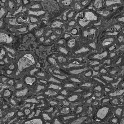 |
Focused ion beam-scanning electron microscopy links pathological myelin outfoldings to axonal changes in mice lacking Plp1 or Mag [940 micrographs in TIFF format] | Steyer AM, Möbius W [Pubmed: 36354016] [DOI: 10.1002/glia.24290] |
12.5 GB | — |
| 2021-04-23 | 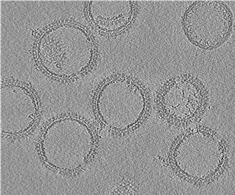 |
Cryo-electron tomography of HIV-1 GagdeltaMASP1T8I assemblies [5 tilt series in MRC format] | Ni T, Frosio T, Mendonça L, Sheng Y, Clare D, Himes BA, Zhang P [Pubmed: 35022621] [DOI: 10.1038/s41596-021-00648-5] |
12.8 GB | 4.5 Å |
| 2019-11-18 | 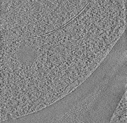 |
cryo-ET of cryo-FIB milled yeast cell in which scs2/22 ist2 are deleted [multiple data sets in MRC format] | Hoffmann PC, Bharat TAM, Wozny MR, Boulanger J, Miller EA, Kukulski W [Pubmed: 31743663] [DOI: 10.1016/j.devcel.2019.09.019] |
12.8 GB | — |
| 2016-06-28 |  |
Designer nanoscale DNA assemblies programmed from the top down [177 micrographs in MRC format] | Veneziano R, Ratanalert S, Zhang K, Zhang F, Yan H, Chiu W, Bathe M [Pubmed: 27229143] [DOI: 10.1126/science.aaf4388] |
13.0 GB | 20.0 Å |
| 2021-09-01 | 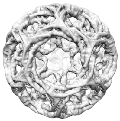 |
Multi-modal adaptor-clathrin contacts drive coated vesicle assembly [stack of 4782 particles in MRCS format] | Smith SM, Larocque G, Wood KM, Morris KL, Roseman AM, Sessions RB, Royle SJ, Smith CJ [Pubmed: 34487371] [DOI: 10.15252/embj.2021108795] |
13.0 GB | 9.1 - 15.0 Å |
| 2024-11-21 | 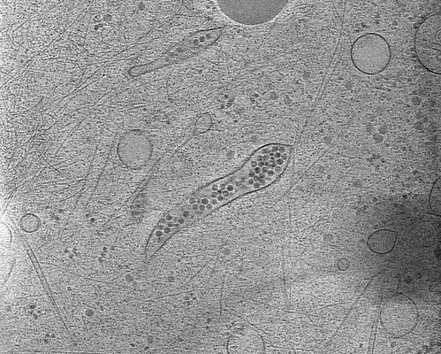 |
Cryo electron tomography of Echovirus 18-infected cos-7 cells [41 tilt series in MRC format] | Ishemgulova A, Mukhamedova L, Trebichalská Z, Füzik T [Pubmed: 30850609] [DOI: 10.1038/s41467-019-09132-x] |
13.1 GB | 3.16 Å |
| 2025-06-09 | 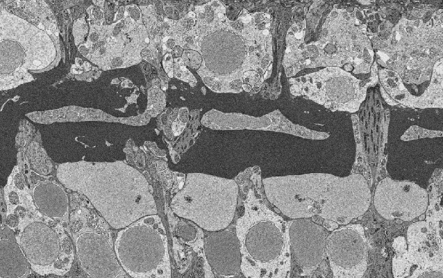 |
Horizontal cell connectivity in the anchovy retina - a 3D electron microscopic study -Scan 2 [677 micrographs in TIFF format] | Heß M [Pubmed: 40390032] [DOI: 10.1186/s12915-025-02242-7] |
13.5 GB | — |
| 2016-01-27 | 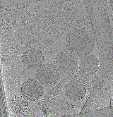 |
Cryo-electron tomogram of Chlamydia trachomatis with type III secretion system in contact with HeLa cell [1 class averages in MRC format] | Nans A, Kudryashev M, Saibil HR, Hayward RD [Pubmed: 26656452] [DOI: 10.1038/ncomms10114] |
13.6 GB | 38.0 Å |
| 2022-12-13 | 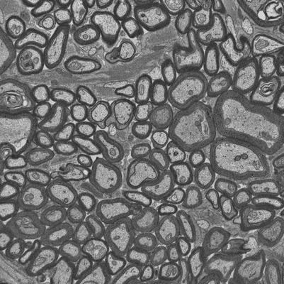 |
Focused ion beam-scanning electron microscopy links pathological myelin outfoldings to axonal changes in mice lacking Plp1 or Mag [910 micrographs in TIFF format] | Steyer AM, Möbius W [Pubmed: 36354016] [DOI: 10.1002/glia.24290] |
13.7 GB | — |
| 2018-04-27 | 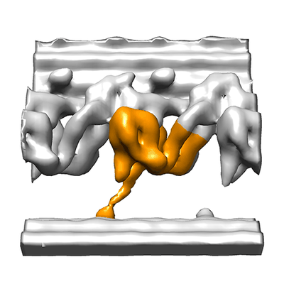 |
Cryo electron tomography of immotile sea urchin sperm flagella [27 tilt series in MRC format] | Lin J, Nicastro D [Pubmed: 29700238] [DOI: 10.1126/science.aar1968] |
13.8 GB | 33.0 - 41.0 Å |
| 2017-11-30 | 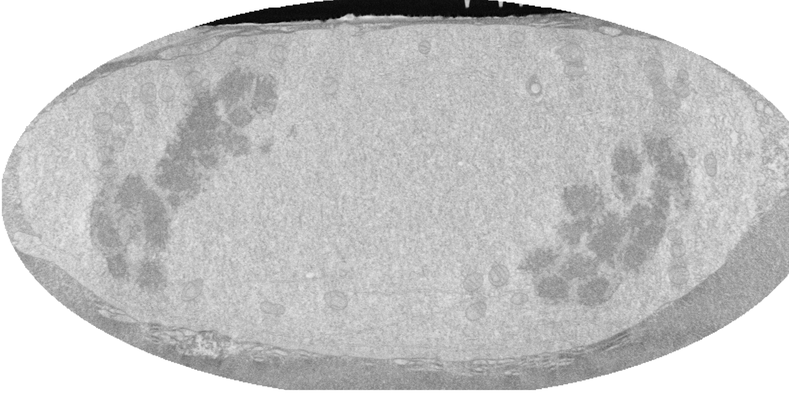 |
FIB-SEM of a dividing cell at 4.3 min after anaphase onset [1358 multi-frame micrographs composed of 1 frames each in TIFF format] | Otsuka S, Steyer AM, Schorb M, Hériché JK, Hossain MJ, Sethi S, Kueblbeck M, Schwab Y, Beck M, Ellenberg J [Pubmed: 29323269] [DOI: 10.1038/s41594-017-0001-9] |
14.0 GB | — |
| 2019-11-18 | 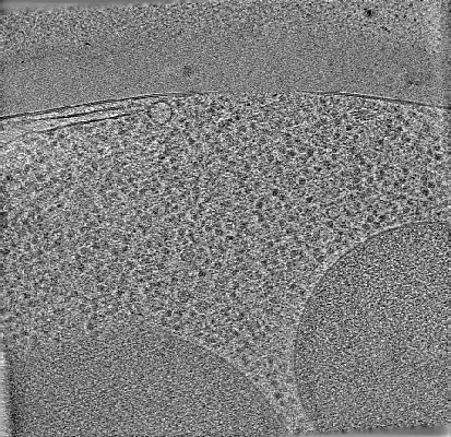 |
cryo-ET of cryo-FIB milled yeast cell in which scs2/22 ist2 are deleted with high intracellular calcium [121 tilt series in MRC format] | Hoffmann PC, Bharat TAM, Wozny MR, Boulanger J, Miller EA, Kukulski W [Pubmed: 31743663] [DOI: 10.1016/j.devcel.2019.09.019] |
14.3 GB | — |
| 2022-07-26 |  |
In situ cryo-electron tomography of P. chlororaphis infected by 201phi2-1 [269 multi-frame micrographs composed of 12 frames each in TIFF format] | Laughlin TG, Deep A, Prichard AM, Seitz C, Gu Y, Enustun E, Suslov S, Khanna K, Birkholz EA, Armbruster E, McCammon JA, Amaro RE, Pogliano J, Corbett KD, Villa E [Pubmed: 35922510] [DOI: 10.1038/s41586-022-05013-4] |
14.5 GB | 10.2 - 24.0 Å |
| 2023-07-18 |  |
Targeted volume Correlative Light and Electron Microscopy of environmental marine microorganisms [1937 micrographs in TIFF format] | Mocaer K, Mizzon G, Gunkel M, Halavatyi A, Steyer AM, Oorschot V, Schorb M, Le Kieffre C, Yee DP, Chevalier F, Gallet B, Decelle J, Schwab Y, Ronchi P [DOI: 10.1101/2023.01.27.525698] |
14.5 GB | — |
| 2021-11-01 |  |
Cryo Soft X-ray data for tetraspeck correlation [multiple data sets in MRC format] | Groen J, Pereiro E [Pubmed: 33990802] [DOI: 10.1038/s41596-021-00522-4] |
15.1 GB | — |
| 2024-11-21 |  |
Human platelets in presence of SARS-CoV-2 spike protein [6 reconstructed volumes in MRC format] | Basnet N, Bodakuntla S, Nichols S, Martinez-Sanchez A, Agostini L, Soh YM, Takagi J, Biertumpfel C, Mizuno N [Pubmed: 36739444] [DOI: 10.1038/s41467-023-36279-5] |
15.3 GB | — |
| 2020-12-11 | 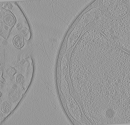 |
Cryo electron tomography after FIB-milling of Planctomycetes species Tuwongella immobilis (33k magnification) [multiple data sets in MRC format] | Seeger C, Andersson SG [Pubmed: 32761170] [DOI: 10.1093/gbe/evaa159] |
15.5 GB | — |
| 2020-08-06 | 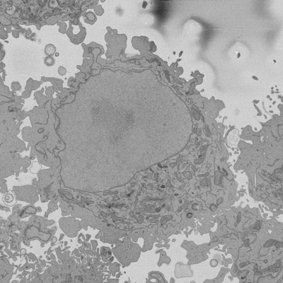 |
Cropped regions from Serial Block Face SEM of HeLa cell pellet with 10 nm pixels and 50 nm slices (benchmark dataset) [18 multi-frame micrographs composed of 300 frames each in TIFF format] | Peddie CP, Jones ML, Collinson LM | 15.6 GB | — |
| 2015-01-16 | 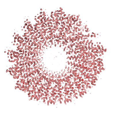 |
Tobacco Mosaic Virus K2 Summit dataset including manually boxed helix coordinates [14 multi-frame micrographs composed of 22 frames each in MRC format] | Fromm SA, Bharat TAM, Jakobi AJ, Hagen WJH, Sachse C [Pubmed: 25528571] [DOI: 10.1016/j.jsb.2014.12.002] |
15.7 GB | 4.0 Å |
| 2018-04-27 | 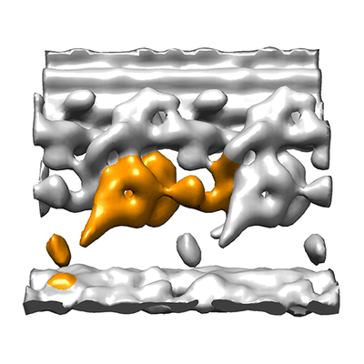 |
Cryo electron tomography of sea urchin sperm flagella [31 tilt series in MRC format] | Lin J, Nicastro D [Pubmed: 29700238] [DOI: 10.1126/science.aar1968] |
15.8 GB | 30.0 - 31.0 Å |
| 2022-01-12 | 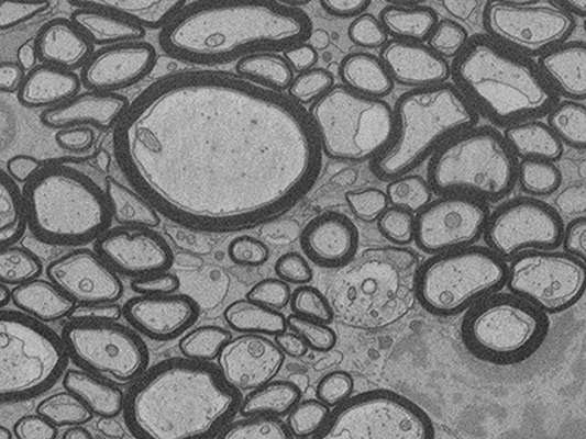 |
FIB-SEM of mouse optic nerve of an inducible conditional Mbp knock-out 16 weeks after induction [696 micrographs in TIFF format] | Meschkat M, Steyer AM, Ruhwedel T, Möbius W [Pubmed: 35246535] [DOI: 10.1038/s41467-022-28720-y] |
15.9 GB | — |
| 2020-02-18 | 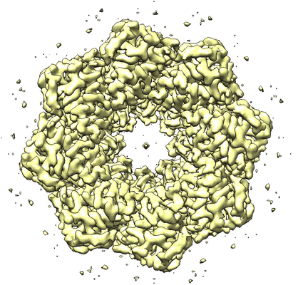 |
Structure of an undocked hemichannel of the N-terminal-deleted INX-6 in a nanodisc [300 micrographs in MRC format] | Burendei B, Shinozaki R, Watanabe M, Terada T, Tani K, Fujiyoshi Y, Oshima A [Pubmed: 32095518] [DOI: 10.1126/sciadv.aax3157] |
15.9 GB | 3.6 Å |
| 2023-06-02 | 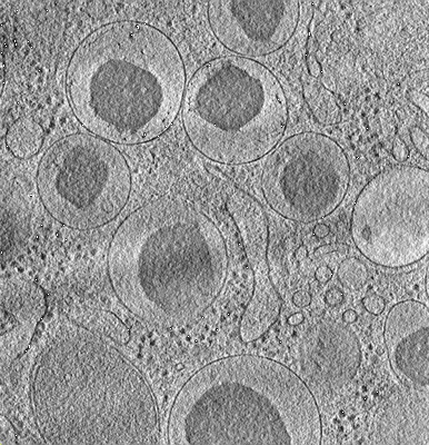 |
Cryo-ET tilt series from mouse islets lift-out sample [multiple data sets in TIFF format] | Wu Y, Qin C, Du W, Guo Z, Chen L, Guo Q [Pubmed: 37201639] [DOI: 10.1016/j.jsb.2023.107971] |
16.0 GB | — |
| 2021-01-22 | 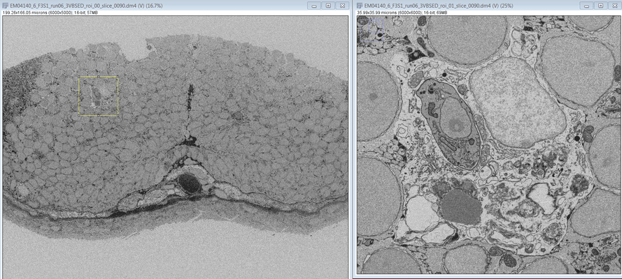 |
SBF SEM images of a Zebrafish hindbrain macrophage containing 2 Toxoplasma gondii tachizoites [multiple data sets in DM4 format] | Peddie CJ, Domart MC, Collinson L [Pubmed: 32461265] [DOI: 10.1242/dmm.043091] |
16.5 GB | — |
| 2020-12-21 |  |
CEM500K - A large-scale heterogeneous unlabeled cellular electron microscopy image dataset for deep learning. [496544 micrographs in TIFF format] | Conrad RW, Narayan K [DOI: 10.1101/2020.12.11.421792] |
16.6 GB | — |
