Electron Microscopy Public Image Archive
Electron Microscopy Public Image Archive
Our EMPIAR-PDBj will suspend the following operations from August 10, 2024 (Saturday) to August 18, 2024 (Sunday):
Web servers and data downloads will remain available during this period.
Additionally, please be aware that EMPIAR-EBI will also be closed for annotation operations from July 25, 2024 (Thursday) to August 18, 2024 (Sunday).
Thank you.
大阪大学的EMPIAR-PDBj团队为亚洲EM研究人员向EMPIAR传送大型EM图像数据提供服务。 除了通过互联网将数据直接传送到EBI(UK),研究人员还可以通过邮政或快递服务将数据硬盘发送到大阪大学,或者通过互联网传送到设置于大阪大学的服务器,然后由我们代为传送至数据登录网站。 如果您想使用此项服务,请先通过 电子邮件 与我们联系。
| Release date | Imageset | Title | Authors and references | Size | Resolution |
|---|---|---|---|---|---|
| 2023-05-22 | 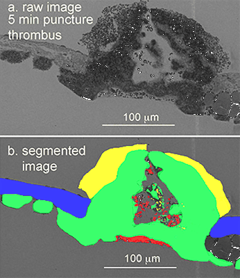 |
Venous puncture wound thrombi, 1, 5, 20 min post-puncture, 100 nm XY raw images, 20 nm XY pixels every 20 micons, 3 nm wide area TEM montages at selected depth [15 multi-frame micrographs composed of 1 frames each in DM4 format] | Storrie B, Leapman RD [Pubmed: 34531522] [DOI: 10.1038/s42003-021-02615-y] |
381.2 GB | — |
| 2022-10-07 |  |
Tilt series of SARS-CoV-2 spike-bearing virus-like particles (VLPs) interacting with hACE2-bearing extracellular vesicles (tEVs), showing various intermediate states of the SARS-CoV-2 spike protein [6 tilt series in MRC format] | Marcink TC, Porotto M, des Georges A, Moscona A [Pubmed: 35984891] [DOI: 10.1126/sciadv.abo3153] |
9.6 GB | — |
| 2020-11-18 |  |
SARS-CoV-2 infection in human adult lung alveolar stem cells [multiple data sets in TIFF format] | Youk J, Kim T, Evans KV, Jeong YI, Hur Y, Hong SP, Kim JH, Yi K, Kim SY, Na KJ, Bleazard T, Kim HM, Fellows M, Mahbubani KT, Saeb-Parsy K, Kim SY, Kim YT, Koh GY, Choi BS, Ju YS, Lee JH [Pubmed: 33142113] [DOI: 10.1016/j.stem.2020.10.004] |
20.8 GB | — |
| 2021-11-08 |  |
Monitoring reversion of hepatitis C virus-induced cellular alterations by Direct-Acting Antivirals using cryo Soft X-ray Tomography and Infrared Microscopy [7 reconstructed volumes in TIFF format] | Perez-Berna AJ, Benseny-Cases N, Rodríguez MJ, Valcarcel R, Carrascosa JL, Gastaminzab P, Pereiroa E [Pubmed: 34726165] [DOI: 10.1107/S2059798321009955] |
260.2 MB | — |
| 2022-01-14 |  |
High resolution 3D imaging of liver subcellular architecture and its link to metabolic function [multiple data sets in TIFF format] | Parlakgul G, Hotamisligil GS [Pubmed: 35264794] [DOI: 10.1038/s41586-022-04488-5] |
2.1 TB | — |
| 2024-06-04 |  |
Cryo-electron tomogram of mitochondria in cryo-FIB milled s-Opa1* mouse embryonic fibroblasts [284 multi-frame micrographs composed of 6 frames each in TIFF format] | Fry MY, Navarro PP, Ananda VY, Ge Y, McDonald JL, Hakim P, Luce BE, Lugo CM, Chao LH [Pubmed: 38225406] [DOI: 10.1038/s44318-024-00027-2] |
21.4 GB | — |
| 2024-06-04 |  |
Cryo-electron tomogram of mitochondria in cryo-FIB milled mouse embryonic fibroblasts with OPA1 deletion [278 multi-frame micrographs composed of 6 frames each in TIFF format] | Fry MY, Navarro PP, Ananda VY, Ge Y, McDonald JL, Hakim P, Luce BE, Lugo CM, Chao LH [Pubmed: 38225406] [DOI: 10.1038/s44318-024-00027-2] |
18.5 GB | — |
| 2024-06-04 |  |
Cryo-electron tomogram of mitochondria in cryo-FIB milled mouse embryonic fibroblasts with OPA1 overexpression [258 multi-frame micrographs composed of 4 frames each in TIFF format] | Fry MY, Navarro PP, Ananda VY, Ge Y, McDonald JL, Hakim P, Luce BE, Lugo CM, Chao LH [Pubmed: 38225406] [DOI: 10.1038/s44318-024-00027-2] |
8.8 GB | — |
| 2024-06-05 | 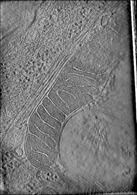 |
Cryo-electron tomogram of mitochondria in cryo-FIB milled l-Opa1* mouse embryonic fibroblasts [245 multi-frame micrographs composed of 6 frames each in TIFF format] | Fry MY, Navarro PP, Ananda VY, Ge Y, McDonald JL, Hakim P, Luce BE, Lugo CM, Chao LH [Pubmed: 38225406] [DOI: 10.1038/s44318-024-00027-2] |
20.2 GB | — |
| 2024-06-05 | 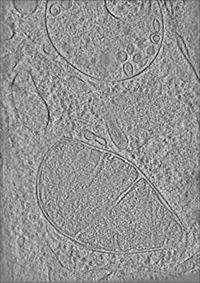 |
Cryo-electron tomogram of mitochondria in cryo-FIB milled mouse embryonic fibroblasts [228 multi-frame micrographs composed of 5 frames each in MRC format] | Fry MY, Navarro PP, Ananda VY, Ge Y, McDonald JL, Hakim P, Luce BE, Lugo CM, Chao LH [Pubmed: 38225406] [DOI: 10.1038/s44318-024-00027-2] |
96.1 GB | — |
| 2015-06-18 | 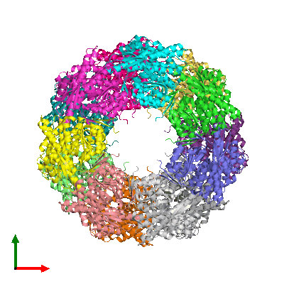 |
A simulated cryoEM data set of GroEL particles [stack of 10000 particles in MRC format] | Deng Y, Sun F | 1.5 GB | — |
| 2022-06-01 |  |
Crosshair, semi-automated targeting for electron microscopy with a motorised ultramicrotome [multiple data sets in TIFF and PNG formats] | Meechan K, Guan W, Riedinger A, Stankova V, Yoshimura A, Pipitone R, Milberger A, Schaar H, Romero-Brey I, Templin R, Peddie C J, Schieber N L, Jones M L, Collinson L, Schwab Y | 151.0 GB | — |
| 2022-12-19 | 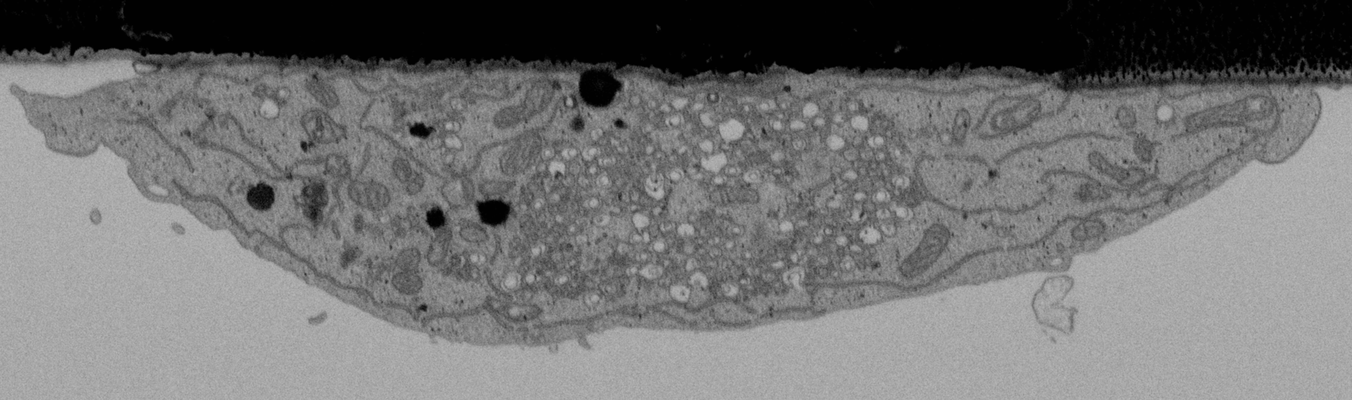 |
CLEMSite, a software for automated phenotypic screens using light microscopy and FIB-SEM. [multiple data sets in TIFF format] | Lleti JMSL, Steyer AMS, Schwab YS | 19.7 GB | — |
| 2024-02-09 | 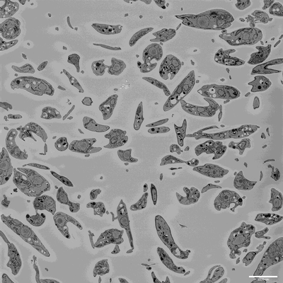 |
SBF-SEM imaging of Leishmania mexicana culture derived promastigotes [708 multi-frame micrographs composed of 1 frames each in MRC format] | Hair M [DOI: 10.1101/2023.11.28.568992] |
131.9 GB | — |
| 2021-11-01 | 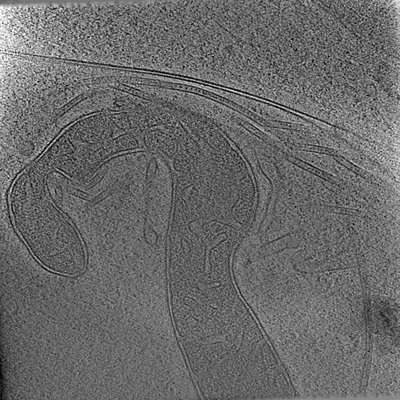 |
electron cryo-tomograms of axons from human cerebral organoids, expressing GFP-ESYT1 [multiple data sets in MRC and TIFF formats] | Hoffmann P, Giandomenico S, Ganeva I, Wozny M, Sutcliffe M, Lancaster M, Kukulski W [Pubmed: 34698018] [DOI: 10.7554/eLife.70269] |
40.9 GB | — |
| 2015-10-09 | 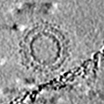 |
Cryo-electron tomography and subtomogram averaging of Rous-Sarcoma-Virus deltaMBD virus-like particles [1 class averages in MRC format] | Schur FKM, Dick RA, Hagen WJH, Vogt VM, Briggs JAG [Pubmed: 26223638] [DOI: 10.1128/JVI.01502-15] |
248.1 MB | — |
| 2020-02-24 |  |
Electron energy-filtered diffraction (eEFD) of catalase 3D crystal with CRYO ARM 300 [84 micrographs in MRC format] | Yonekura K, Ishikawa T, Maki-Yonekura S [Pubmed: 30928615] [DOI: 10.1016/j.jsb.2019.03.009] |
5.3 GB | — |
| 2021-11-01 |  |
electron cryo-tomograms of axons from human cerebral organoids, expressing L1CAM-GFP [multiple data sets in MRC and TIFF formats] | Hoffmann P, Giandomenico S, Ganeva I, Wozny M, Sutcliffe M, Lancaster M, Kukulski W [Pubmed: 34698018] [DOI: 10.7554/eLife.70269] |
30.5 GB | — |
| 2021-11-01 |  |
electron cryo-tomograms of axons from human cerebral organoids, expressing membrane-targeted GFP [multiple data sets in MRC and MRCS formats] | Hoffmann P, Giandomenico S, Ganeva I, Wozny M, Sutcliffe M, Lancaster M, Kukulski W [Pubmed: 34698018] [DOI: 10.7554/eLife.70269] |
181.4 GB | — |
| 2024-04-09 |  |
In situ cryo-ET dataset of Chlamydomonas reinhardtii prepared using cryo-plasmaFIB milling [multiple data sets in EER and MRC formats] | Kelley R, Khavnekar S, Zhang X, Obr M, Chakraborty S, Koh AF, Heebner J, Righetto R, Waltz F, McCafferty C, Van den Hoek H, Wietrzynski W, Van Der Stappen P, Michael A, Van Dorst S, Tagiltsev G, Beck F, Zhong E, Wan W, Briggs J, Plitzko J, Engel B, Kotecha A [Pubmed: 37613825] [DOI: 10.1093/micmic/ozad067.480] |
27.7 TB | — |
| 2023-02-01 | 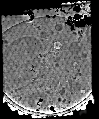 |
Cryo-EM data and 2DTM results of entire sections of differentiated ER-HoxB8 cells [multiple data sets in TIFF and MRC formats] | Elferich JE, Schiroli GS, Scadden DS, Grigorieff NG [Pubmed: 36382886] [DOI: 10.7554/elife.80980] |
1.3 TB | — |
| 2024-02-13 | 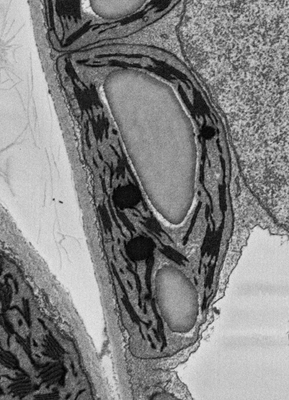 |
Plant SBF-SEM - Tobacco Leaf Chloroplast [130 micrographs in TIFF format] | Wickramanayake JS, Czymmek KJ [Pubmed: 37451777] [DOI: 10.1016/bs.mcb.2023.04.008] |
544.4 MB | — |
| 2021-04-30 | 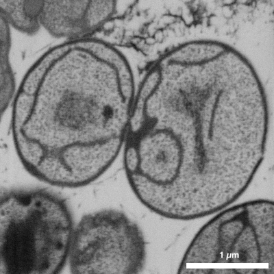 |
FIB-SEM of Gemmata obscuriglobus - a species of the Planctomycetes phylum [1 stitched maps in TIFF format] | Seeger C, Dyrhage K, Mahajan M, Odelgard A, Bergström Lind S, Andersson SGE [DOI: 10.3389/fmicb.2021.643045] |
1.6 GB | — |
| 2021-04-30 |  |
FIB-SEM of Tuwongella immobilis - a species of the Planctomycetes phylum [1 multi-frame micrographs composed of 464 frames each in TIFF format] | Andersson SGE, Odelgard A, Dyrhage K, Mahajan M, Seeger C [DOI: 10.3389/fmicb.2021.643045] |
6.7 GB | — |
| 2023-03-15 | 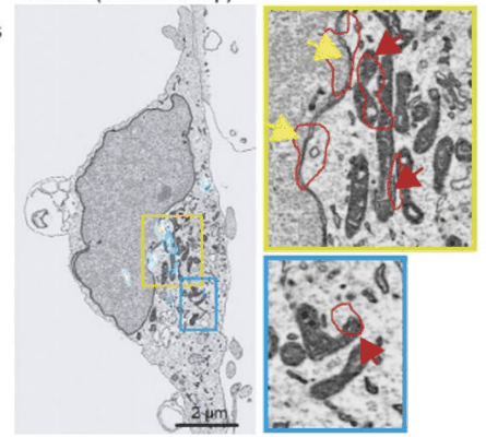 |
Performing Correlative Light and Electron Microscopy to reveal the structural organization and location of alpha-synuclein aggregation hotspots inside the neuron. [multiple data sets in DM4 and TIFF formats] | Choi ML, Chappard A, Singh BP, Maclachlan C, Rodrigues M, Fedotova E, Berezhnov AV, De S, Peddie C, Athauda D, Viridi GS, Zhang W, Evans JR, Wernick A, Zanjani ZS, Angelova PR, Esteras N, Vinikurov A, Morris K, Jeacock K, Tosatto L, Little D, Gissen P, Collinson L, Clarke DJ, Kunath T, Klenerman D, Abramov AY, Horrocks MH, Gandhi S [DOI: 10.1101/2022.06.07.494932] |
88.4 GB | — |
