Electron Microscopy Public Image Archive
Electron Microscopy Public Image Archive
大阪大学的EMPIAR-PDBj团队为亚洲EM研究人员向EMPIAR传送大型EM图像数据提供服务。 除了通过互联网将数据直接传送到EBI(UK),研究人员还可以通过邮政或快递服务将数据硬盘发送到大阪大学,或者通过互联网传送到设置于大阪大学的服务器,然后由我们代为传送至数据登录网站。 如果您想使用此项服务,请先通过 电子邮件 与我们联系。
| Release date | Imageset | Title | Authors and references | Size | Resolution |
|---|---|---|---|---|---|
| 2023-10-03 | 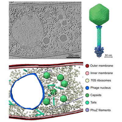 |
In situ cryo-electron tomography of E. amylovora cells infected by the jumbo bacteriophage RAY [multiple data sets in TIFF format] | Prichard A, Lee J, Laughlin TG, Lee A, Thomas KP, Sy A, Spencer T, Asavavimol A, Cafferata A, Cameron M, Chiu N, Davydov D, Desai I, Diaz G, Guereca M, Hearst K, Huang L, Jacobs E, Johnson A, Kahn S, Koch R, Martinez A, Norquist M, Pau T, Prasad G, Saam K, Sandhu M, Sarabria AJ, Schumaker S, Sonin S, Sonin A, Uyeno A, Zhao A, Corbett K, Pogliano K, Meyer J, Grose JH, Villa E, Dutton R, Pogliano J [Pubmed: 36865095] [DOI: 10.1101/2023.02.24.529968] |
244.3 GB | 8.9 - 38.0 Å |
| 2018-01-22 |  |
Cryo-EM structure of the TMEM16A in LMNG [multiple data sets in MRCS format] | Dang S, Cheng Y [Pubmed: 29236684] [DOI: 10.1038/nature25024] |
244.2 GB | 3.8 Å |
| 2024-11-19 |  |
Raw data used to generate the structure of WT ACTG2 F-actin [526 multi-frame micrographs composed of 36 frames each in TIFF format] | Ceron RH, Baez-Cruz FA, Palmer NJ, Carman PJ, Boczkowska M, Heuckeroth RO, Ostap EM, Dominguez R, Barrie KR [Pubmed: 38820162] [DOI: 10.1126/sciadv.adn6615] |
243.0 GB | 2.45 Å |
| 2025-01-24 |  |
Cryo-ET of Dictyostelium discoideum in distilled H2O (hypoosmotic stress) [59 tilt series in MRC format] | Hoffmann PC, Beck M [Pubmed: 39729993] [DOI: 10.1016/j.molcel.2024.11.038] |
241.1 GB | 37.0 Å |
| 2021-05-07 |  |
Affinity-purified VgaA-LC in complex with 70S ribosomes from Staphylococcus aureus [3348 multi-frame micrographs composed of 20 frames each in TIFF format] | Crowe-McAuliffe CT, Murina V, Hauryliuk V, Wilson DN [Pubmed: 34117249] [DOI: 10.1038/s41467-021-23753-1] |
241.0 GB | 3.1 Å |
| 2023-04-26 |  |
Cryo-electron tomography of ChAdOx spikes HexaPro mutant [68 tilt series in MRC format] | Ni T, Mendonca L, Zhu Y, Howe A, Radecke J, Sheng Y, Krebs AS, Shah P, Allen E, Spencer A, Morris S, Stuart D, Gilbert S, Zhang P | 240.3 GB | 9.0 - 10.6 Å |
| 2025-01-29 |  |
Cryo-ET tilt series of FIB-milled wild-type mouse neural progenitor cells [96 tilt series in MRC format] | Taniguchi R, Orniacki C, Kreysing JP, Zila V, Zimmerli CE, Böhm S, Turoňová B, Kräusslich H-G, Doye V, Beck M | 239.8 GB | 30.7 - 31.8 Å |
| 2024-11-06 |  |
Comparative 3D ultrastructure of Plasmodium falciparum gametocytes [multiple data sets in TIFF format] | Evers F, Roverts R, Boshoven C, Lindert MK, Sinden RE, Akiva A, Kooij TWA, Verhoef JMJ, Sommerdijk N [DOI: 10.1101/2023.03.10.531920] |
239.1 GB | — |
| 2020-09-25 |  |
A "drug sweeping" state of the TriABC triclosan efflux pump from Pseudomonas aeruginosa [multiple data sets in MRCS format] | Fabre L, Abigail LT, Ntreh AT, Amira A, Yazidi A, Inga IV, Weeks JW, Leus IV, Jon JW, Sudipta S, Ruickoldt J, Jakob J, Rouiller I, Zgurskaya HI, Isabelle I, Sygusch J, Helen HI, Jurgen J [Pubmed: 32966762] [DOI: 10.1016/j.str.2020.09.001] |
238.7 GB | 4.3 - 20.0 Å |
| 2018-07-06 |  |
Structure of the herpes-simplex virus portal-vertex [3818 micrographs in MRC format] | McElwee M, Vijayakrishnan S, Rixon FJ, Bhella D [Pubmed: 29924793] [DOI: 10.1371/journal.pbio.2006191] |
238.6 GB | 7.7 Å |
| 2018-02-07 |  |
Raw 2d tomographic tilt series of a dividing cell [65 tilt series in ST format] | Otsuka S [Pubmed: 29323269] [DOI: 10.1038/s41594-017-0001-9] |
237.8 GB | — |
| 2017-02-20 |  |
Cryo-EM structure of haemoglobin at 3.2 Å determined with the Volta phase plate [2261 multi-frame micrographs composed of 40 frames each in TIFF format] | Khoshouei M, Radjainia M, Baumeister W, Danev R [Pubmed: 28665412] [DOI: 10.1038/ncomms16099] |
237.1 GB | 3.2 Å |
| 2023-03-22 |  |
Electron cryo-tomography data on the ER-mitochondria encounter structure in cryo-FIB milled yeast cells [multiple data sets in MRC and TIFF formats] | Wozny MR, Di Luca A, Morado DR, Picco A, Khaddaj R, Campomanes P, Ivanovic L, Hoffmann PC, Miller EA, Vanni S, Kukulski W [Pubmed: 37165187] [DOI: 10.1038/s41586-023-06050-3] |
236.6 GB | — |
| 2017-12-18 |  |
CryoET of T20S proteasome single particle [multiple data sets in MRC format] | Noble AJ, Dandey VP, Wei H, Brasch J, Chase J, Acharya P, Tan YZ, Zhang Z, Kim LY, Scapin G, Rapp M, Eng ET, Rice WJ, Cheng A, Negro CJ, Shapiro L, Kwong PD, Jeruzalmi D, des Georges A, Potter CS, Carragher B [Pubmed: 29809143] [DOI: 10.7554/eLife.34257] |
235.3 GB | — |
| 2023-02-01 |  |
Cryo-EM data of alpha-synuclein A53T fibril [2663 micrographs in MRC format] | Wu KP, Huang JYC | 233.8 GB | 3.4 Å |
| 2024-09-26 |  |
SBF SEM images of a notoroid in longitudinal orientation containing TNE-ZsGreen+ notochord cells (Fig S26 part 2) [multiple data sets in DM4 and TIFF formats] | Marie-Charlotte Domart MCD, Lucy M Collinson LMC [DOI: 10.1038/s41586-024-08332-w] |
232.5 GB | — |
| 2023-07-10 |  |
Cryo electron tomography of Cytochalasin D-induced protrusions of Drosophila S2 alpha-tubulin acetyltransferase knock-out (dTAT KO) cells - Dataset 8 [multiple data sets in TIFF and MRC formats] | Ventura Santos C, Carter AP, Rogers SL [Pubmed: 37034688] [DOI: 10.1101/2023.03.31.535077] |
231.5 GB | — |
| 2021-03-19 |  |
GluK2/K5 apo [970 multi-frame micrographs composed of 40 frames each in TIFF format] | Khanra N, Meyerson J [Pubmed: 33724189] [DOI: 10.7554/eLife.66097] |
230.4 GB | 7.5 Å |
| 2020-10-09 |  |
Human delta protocadherin 1 full ectodomains on membranes, tomogram 2 [multiple data sets in TIFF, JPEG and MRC formats] | Harrison OJ, Brasch J, Katsamba PS, Ahlsen G, Noble AJ, Dan H, Sampogna R, Potter CS, Carragher B, Honig B, Shapiro L [Pubmed: 32101743] [DOI: 10.1016/j.celrep.2020.02.003] |
230.3 GB | — |
| 2023-06-23 |  |
Unaligned and aligned cryo-EM micrographs of 82-kDa malate synthase G [multiple data sets in TIFF format] | Wu K.-P. [Pubmed: 36997036] [DOI: 10.1016/j.jsb.2023.107958] |
227.3 GB | 2.89 - 4.14 Å |
| 2022-09-20 |  |
Cryo-EM dataset of Candida albicans CIII, inhibitor free [3634 micrographs in MRC format] | Di Trani J, Rubinstein JL [Pubmed: 34525326] [DOI: 10.1016/j.str.2021.08.006] |
227.1 GB | 3.0 Å |
| 2018-01-22 |  |
Cryo-EM structure of the TMEM16A in Nanodisc [stack of 3149 particles in MRCS format] | Dang S, Cheng Y [Pubmed: 29236684] [DOI: 10.1038/nature25024] |
226.7 GB | 3.8 Å |
| 2021-08-13 |  |
Human apo ferritin frozen on TEM grid with amorphous carbon supporting film [743 multi-frame micrographs composed of 32 frames each in TIFF format] | Huang X, Zhang L, Wen Z, Chen H, Li S, Ji G, Yin CC, Sun F [Pubmed: 32758492] [DOI: 10.1016/j.pbiomolbio.2020.07.009] |
226.5 GB | 2.6 Å |
| 2023-10-13 |  |
SBF-SEM micrographs of A. algerae microsporidia spores, 5 min germination [1215 micrographs in TIFF format] | Davydov A, Jaroenlak P, Ekiert D, Bhabha G [DOI: 10.7554/eLife.86638.1] |
226.3 GB | — |
| 2021-03-05 |  |
3.2 Å resolution structure of a functional monomeric Photosystem I from Thermosynechococcus elongatus BP-1 by single particle cryo-EM with a 200 kV CRYO ARM electron microscope [904 multi-frame micrographs composed of 60 frames each in TIFF format] | Çoruh O, Frank A, Tanaka H, Kawamoto A, El-Mohsnawy E, Kato T, Namba K, Gerle C, Nowaczyk MM, Kurisu G [Pubmed: 33686186] [DOI: 10.1038/s42003-021-01808-9] |
226.1 GB | 3.2 Å |
