Electron Microscopy Public Image Archive
Electron Microscopy Public Image Archive
오사카 대학의 EMPIAR-PDBj 팀은, 아시아의 EM 연구자가 용량이 큰 EM 이미지를 EMPIAR 데이터베이스에 전송하는 것을 돕고 있습니다. 인터넷을 통하여 EBI (UK)에 직>접 데이터를 전송하는 대신, 이용자는 우편이나 택배를 통하여 하드 디스크를 오사카 대학으로 보내실 수 있습니다. 혹은 인터넷을 이용하여 오사카 대학의 서버로 전>송 하실 수 있습니다. 오사카 대학에 데이터 전송 서비스를 희망하시는 분은 데이터를 보내시기 전에 먼저 이메일 통하여 등록하시고 싶은 EM데이터에 관하여 상담하십시오.
| Release date | Imageset | Title | Authors and references | Size | Resolution |
|---|---|---|---|---|---|
| 2022-09-26 |  |
In situ cryo-electron tomography of autophagic structures in S. cerevisiae [84 tilt series in MRC format] | Bieber A, Capitanio C, Erdmann PS, Schulman BA, Baumeister W, Wilfling F [Pubmed: 36122245] [DOI: 10.1073/pnas.2209823119] |
249.0 GB | — |
| 2020-07-24 |  |
Human delta protocadherin 1 full ectodomains on membranes, tomogram 1 [multiple data sets in MRC format] | Harrison OJ, Brasch J, Katsamba PS, Ahlsen G, Noble AJ, Dan H, Sampogna R, Potter CS, Carragher B, Honig B, Shapiro L [Pubmed: 32101743] [DOI: 10.1016/j.celrep.2020.02.003] |
247.7 GB | — |
| 2018-05-11 |  |
Rabbit muscle aldolase movies obtained using Talos Arctica operating at 200 kV equipped with a K2 [663 multi-frame micrographs composed of 44 frames each in MRC format] | Herzik Jr MA, Wu M, Lander GC [Pubmed: 28991891] [DOI: 10.1038/nmeth.4461] |
244.7 GB | 2.6 Å |
| 2023-10-03 |  |
In situ cryo-electron tomography of E. amylovora cells infected by the jumbo bacteriophage RAY [multiple data sets in TIFF format] | Prichard A, Lee J, Laughlin TG, Lee A, Thomas KP, Sy A, Spencer T, Asavavimol A, Cafferata A, Cameron M, Chiu N, Davydov D, Desai I, Diaz G, Guereca M, Hearst K, Huang L, Jacobs E, Johnson A, Kahn S, Koch R, Martinez A, Norquist M, Pau T, Prasad G, Saam K, Sandhu M, Sarabria AJ, Schumaker S, Sonin S, Sonin A, Uyeno A, Zhao A, Corbett K, Pogliano K, Meyer J, Grose JH, Villa E, Dutton R, Pogliano J [Pubmed: 36865095] [DOI: 10.1101/2023.02.24.529968] |
244.3 GB | 8.9 - 38.0 Å |
| 2018-01-22 | 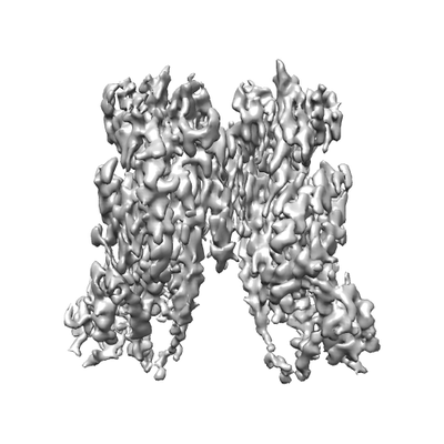 |
Cryo-EM structure of the TMEM16A in LMNG [multiple data sets in MRCS format] | Dang S, Cheng Y [Pubmed: 29236684] [DOI: 10.1038/nature25024] |
244.2 GB | 3.8 Å |
| 2021-05-07 | 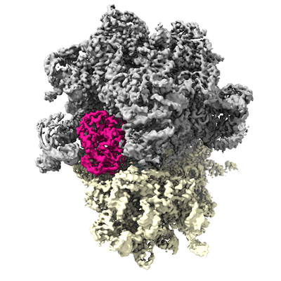 |
Affinity-purified VgaA-LC in complex with 70S ribosomes from Staphylococcus aureus [3348 multi-frame micrographs composed of 20 frames each in TIFF format] | Crowe-McAuliffe CT, Murina V, Hauryliuk V, Wilson DN [Pubmed: 34117249] [DOI: 10.1038/s41467-021-23753-1] |
241.0 GB | 3.1 Å |
| 2023-04-26 | 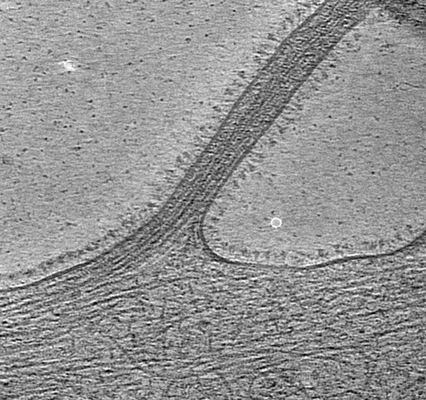 |
Cryo-electron tomography of ChAdOx spikes HexaPro mutant [68 tilt series in MRC format] | Ni T, Mendonca L, Zhu Y, Howe A, Radecke J, Sheng Y, Krebs AS, Shah P, Allen E, Spencer A, Morris S, Stuart D, Gilbert S, Zhang P | 240.3 GB | 9.0 - 10.6 Å |
| 2020-09-25 | 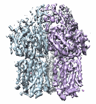 |
A "drug sweeping" state of the TriABC triclosan efflux pump from Pseudomonas aeruginosa [multiple data sets in MRCS format] | Fabre L, Abigail LT, Ntreh AT, Amira A, Yazidi A, Inga IV, Weeks JW, Leus IV, Jon JW, Sudipta S, Ruickoldt J, Jakob J, Rouiller I, Zgurskaya HI, Isabelle I, Sygusch J, Helen HI, Jurgen J [Pubmed: 32966762] [DOI: 10.1016/j.str.2020.09.001] |
238.7 GB | 4.3 - 20.0 Å |
| 2018-07-06 |  |
Structure of the herpes-simplex virus portal-vertex [3818 micrographs in MRC format] | McElwee M, Vijayakrishnan S, Rixon FJ, Bhella D [Pubmed: 29924793] [DOI: 10.1371/journal.pbio.2006191] |
238.6 GB | 7.7 Å |
| 2018-02-07 |  |
Raw 2d tomographic tilt series of a dividing cell [65 tilt series in ST format] | Otsuka S [Pubmed: 29323269] [DOI: 10.1038/s41594-017-0001-9] |
237.8 GB | — |
| 2017-02-20 |  |
Cryo-EM structure of haemoglobin at 3.2 Å determined with the Volta phase plate [2261 multi-frame micrographs composed of 40 frames each in TIFF format] | Khoshouei M, Radjainia M, Baumeister W, Danev R [Pubmed: 28665412] [DOI: 10.1038/ncomms16099] |
237.1 GB | 3.2 Å |
| 2023-03-22 |  |
Electron cryo-tomography data on the ER-mitochondria encounter structure in cryo-FIB milled yeast cells [multiple data sets in MRC and TIFF formats] | Wozny MR, Di Luca A, Morado DR, Picco A, Khaddaj R, Campomanes P, Ivanovic L, Hoffmann PC, Miller EA, Vanni S, Kukulski W [Pubmed: 37165187] [DOI: 10.1038/s41586-023-06050-3] |
236.6 GB | — |
| 2017-12-18 |  |
CryoET of T20S proteasome single particle [multiple data sets in MRC format] | Noble AJ, Dandey VP, Wei H, Brasch J, Chase J, Acharya P, Tan YZ, Zhang Z, Kim LY, Scapin G, Rapp M, Eng ET, Rice WJ, Cheng A, Negro CJ, Shapiro L, Kwong PD, Jeruzalmi D, des Georges A, Potter CS, Carragher B [Pubmed: 29809143] [DOI: 10.7554/eLife.34257] |
235.3 GB | — |
| 2023-02-01 |  |
Cryo-EM data of alpha-synuclein A53T fibril [2663 micrographs in MRC format] | Wu KP, Huang JYC | 233.8 GB | 3.4 Å |
| 2023-07-10 |  |
Cryo electron tomography of Cytochalasin D-induced protrusions of Drosophila S2 alpha-tubulin acetyltransferase knock-out (dTAT KO) cells - Dataset 8 [multiple data sets in TIFF and MRC formats] | Ventura Santos C, Carter AP, Rogers SL [Pubmed: 37034688] [DOI: 10.1101/2023.03.31.535077] |
231.5 GB | — |
| 2021-03-19 |  |
GluK2/K5 apo [970 multi-frame micrographs composed of 40 frames each in TIFF format] | Khanra N, Meyerson J [Pubmed: 33724189] [DOI: 10.7554/eLife.66097] |
230.4 GB | 7.5 Å |
| 2020-10-09 |  |
Human delta protocadherin 1 full ectodomains on membranes, tomogram 2 [multiple data sets in TIFF, JPEG and MRC formats] | Harrison OJ, Brasch J, Katsamba PS, Ahlsen G, Noble AJ, Dan H, Sampogna R, Potter CS, Carragher B, Honig B, Shapiro L [Pubmed: 32101743] [DOI: 10.1016/j.celrep.2020.02.003] |
230.3 GB | — |
| 2023-06-23 |  |
Unaligned and aligned cryo-EM micrographs of 82-kDa malate synthase G [multiple data sets in TIFF format] | Wu K.-P. [Pubmed: 36997036] [DOI: 10.1016/j.jsb.2023.107958] |
227.3 GB | 2.89 - 4.14 Å |
| 2022-09-20 |  |
Cryo-EM dataset of Candida albicans CIII, inhibitor free [3634 micrographs in MRC format] | Di Trani J, Rubinstein JL [Pubmed: 34525326] [DOI: 10.1016/j.str.2021.08.006] |
227.1 GB | 3.0 Å |
| 2018-01-22 |  |
Cryo-EM structure of the TMEM16A in Nanodisc [stack of 3149 particles in MRCS format] | Dang S, Cheng Y [Pubmed: 29236684] [DOI: 10.1038/nature25024] |
226.7 GB | 3.8 Å |
| 2021-08-13 | 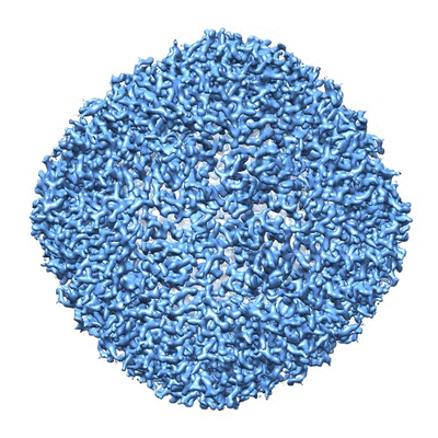 |
Human apo ferritin frozen on TEM grid with amorphous carbon supporting film [743 multi-frame micrographs composed of 32 frames each in TIFF format] | Huang X, Zhang L, Wen Z, Chen H, Li S, Ji G, Yin CC, Sun F [Pubmed: 32758492] [DOI: 10.1016/j.pbiomolbio.2020.07.009] |
226.5 GB | 2.6 Å |
| 2023-10-13 | 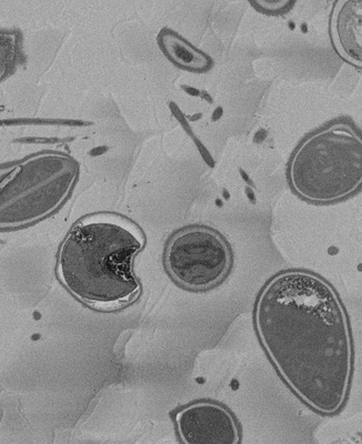 |
SBF-SEM micrographs of A. algerae microsporidia spores, 5 min germination [1215 micrographs in TIFF format] | Davydov A, Jaroenlak P, Ekiert D, Bhabha G [DOI: 10.7554/eLife.86638.1] |
226.3 GB | — |
| 2021-03-05 |  |
3.2 Å resolution structure of a functional monomeric Photosystem I from Thermosynechococcus elongatus BP-1 by single particle cryo-EM with a 200 kV CRYO ARM electron microscope [904 multi-frame micrographs composed of 60 frames each in TIFF format] | Coruh O, Frank A, Tanaka H, Kawamoto A, El-Mohsnawy E, Kato T, Namba K, Gerle C, Nowaczyk MM, Kurisu G [Pubmed: 33686186] [DOI: 10.1038/s42003-021-01808-9] |
226.1 GB | 3.2 Å |
| 2019-05-08 | 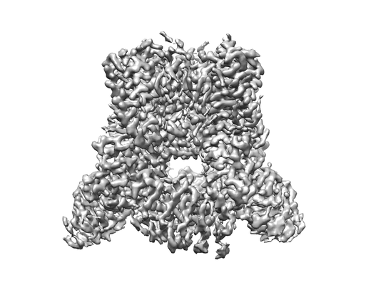 |
Cryo-EM structure of TRPV5 1-660 in nanodisc [stack of 2968 particles in MRCS format] | Dang S, van Goor MK, Asarnow D, Wang Y, Julius D, Cheng Y, van der Wijst J [Pubmed: 30975749] [DOI: 10.1073/pnas.1820323116] |
224.3 GB | 2.9 Å |
| 2022-05-20 |  |
Parallel cryo electron tomography (PACE-tomo) of 70S ribosomes (200 kV, side-entry holder) [multiple data sets in MRC format] | Eisenstein F, Danev R [Pubmed: 36456783] [DOI: 10.1038/s41592-022-01690-1] |
222.8 GB | 5.8 - 6.5 Å |
