Electron Microscopy Public Image Archive
Electron Microscopy Public Image Archive
오사카 대학의 EMPIAR-PDBj 팀은, 아시아의 EM 연구자가 용량이 큰 EM 이미지를 EMPIAR 데이터베이스에 전송하는 것을 돕고 있습니다. 인터넷을 통하여 EBI (UK)에 직>접 데이터를 전송하는 대신, 이용자는 우편이나 택배를 통하여 하드 디스크를 오사카 대학으로 보내실 수 있습니다. 혹은 인터넷을 이용하여 오사카 대학의 서버로 전>송 하실 수 있습니다. 오사카 대학에 데이터 전송 서비스를 희망하시는 분은 데이터를 보내시기 전에 먼저 이메일 통하여 등록하시고 싶은 EM데이터에 관하여 상담하십시오.
| Release date | Imageset | Title | Authors and references | Size | Resolution |
|---|---|---|---|---|---|
| 2021-03-12 | 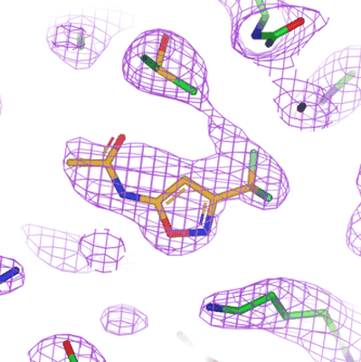 |
PKM2 in complex with Compound 6 [590 multi-frame micrographs composed of 75 frames each in TIFF format] | Saur M, Hartshorn MJ, Dong J, Reeks J, Bunkoczi G, Jhoti H, Williams PA [Pubmed: 31877353] [DOI: 10.1016/j.drudis.2019.12.006] |
768.0 GB | 2.5 Å |
| 2021-03-12 | 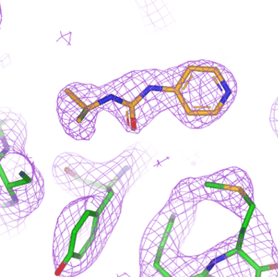 |
PKM2 in complex with Compound 10 [572 multi-frame micrographs composed of 75 frames each in TIFF format] | Saur M, Hartshorn MJ, Dong J, Reeks J, Bunkoczi G, Jhoti H, Williams PA [Pubmed: 31877353] [DOI: 10.1016/j.drudis.2019.12.006] |
745.1 GB | 2.7 Å |
| 2021-03-12 | 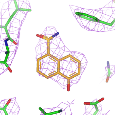 |
PKM2 in complex with Compound 5 [707 multi-frame micrographs composed of 60 frames each in TIFF format] | Saur M, Hartshorn MJ, Dong J, Reeks J, Bunkoczi G, Jhoti H, Williams PA [Pubmed: 31877353] [DOI: 10.1016/j.drudis.2019.12.006] |
773.8 GB | 3.2 Å |
| 2021-03-12 | 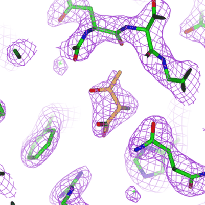 |
PKM2 in complex with L-threonine [476 multi-frame micrographs composed of 75 frames each in TIFF format] | Saur M, Hartshorn MJ, Dong J, Reeks J, Bunkoczi G, Jhoti H, Williams PA [Pubmed: 31877353] [DOI: 10.1016/j.drudis.2019.12.006] |
620.8 GB | 2.6 Å |
| 2021-03-12 | 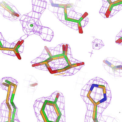 |
Beta-galactosidase in complex with L-ribose [517 multi-frame micrographs composed of 75 frames each in TIFF format] | Saur M, Hartshorn MJ, Dong J, Reeks J, Bunkoczi G, Jhoti H, Williams PA [Pubmed: 31877353] [DOI: 10.1016/j.drudis.2019.12.006] |
669.1 GB | 2.3 Å |
| 2021-03-12 | 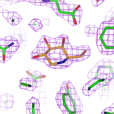 |
Beta-galactosidase in complex with deoxygalacto-nojirimycin [598 multi-frame micrographs composed of 75 frames each in TIFF format] | Saur M, Hartshorn MJ, Dong J, Reeks J, Bunkoczi G, Jhoti H, Williams PA [Pubmed: 31877353] [DOI: 10.1016/j.drudis.2019.12.006] |
765.8 GB | 2.3 Å |
| 2021-03-12 | 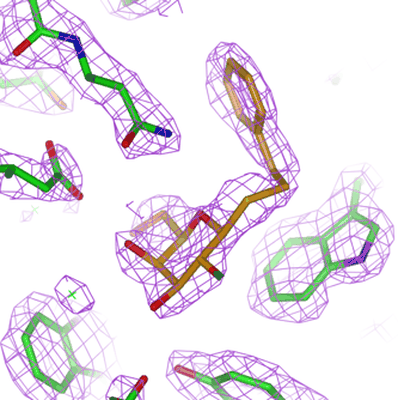 |
Beta-galactosidase in complex with PETG [562 multi-frame micrographs composed of 75 frames each in TIFF format] | Saur M, Hartshorn MJ, Dong J, Reeks J, Bunkoczi G, Jhoti H, Williams PA [Pubmed: 31877353] [DOI: 10.1016/j.drudis.2019.12.006] |
720.1 GB | 2.2 Å |
| 2021-04-23 | 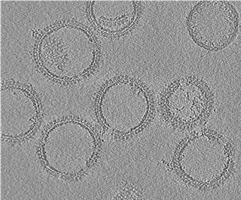 |
Cryo-electron tomography of HIV-1 GagdeltaMASP1T8I assemblies [5 tilt series in MRC format] | Ni T, Frosio T, Mendonça L, Sheng Y, Clare D, Himes BA, Zhang P [Pubmed: 35022621] [DOI: 10.1038/s41596-021-00648-5] |
25.6 GB | 4.5 Å |
| 2021-06-11 | 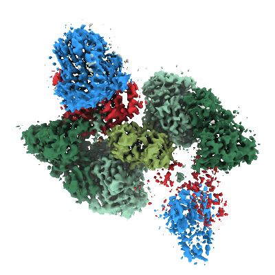 |
Cas6-reverse transcriptase-Cas1—Cas2 CRISPR integrase complex [3330 multi-frame micrographs composed of 50 frames each in TIFF format] | Hoel CM, Wang JY, Doudna JA, Brohawn SG [Pubmed: 33958590] [DOI: 10.1038/s41467-021-22900-y] |
2.2 TB | 3.4 - 3.9 Å |
| 2021-03-19 | 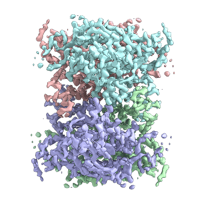 |
1.93 A cryo-EM structure of streptavidin [2277 multi-frame micrographs composed of 70 frames each in TIFF format] | Hiraizumi M, Yamashita K, Nisihzawa T, Kikkawa M, Nureki O | 373.1 GB | 1.93 Å |
| 2021-03-05 | 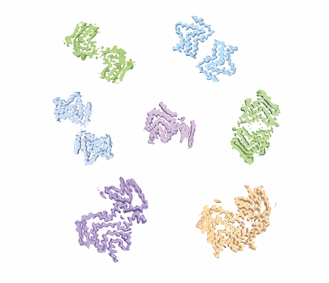 |
Cryo-EM of Multiple System Atrophy seeded assembly of alpha-synuclein filaments [multiple data sets in TIFF and MRCS formats] | Lövestam S, Goedert M, Scheres S [Pubmed: 33548114] [DOI: 10.1002/2211-5463.13110] |
1.1 TB | 3.18 - 4.23 Å |
| 2021-06-18 | 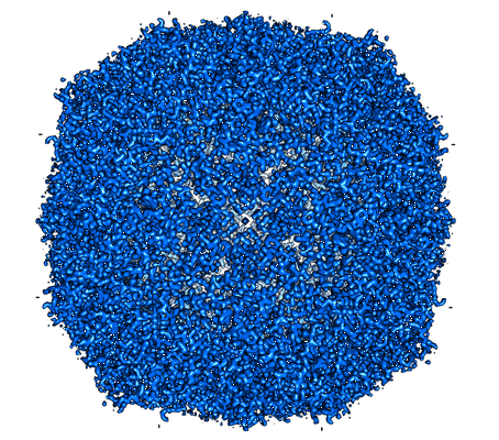 |
Single particle cryo-EM dataset of mouse heavy chain apoferritin collected on cryoARM300 with beam-image shift of 7 um [3125 multi-frame micrographs composed of 59 frames each in TIFF format] | Efremov R, Stroobants A [Pubmed: 33950012] [DOI: 10.1107/S2059798321002151] |
695.6 GB | 1.7 Å |
| 2021-05-07 | 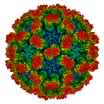 |
The cryo-EM structure of vesivirus 2117 highlights functional variations in entry pathways for viruses in different clades of the vesivirus genus. [2000 multi-frame micrographs composed of 50 frames each in MRC format] | Sutherland H, Conley MJ, Emmott E, Streetley J, Goodfellow IG, Bhella D [Pubmed: 33853966] [DOI: 10.1128/JVI.00282-21] |
24.4 TB | 3.65 Å |
| 2021-04-06 | 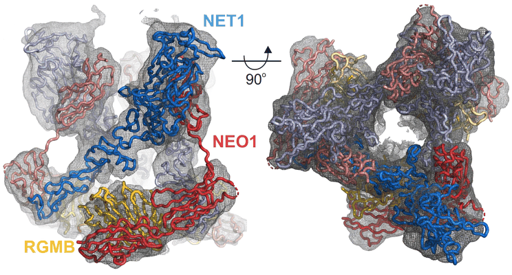 |
Cryo-EM structure of the ternary Netrin 1-Neogenin 1-Repulsive Guidance Molecule B complex [1635 multi-frame micrographs composed of 40 frames each in TIFF format] | Robinson RA, Griffiths SC, van de Haar LL, Malinauskas T, van Battum EY, Zelina P, Schwab RA, Karia D, Malinauskaite L, Brignani S, van den Munkhof M, Dudukcu O, De Ruiter AA, Van den Heuvel DMA, Bishop B, Elegheert J, Aricescu AR, Pasterkamp RJ, Siebold C [Pubmed: 33740419] [DOI: 10.1016/j.cell.2021.02.045] |
1.2 TB | 5.98 Å |
| 2021-09-10 | 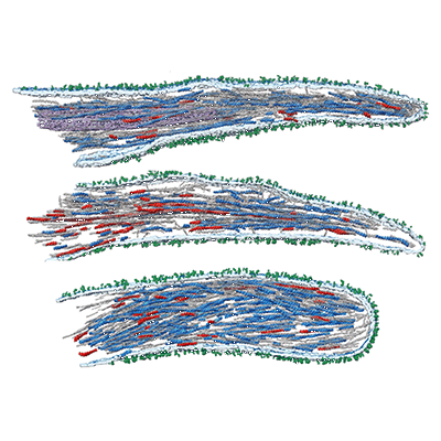 |
Structural analysis of receptors and actin polarity in platelet protrusions [3 reconstructed volumes in EM format] | Sorrentino S, Conesa JJ, Cuervo A, Melero R, Martins B, Fernandez-Gimenez E, de Isidro-Gomez FP, de la Morena J, Studt JD, Sorzano COS, Eibauer M, Carazo JM, Medalia O [Pubmed: 34504018] [DOI: 10.1073/pnas.2105004118] |
6.0 GB | 26.6 Å |
| 2021-02-19 | 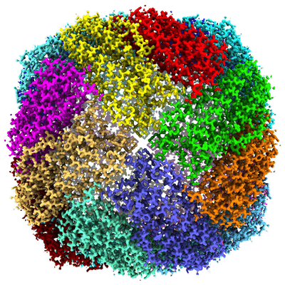 |
Apoferritin structure at 1.36 angstrom resolution determined from a 300 kV Titan Krios G3i electron microscope with Falcon4 detector [7734 multi-frame micrographs composed of 40 frames each in MRC format] | Zhang K, Pintilie GD, Li S, Schmid MF, Chiu W [Pubmed: 33139928] [DOI: 10.1038/s41422-020-00432-2] |
3.7 TB | 1.36 Å |
| 2021-02-19 |  |
CryoEM structure of the LCD of hnRNPA2 amyloid-like fibrils [3937 multi-frame micrographs composed of 2 frames each in MRC format] | Lu J, Cao Q, Hughes MP, Sawaya MR, Boyer DR, Cascio D, Eisenberg DS [Pubmed: 32796831] [DOI: 10.1038/s41467-020-17905-y] |
1.2 TB | 3.1 Å |
| 2021-04-14 |  |
Cryo-electron tomography of the metazoan membrane-assembled retromer:SNX3 coat containing Wls cargo motif [multiple data sets in TIFF and MRC formats] | Leneva N, Kovtun O, Morado DR, Briggs JAG, Owen DJ [Pubmed: 33762348] [DOI: 10.1126/sciadv.abf8598] |
764.9 GB | 8.9 - 9.5 Å |
| 2021-03-03 |  |
Cryo electron microscopy of alpha-synuclein H50Q fibrils [3577 multi-frame micrographs composed of 30 frames each in MRC format] | Boyer DR, Li B, Sun C, Fan W, Sawaya MR, Jiang L, Eisenberg DS [Pubmed: 31695184] [DOI: 10.1038/s41594-019-0322-y] |
595.3 GB | 3.3 Å |
| 2021-07-09 |  |
Cryo-electron tomography of the fungal membrane-assembled retromer:Grd19 coat containing Kex2 cargo motif [multiple data sets in TIFF and MRC formats] | Leneva N, Kovtun O, Morado DR, Briggs JAG, Owen DJ [Pubmed: 33762348] [DOI: 10.1126/sciadv.abf8598] |
347.7 GB | 9.2 - 9.5 Å |
| 2021-11-23 |  |
CryoEM structure of disease related M854K MDA5-dsRNA filament in complex with ATP [7176 multi-frame micrographs composed of 40 frames each in TIFF format] | Yu Q, Herrero Del Valle A, Singh R, Modis Y [Pubmed: 34795277] [DOI: 10.1038/s41467-021-27062-5] |
1.2 TB | 2.8 Å |
| 2021-02-10 |  |
Cryo-EM structure of K+-bound hERG channel in the presence of astemizole [1865 multi-frame micrographs composed of 50 frames each in TIFF format] | Asai T, Adachi N, Moriya T, Kawasaki M, Suzuki K, Senda T, Murata T [Pubmed: 33450182] [DOI: 10.1016/j.str.2020.12.007] |
1.8 TB | 3.7 Å |
| 2021-02-10 |  |
Cryo-EM structure of K+-bound hERG channel [1496 multi-frame micrographs composed of 50 frames each in TIFF format] | Asai T, Adachi N, Moriya T, Kawasaki M, Suzuki K, Senda T, Murata T [Pubmed: 33450182] [DOI: 10.1016/j.str.2020.12.007] |
1.4 TB | 3.9 Å |
| 2021-11-16 |  |
Poly-alanine backbone model of E. coli BcsA bound to BcsB [multiple data sets in TIFF format] | Acheson JF, Ho R, Goularte NF, Cegelski L, Zimmer J [Pubmed: 33712813] [DOI: 10.1038/s41594-021-00569-7] |
3.2 TB | 3.4 - 4.2 Å |
| 2021-11-16 |  |
Cryo EM Structure of the E. coli BcsB Hexamer [multiple data sets in TIFF format] | Acheson JF, Ho R, Goularte NF, Cegelski L, Zimmer J [Pubmed: 33712813] [DOI: 10.1038/s41594-021-00569-7] |
837.2 GB | 3.4 Å |
