Electron Microscopy Public Image Archive
Electron Microscopy Public Image Archive
The EMPIAR-PDBj team at Osaka University assists Asian EM researchers with the transfer of big EM image data to EMPIAR. Instead of sending the data directly to the EBI (UK) via the internet, hard drives can also be sent to Osaka University by postal mail or via a courier service. As an alternative, internet transfer to our server in Osaka is also available. If you would like to take advantage of our submission services, please contact us first by e-mail before sending the data to us.
| Release date | Imageset | Title | Authors and references | Size | Resolution |
|---|---|---|---|---|---|
| 2024-11-30 | 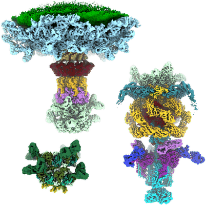 |
Cryo-electron microscopy of mycobacteriophage BaDAS-1 particles [5571 multi-frame micrographs composed of 30 frames each in TIFF format] | Freeman KF, Podgorski J, White SJ, Hatfull GF | 1.4 TB | 2.67 - 4.0 Å |
| 2022-09-09 | 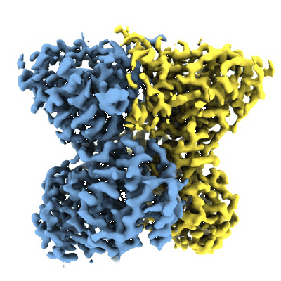 |
Cryo-electron microscopy of human exostosin-like 3 (EXTL3) in the presence of UDP [2573 multi-frame micrographs composed of 1 frames each in MRC format] | Wilson LFL, Dendooven T, Hardwick SW, Echevarría-Poza A, Tryfona T, Krogh KBRM, Chirgadze DY, Luisi BF, Logan DT, Mani K, Dupree P [Pubmed: 35676258] [DOI: 10.1038/s41467-022-31048-2] |
669.8 GB | 2.93 Å |
| 2022-09-09 | 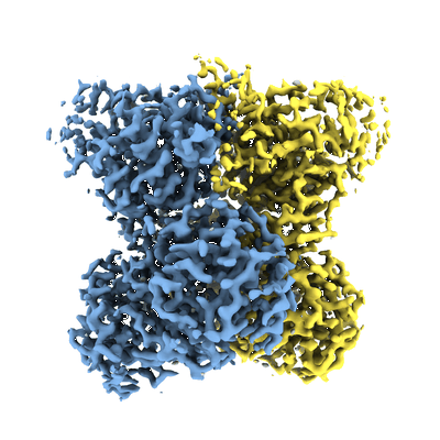 |
Cryo-electron microscopy of human exostosin-like 3 (EXTL3) [1626 multi-frame micrographs composed of 70 frames each in MRC format] | Wilson LFL, Dendooven T, Hardwick SW, Echevarría-Poza A, Tryfona T, Krogh KBRM, Chirgadze DY, Luisi BF, Logan DT, Mani K, Dupree P [Pubmed: 35676258] [DOI: 10.1038/s41467-022-31048-2] |
3.5 TB | 2.43 Å |
| 2022-11-16 | 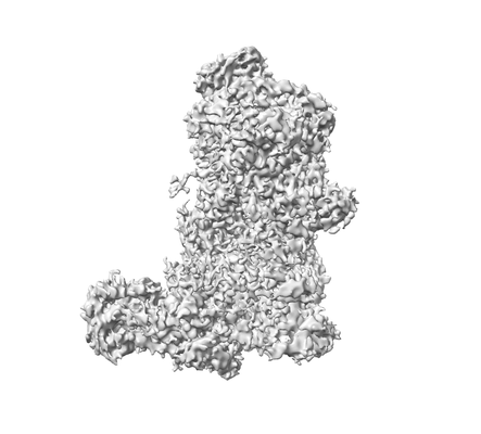 |
Cryo-electron microscopy of Dicer-1 and Its Partner Protein Loqs-PB complex with model pre-miRNA in presence of Mg2+ ions [8128 multi-frame micrographs composed of 38 frames each in TIFF format] | Jouravleva K, Golovenko D, Demo G, Dutcher RC, Hall TMT, Zamore PD, Korostelev AA [Pubmed: 36182693] [DOI: 10.1016/j.molcel.2022.09.002] |
2.9 TB | 3.26 - 3.73 Å |
| 2022-11-14 | 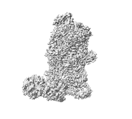 |
Cryo-electron microscopy of Dicer-1 and Its Partner Protein Loqs-PB complex with model pre-miRNA in presence of Ca2+ ions [3057 multi-frame micrographs composed of 40 frames each in TIFF format] | Jouravleva K, Golovenko D, Demo G, Dutcher RC, Hall TMT, Zamore PD, Korostelev AA [Pubmed: 36182693] [DOI: 10.1016/j.molcel.2022.09.002] |
1.4 TB | 3.06 Å |
| 2022-11-16 |  |
Cryo-electron microscopy of Dicer-1 and Its Partner Protein Loqs-PB complex [2849 multi-frame micrographs composed of 30 frames each in TIFF format] | Jouravleva K, Golovenko D, Demo G, Dutcher RC, Hall TMT, Zamore PD, Korostelev AA [Pubmed: 36182693] [DOI: 10.1016/j.molcel.2022.09.002] |
1.1 TB | 3.94 - 4.02 Å |
| 2024-11-08 | 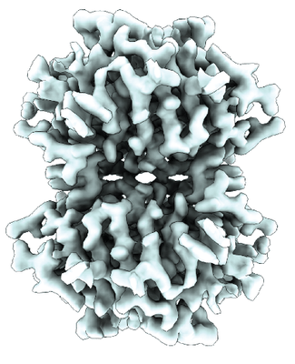 |
Cryo-electron microscopy of Avidin [14206 multi-frame micrographs composed of 623 frames each in EER format] | Nazarov S, Beckert Dominique B, Myasnikow A | 3.4 TB | 2.08 Å |
| 2025-06-19 | 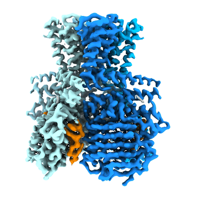 |
Cryo-electron microscopy micrographs of Hailong HalA in complex with oligodeoxyadenylate [10951 multi-frame micrographs composed of 55 frames each in TIFF format] | Tan JMJ, Cofsky JC, Kruse AC, Kranzusch PJ [Pubmed: 40306316] [DOI: 10.1038/s41586-025-09058-z] |
1.9 TB | 1.98 Å |
| 2018-10-23 | 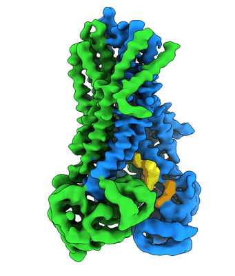 |
Cryo-electron microscopy data of thermostabilized avian CFTR [stack of 2 particles in MRCS format] | Fay JF [Pubmed: 30281975] [DOI: 10.1021/acs.biochem.8b00763] |
19.3 GB | 4.3 - 6.6 Å |
| 2024-03-20 | 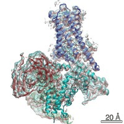 |
Cryo-electron microscopy Structure of the Human Cannabinoid Receptor CB2-Gi Signaling Complex [multiple data sets in TIFF format] | Xing C, Zhuang Y, Xu TH, Feng Z, Zhou XE, Chen M, Wang L, Meng X, Xue Y, Wang J, Liu H, McGuire TF, Zhao G, Melcher K, Zhang C, Xu HE, Xie XQ [Pubmed: 32004460] [DOI: 10.1016/j.cell.2020.01.007] |
2.5 TB | 3.2 Å |
| 2023-06-30 | 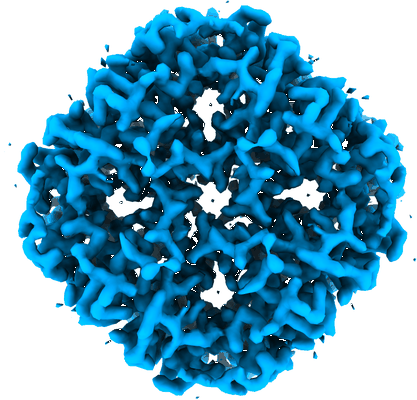 |
Cryo-electron micrographs of hAQP2 in DDM [6592 multi-frame micrographs composed of 40 frames each in TIFF format] | Kamegawa A, Suzuki H, Fujiyoshi Y [Pubmed: 37315821] [DOI: 10.1016/j.jsb.2023.107984] |
1.2 TB | 2.89 Å |
| 2024-07-16 | 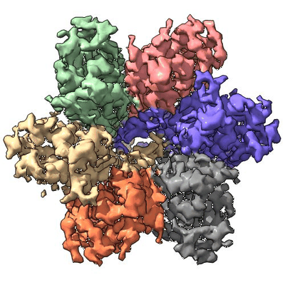 |
Cryo-electron micrographs of Proteorhodopsin A18L [27712 multi-frame micrographs composed of 40 frames each in TIFF format] | Hirschi S, Lemmin T, Fotiadis D | 3.8 TB | 2.82 - 3.54 Å |
| 2023-06-07 | 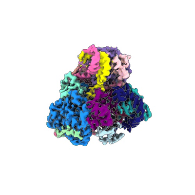 |
Cryo-electron micrographs of Hantaan virus polymerase in pre-initiation state [3261 multi-frame micrographs composed of 50 frames each in TIFF format] | Durieux Trouilleton Q, Arragain B, Malet H [Pubmed: 37221161] [DOI: 10.1038/s41467-023-38555-w] |
776.9 GB | 3.36 Å |
| 2023-06-07 |  |
Cryo-electron micrographs of Hantaan virus polymerase in its apo state [2865 multi-frame micrographs composed of 50 frames each in TIFF format] | Durieux Trouilleton Q, Arragain B, Malet H [Pubmed: 37221161] [DOI: 10.1038/s41467-023-38555-w] |
660.7 GB | 3.27 Å |
| 2023-06-07 |  |
Cryo-electron micrographs of Hantaan virus polymerase in elongation state [3785 multi-frame micrographs composed of 50 frames each in TIFF format] | Durieux Trouilleton Q, Arragain B, Malet H [Pubmed: 37221161] [DOI: 10.1038/s41467-023-38555-w] |
927.5 GB | 3.14 Å |
| 2023-06-07 |  |
Cryo-electron micrographs of Hantaan virus polymerase bound to its 5' viral RNA [2547 multi-frame micrographs composed of 50 frames each in TIFF format] | Durieux Trouilleton Q, Arragain B, Malet H [Pubmed: 37221161] [DOI: 10.1038/s41467-023-38555-w] |
608.2 GB | 3.23 Å |
| 2025-03-10 |  |
Cryo-electron micrographs of Cholinephosphotransferase in complex with diacylglycerol [multiple data sets in TIFF and MRC formats] | Roberts JR [Pubmed: 39747155] [DOI: 10.1038/s41467-024-55673-1] |
1.2 TB | 3.2 Å |
| 2022-01-12 |  |
Cryo-FIB-SEM volume in a Sum159 human cell line [20 micrographs in TIFF format] | Klumpe S, Fung HKH, Goetz SK, Zagoriy I, Hampoelz B, Zhang X, Erdmann PS, Baumbach J, Müller CW, Beck M, Plitzko JM, Mahamid J [Pubmed: 34951584] [DOI: 10.7554/elife.70506] |
120.1 MB | — |
| 2022-01-11 |  |
Cryo-FIB-SEM data on Chlamydomonas reinhardtii cells [37 micrographs in TIFF format] | Klumpe S [Pubmed: 34951584] [DOI: 10.7554/elife.70506] |
888.2 MB | — |
| 2023-05-16 |  |
Cryo-Electron microscopy of the antimicrobial peptide Drosocin from Drosophila melanogaster bound to the E.coli 70S ribosome. [8861 multi-frame micrographs composed of 1532 frames each in EER format] | Koller TO, Morici M, Berger M, Safdari HA, Beckert B, Wilson DN [Pubmed: 36997646] [DOI: 10.1038/s41589-023-01293-7] |
7.9 TB | 2.1 - 2.8 Å |
| 2024-04-23 |  |
Cryo-Electron Tomography of Zebra Fish Muscle Tissue Biopsy [1 tilt series in MRC format] | Gervinskas G, Trepout S, Venugopal H, Ramm G | 37.6 GB | — |
| 2024-04-23 |  |
Cryo-Electron Tomography of Mouse Kidney Tissue Biopsy [1 tilt series in MRC format] | Gervinskas G, Trepout S, Venugopal H, Ramm G | 31.8 GB | — |
| 2024-04-23 |  |
Cryo-Electron Tomography of Mouse Brain Tissue Biopsy [1 tilt series in MRC format] | Gervinskas G, Trepout S, Venugopal H, Ramm G | 38.5 GB | — |
| 2024-04-23 |  |
Cryo-Electron Tomography of Golgi Apparatus from a Zebra Fish Embryo [1 tilt series in MRC format] | Gervinskas G, Trepout S, Venugopal H, Ramm G | 27.6 GB | — |
| 2025-06-30 |  |
Cryo-Electron Tomography Data and Comprehensive Annotation of C. elegans Centrosomes in Interphase and Mitosis [multiple data sets in MRC and TIFF formats] | Tollervey F [Pubmed: 39721584] [DOI: 10.1016/j.devcel.2024.12.002] |
111.4 GB | 26.0 - 44.0 Å |
