Electron Microscopy Public Image Archive
Electron Microscopy Public Image Archive
The EMPIAR-PDBj team at Osaka University assists Asian EM researchers with the transfer of big EM image data to EMPIAR. Instead of sending the data directly to the EBI (UK) via the internet, hard drives can also be sent to Osaka University by postal mail or via a courier service. As an alternative, internet transfer to our server in Osaka is also available. If you would like to take advantage of our submission services, please contact us first by e-mail before sending the data to us.
| Release date | Imageset | Title | Authors and references | Size | Resolution |
|---|---|---|---|---|---|
| 2018-08-09 |  |
Three-dimensional nanostructure of an intact microglia cell [multiple data sets in TIFF and IMOD formats] | Bolasco G, Weinhard L, Boissonnet T, Neujahr R, Gross CT [DOI: 10.3389/fnana.2018.00105] |
7.4 GB | — |
| 2020-02-28 | 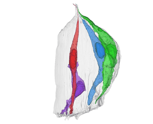 |
Three-Dimensional Reconstructions of Mouse Circumvallate Taste Buds Using Serial Blockface Scanning Electron Microscopy: I. Cell Types and the Apical Region of the Taste Bud [1194 multi-frame micrographs composed of 1 frames each in TIFF format] | Yang R, Dzowo YK, Wilson CE, Russell RL, Kidd GJ, Salcedo E, Lasher RS, Kinnamon JC, Finger TE [Pubmed: 31587284] [DOI: 10.1002/cne.24779] |
184.9 GB | — |
| 2023-04-14 | 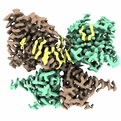 |
Thermostabilized human prestin in complex with chloride, sulfate or salicylate [multiple data sets in TIFF format] | Futamata H, Fukuda M, Umeda R, Yamashita K, Tomita A, Takahashi S, Shikakura T, Hayashi S, Kusakizako T, Nishizawa T, Homma K, Nureki O [Pubmed: 36266333] [DOI: 10.1038/s41467-022-34017-x] |
3.5 TB | 3.52 - 3.63 Å |
| 2018-10-25 | 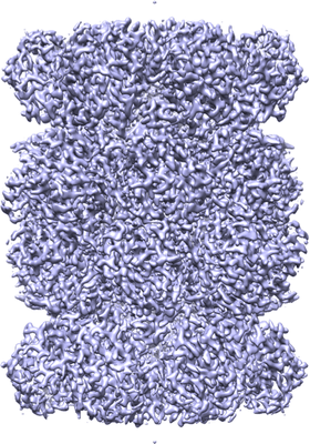 |
Thermoplasma acidophilum 20S [1175 multi-frame micrographs composed of 50 frames each in MRC format] | Eng ET, Kopylov M, Negro CJ, Dallaykan S, Rice WJ, Jordan KJ, Kelley K, Carragher BO, Potter CS | 1.5 TB | 2.1 Å |
| 2020-10-28 | 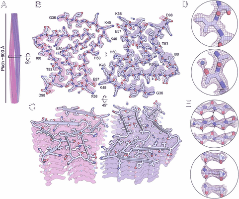 |
The α-synuclein hereditary mutation E46K unlocks a more stable, pathogenic fibril structure [4919 multi-frame micrographs composed of 30 frames each in TIFF format] | Boyer DR, Li B, Sun C, Fan W, Zhou K, Hughes MP, Sawaya MR, Jiang L, Eisenberg DS [Pubmed: 32015135] [DOI: 10.1073/pnas.1917914117] |
897.9 GB | 2.5 Å |
| 2022-02-28 |  |
The structure of natively iodinated bovine thyroglobulin [5016 multi-frame micrographs composed of 50 frames each in TIFF format] | Kim K, Kopylov M, Bobe D, Kelley K, Eng ET, Arvan P, Clarke OB [Pubmed: 34726172] [DOI: 10.1107/S2059798321010056] |
1.2 TB | 2.61 Å |
| 2021-06-25 |  |
The structure of human CST reveals a decameric assembly bound to telomeric DNA [multiple data sets in TIFF and MRC formats] | Lim C, Barbour AT, Zaug AJ, Goodrich KJ, McKay AE, Wuttke DS, Cech TR [Pubmed: 32499435] [DOI: 10.1126/science.aaz9649] |
14.3 TB | 3.0 Å |
| 2022-10-14 |  |
The structure of hemolysin A secretion system, wild-type HlyB/D complex without nucleotide. [multiple data sets in TIFF and MRC formats] | Zhao H, Chen J [Pubmed: 36055198] [DOI: 10.1016/j.cell.2022.07.017] |
3.5 TB | 2.9 Å |
| 2022-10-14 |  |
The structure of hemolysin A secretion system, HlyB(E631Q)/D complex with ATPMg. [12543 multi-frame micrographs composed of 40 frames each in TIFF format] | Zhao H, Chen J [Pubmed: 36055198] [DOI: 10.1016/j.cell.2022.07.017] |
5.4 TB | 3.4 Å |
| 2022-11-14 |  |
The structure of PldA-PA3488 complex [3732 micrographs in MRC format] | Yang X, Li Z, Zhao L, She Z, Gao Z, Sui SF, Dong Y, Li Y [Pubmed: 36216841] [DOI: 10.1038/s41467-022-33690-2] |
327.7 GB | 3.05 Å |
| 2022-02-04 |  |
The pump-like chanelrhodopsin ChRmine [3528 multi-frame micrographs composed of 48 frames each in TIFF format] | Kishi KE, Kim YS, Fukuda M, Yamahita K [Pubmed: 35114111] [DOI: 10.1016/j.cell.2022.01.007] |
850.8 GB | 2.02 Å |
| 2023-09-25 |  |
The potassium-selective channelrhodopsin HcKCR1 and HcKCR2 in lipid nanodisc [multiple data sets in TIFF format] | Tajima S, Kim YS, Fukuda M, Nakamura S, Yamashita K, Deisseroth K [Pubmed: 37652010] [DOI: 10.1016/j.cell.2023.08.009] |
4.1 TB | 2.53 - 2.66 Å |
| 2016-11-24 |  |
The pathway to GTPase activation of elongation factor SelB on the ribosome [multiple data sets in MRC format] | Fischer N, Neumann P, Bock LV, Maracci C, Wang Z, Paleskava A, Konevega AL, Schroeder GF, Grubmueller H, Rodnina MV, Stark H [Pubmed: 27842381] [DOI: 10.1038/nature20560] |
1.0 TB | 3.4 - 5.3 Å |
| 2023-11-07 |  |
The lipid linked oligosaccharide polymerase Wzy and its regulating co-polymerase Wzz form a complex in vivo and in vitro [2351 multi-frame micrographs composed of 20 frames each in MRC format] | Weckener M, Woodward LS, Clarke BR, Liu H, Ward PN, Le Bas A, Bhella D, Whitfield C, Naismith JH [Pubmed: 36944376] [DOI: 10.1098/rsob.220373] |
273.6 GB | 3.6 Å |
| 2018-10-22 |  |
The in situ structures of mono-, di-, and trinucleosomes in human heterochromatin [59 tilt series in MRC format] | Cai S, Böck D, Pilhofer M, Gan L [Pubmed: 30091658] [DOI: 10.1091/mbc.E18-05-0331] |
1.9 GB | 21.0 - 24.0 Å |
| 2018-08-15 |  |
The first reconstruction of beta-galactosidase solved by cryoARM200 [1338 multi-frame micrographs composed of 49 frames each in TIFF format] | Kato T, Terehara N, Namba K | 321.4 GB | 2.6 Å |
| 2021-05-07 |  |
The cryo-EM structure of vesivirus 2117 highlights functional variations in entry pathways for viruses in different clades of the vesivirus genus. [2000 multi-frame micrographs composed of 50 frames each in MRC format] | Sutherland H, Conley MJ, Emmott E, Streetley J, Goodfellow IG, Bhella D [Pubmed: 33853966] [DOI: 10.1128/JVI.00282-21] |
24.4 TB | 3.65 Å |
| 2023-10-23 |  |
The cryo-EM structure of the human DNMT3A2-DNMT3B3 complex bound to nucleosome [12438 multi-frame micrographs composed of 40 frames each in TIFF format] | Xu TH, Liu M, Zhou XE, Liang G, Zhao G, Xu HE, Melcher K, Jones PA [Pubmed: 32968275] [DOI: 10.1038/s41586-020-2747-1] |
4.2 TB | 2.94 Å |
| 2023-10-23 |  |
The cryo-EM structure of the human DNMT3A2-DNMT3B3 complex bound to NCP_Kc36me3. [1504 multi-frame micrographs composed of 40 frames each in TIFF format] | Xu TH, Liu M, Zhou EX, Liang G, Zhao G, Xu HE, Melcher K, Jones PA [Pubmed: 32968275] [DOI: 10.1038/s41586-020-2747-1] |
583.5 GB | 4.26 Å |
| 2021-11-16 |  |
The cryo-EM structure of the CENP-A nucleosome in complex with the phosphorylated CENP-C:: CENP-A nucleosome in complex with phosphorylated CENP-C C-terminal domain(601-864) [6533 multi-frame micrographs composed of 50 frames each in TIFF format] | Ariyoshi M, Makino F, Watanabe R, Nakagawa R, Kato T, Namba K, Arimura Y, Fujita R, Kurumizaka H, Okumura EI, Hara M, Fukagawa T [Pubmed: 33463726] [DOI: 10.15252/embj.2020105671] |
1.4 TB | 6.8 Å |
| 2021-11-16 |  |
The cryo-EM structure of the CENP-A nucleosome in complex with the phosphorylated CENP-C:: CENP-A nucleosome in complex with CENP-C motif (655-675) and CENP-N N-terminal domain (1-211) [4630 multi-frame micrographs composed of 50 frames each in TIFF format] | Ariyoshi M, Makino F, Watanabe R, Nakagawa R, Kato T, Namba K, Arimura Y, Fujita R, Kurumizaka H, Okumura EI, Hara M, Fukagawa T [Pubmed: 33463726] [DOI: 10.15252/embj.2020105671] |
1.3 TB | 4.2 Å |
| 2021-11-16 |  |
The cryo-EM structure of the CENP-A nucleosome in complex with the phosphorylated CENP-C: CENP-A nucleosome in complex with phosphorylated CENP-C C-terminal domain (601-864) and CENP-N N-terminal domain (1-211) [8017 multi-frame micrographs composed of 50 frames each in TIFF format] | Ariyoshi M, Makino F, Watanabe R, Nakagawa R, Kato T, Namba K, Arimura Y, Fujita R, Kurumizaka H, Okumura EI, Hara M, Fukagawa T [Pubmed: 33463726] [DOI: 10.15252/embj.2020105671] |
1.6 TB | 4.5 - 7.8 Å |
| 2021-11-01 | 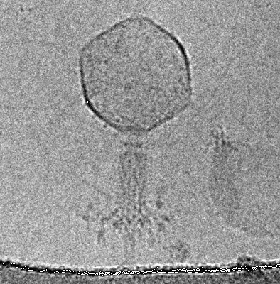 |
The contracted tail of myophage vb_EcoM_CBA120 (CBA120) [535 multi-frame micrographs composed of 20 frames each in MRCS format] | Nazarov S, Leiman P | 674.2 GB | 4.9 Å |
| 2023-08-20 | 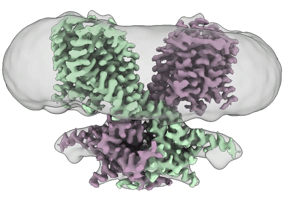 |
The conformational cycle of prestin underlies outer-hair cell electromotility [6796 multi-frame micrographs composed of 40 frames each in TIFF format] | Perozo E [Pubmed: 34695838] [DOI: 10.1038/s41586-021-04152-4] |
3.1 TB | 3.3 Å |
| 2022-05-24 | 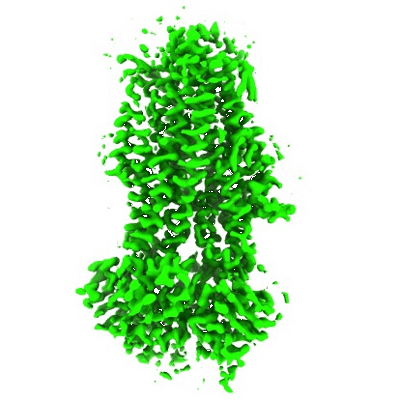 |
The complex of phosphorylated human cystic fibrosis transmembrane conductance regulator (CFTR) with ATP/Mg and Tezacaftor (VX-661) [4257 multi-frame micrographs composed of 50 frames each in TIFF format] | Fiedorczuk K, Chen J [Pubmed: 34995514] [DOI: 10.1016/j.cell.2021.12.009] |
1.8 TB | 3.8 Å |
