Electron Microscopy Public Image Archive
Electron Microscopy Public Image Archive
The EMPIAR-PDBj team at Osaka University assists Asian EM researchers with the transfer of big EM image data to EMPIAR. Instead of sending the data directly to the EBI (UK) via the internet, hard drives can also be sent to Osaka University by postal mail or via a courier service. As an alternative, internet transfer to our server in Osaka is also available. If you would like to take advantage of our submission services, please contact us first by e-mail before sending the data to us.
| Release date | Imageset | Title | Authors and references | Size | Resolution |
|---|---|---|---|---|---|
| 2023-06-07 |  |
Cryo-electron micrographs of Hantaan virus polymerase in its apo state [2865 multi-frame micrographs composed of 50 frames each in TIFF format] | Durieux Trouilleton Q, Arragain B, Malet H [Pubmed: 37221161] [DOI: 10.1038/s41467-023-38555-w] |
660.7 GB | 3.27 Å |
| 2023-06-07 |  |
Cryo-electron micrographs of Hantaan virus polymerase in pre-initiation state [3261 multi-frame micrographs composed of 50 frames each in TIFF format] | Durieux Trouilleton Q, Arragain B, Malet H [Pubmed: 37221161] [DOI: 10.1038/s41467-023-38555-w] |
776.9 GB | 3.36 Å |
| 2023-06-07 |  |
Cryo-electron micrographs of Hantaan virus polymerase bound to its 5' viral RNA [2547 multi-frame micrographs composed of 50 frames each in TIFF format] | Durieux Trouilleton Q, Arragain B, Malet H [Pubmed: 37221161] [DOI: 10.1038/s41467-023-38555-w] |
608.2 GB | 3.23 Å |
| 2023-06-07 |  |
Cryo-electron micrographs of Hantaan virus polymerase in elongation state [3785 multi-frame micrographs composed of 50 frames each in TIFF format] | Durieux Trouilleton Q, Arragain B, Malet H [Pubmed: 37221161] [DOI: 10.1038/s41467-023-38555-w] |
927.5 GB | 3.14 Å |
| 2022-01-12 |  |
Motion corrected micrographs - purified Dot/Icm T4SS particles [3590 micrographs in MRC format] | Durie CL, Sheedlo MJ, Chung JM, Byrne BG, Su M, Knight T, Swanson M, Lacy DB, Ohi MD [Pubmed: 32876045] [DOI: 10.7554/eLife.59530] |
190.4 GB | 3.7 Å |
| 2023-02-28 |  |
Cryo pFIB/SEM of PEG beads (test sample) [193 micrographs in TIFF format] | Dumoux M, Glen T, Smith JLR, Ho EML, Perdigão LMA, Pennington A, Klumpe S, Yee NBY, Farmer DA, Lai PYA, Bowles W, Kelley R, Plitzko JM, Wu L, Basham M, Clare DK, Siebert CA, Darrow MC, Naismith JH, Grange M [Pubmed: 36805107] [DOI: 10.7554/elife.83623] |
5.3 GB | — |
| 2023-02-28 |  |
Cryo serial FIB SEM of mouse brain tissue [59 micrographs in TIFF format] | Dumoux M, Glen T, Smith JLR, Ho EML, Perdigão LMA, Pennington A, Klumpe S, Yee NBY, Farmer DA, Lai PYA, Bowles W, Kelley R, Plitzko JM, Wu L, Basham M, Clare DK, Siebert CA, Darrow MC, Naismith JH, Grange M [Pubmed: 36805107] [DOI: 10.7554/elife.83623] |
3.0 GB | — |
| 2023-02-17 |  |
Cryo serial FIB/SEM of Saccharomyces cerevisiae [109 micrographs in TIFF format] | Dumoux M, Glen T, Smith JLR, Ho EML, Perdigão LMA, Pennington A, Klumpe S, Yee NBY, Farmer DA, Lai PYA, Bowles W, Kelley R, Plitzko JM, Wu L, Basham M, Clare DK, Siebert CA, Darrow MC, Naismith JH, Grange M [Pubmed: 36805107] [DOI: 10.7554/elife.83623] |
4.8 GB | — |
| 2023-02-17 |  |
Cryo serial FIB/SEM of Vero cells [46 micrographs in TIFF format] | Dumoux M, Glen T, Smith JLR, Ho EML, Perdigão LMA, Pennington A, Klumpe S, Yee NBY, Farmer DA, Lai PYA, Bowles W, Kelley R, Plitzko JM, Wu L, Basham M, Clare DK, Siebert CA, Darrow MC, Naismith JH, Grange M [Pubmed: 36805107] [DOI: 10.7554/elife.83623] |
7.1 GB | — |
| 2023-02-17 |  |
Cryo serial FIB/SEM of Rhodospirillum rubrum [43 micrographs in TIFF format] | Dumoux M, Glen T, Smith JLR, Ho EML, Perdigão LMA, Pennington A, Klumpe S, Yee NBY, Farmer DA, Lai PYA, Bowles W, Kelley R, Plitzko JM, Wu L, Basham M, Clare DK, Siebert CA, Darrow MC, Naismith JH, Grange M [Pubmed: 36805107] [DOI: 10.7554/elife.83623] |
5.7 GB | — |
| 2023-02-28 |  |
Cryo serial FIB/SEM of HeLa cells [46 micrographs in TIFF format] | Dumoux M, Glen T, Smith JLR, Ho EML, Perdigão LMA, Pennington A, Klumpe S, Yee NBY, Farmer DA, Lai PYA, Bowles W, Kelley R, Plitzko JM, Wu L, Basham M, Clare DK, Siebert CA, Darrow MC, Naismith JH, Grange M [Pubmed: 36805107] [DOI: 10.7554/elife.83623] |
6.7 GB | — |
| 2023-02-28 |  |
Cryo serial FIB/SEM of mouse heart tissue [136 micrographs in TIFF format] | Dumoux M, Glen T, Smith JLR, Ho EML, Perdigão LMA, Pennington A, Klumpe S, Yee NBY, Farmer DA, Lai PYA, Bowles W, Kelley R, Plitzko JM, Wu L, Basham M, Clare DK, Siebert CA, Darrow MC, Naismith JH, Grange M [Pubmed: 36805107] [DOI: 10.7554/elife.83623] |
18.0 GB | — |
| 2023-02-28 |  |
Cryo serial FIB/SEM of RPE-1 cells [18 micrographs in TIFF format] | Dumoux M, Glen T, Smith JLR, Ho EML, Perdigão LMA, Pennington A, Klumpe S, Yee NBY, Farmer DA, Lai PYA, Bowles W, Kelley R, Plitzko JM, Wu L, Basham M, Clare DK, Siebert CA, Darrow MC, Naismith JH, Grange M [Pubmed: 36805107] [DOI: 10.7554/elife.83623] |
826.6 MB | — |
| 2019-07-25 |  |
Open state structure of the full-length TRPV2 cation channel with a resolved pore turret domain [2447 multi-frame micrographs composed of 50 frames each in MRCS format] | Dosey TL, Wang Z, Fan G [Pubmed: 30598551] [DOI: 10.1038/s41594-018-0168-8] |
934.8 GB | 3.6 Å |
| 2021-11-30 |  |
Cryo-EM dataset of the substrate-engaged human 26S proteasome [44688 micrographs in MRC format] | Dong Y, Zhang S, Wu Z, Wang WL, Mao Y [Pubmed: 30479383] [DOI: 10.1038/s41586-018-0736-4] |
13.9 TB | 2.8 - 3.6 Å |
| 2023-09-05 |  |
Benchmark SBF SEM data of HeLa cells previously imaged by Zeiss LSM900 Airyscan microscopy [multiple data sets in DM4 and TIFF formats] | Domart MC, Collinson LM [DOI: 10.1101/2023.05.11.540445] |
39.2 GB | — |
| 2020-12-09 | 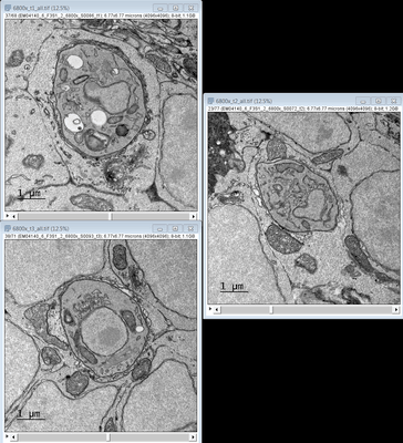 |
TEM images of a Zebrafish hindbrain cells containing Toxoplasma gondii tachizoites [multiple data sets in TIFF format] | Domart MC, Collinson L [Pubmed: 32461265] [DOI: 10.1242/dmm.043091] |
3.4 GB | — |
| 2019-06-19 | 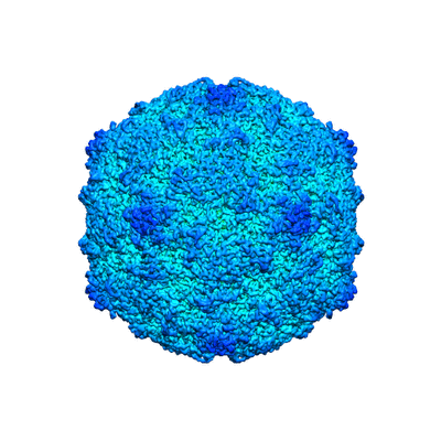 |
Extracellular albumin and endosomal ions prime enterovirus particles for uncoating that can be prevented by fatty acid saturation [multiple data sets in MRC format] | Domanska A, Ruokolainen VP, Pelliccia M, Laajala MA, Marjomäki VS, Butcher SJ [Pubmed: 31189702] [DOI: 10.1128/JVI.00599-19] |
2.4 TB | 3.5 - 3.6 Å |
| 2023-01-20 | 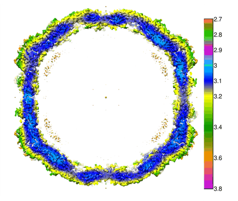 |
Cryogenic electron tomographs of Coxsackievirus A9 treated with endosomal ionic condition buffer [1964 multi-frame micrographs composed of 30 frames each in MRC format] | Domanska A, Plavec Z, Ruokolainen V, Löflund B, Marjomäki V, Butcher SJ [Pubmed: 36448797] [DOI: 10.1128/jvi.01367-22] |
1.8 TB | 3.3 Å |
| 2023-01-19 | 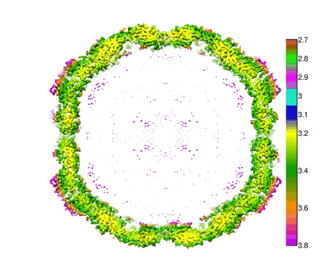 |
Cryogenic electron tomographs of Coxsackievirus A9 treated with fatty-acid-free BSA [2472 multi-frame micrographs composed of 30 frames each in MRC format] | Domanska A, Plavec Z, Ruokolainen V, Löflund B, Marjomäki V, Butcher SJ [Pubmed: 36448797] [DOI: 10.1128/jvi.01367-22] |
2.3 TB | 3.5 Å |
| 2023-01-30 | 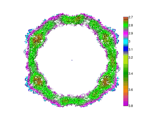 |
Cryogenic electron tomographs of Coxsackievirus A9 [2421 multi-frame micrographs composed of 30 frames each in MRC format] | Domanska A, Plavec Z, Ruokolainen V, Löflund B, Marjomäki V, Butcher SJ [Pubmed: 36448797] [DOI: 10.1128/jvi.01367-22] |
2.1 TB | 2.9 Å |
| 2022-03-22 | 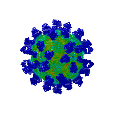 |
High-resolution Cryo-EM of Fab-labeled human parechovirus 3 [6759 multi-frame micrographs composed of 16 frames each in MRCS format] | Domanska A, Flatt JW, Jukonen JJJ, Geraets JA, Butcher SJ [Pubmed: 30463974] [DOI: 10.1128/JVI.01597-18] |
1.5 TB | 2.8 Å |
| 2024-03-25 | 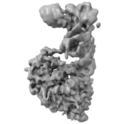 |
Cryo-EM micrographs of Tb ADAT2/3 bound to tRNA [6150 multi-frame micrographs composed of 80 frames each in TIFF format] | Dolce LG, Zimmer AA, Tengo L, Weis F, Rubio MAT, Alfonzo JD, Kowalinski E [Pubmed: 36347890] [DOI: 10.1038/s41467-022-34441-z] |
1.8 TB | 3.6 Å |
| 2023-09-11 | 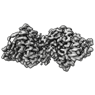 |
Cryo-EM micrographs of RESC1-RESC2 complex [6816 multi-frame micrographs composed of 100 frames each in TIFF format] | Dolce LG, Nesterenko Y, Walther L, Weis F, Kowalinski E [Pubmed: 36999600] [DOI: 10.1093/nar/gkad217] |
1.9 TB | 3.4 Å |
| 2023-09-22 |  |
Cryo-EM micrographs of RESC1-RESC2 complex bound to gRNA [4420 multi-frame micrographs composed of 60 frames each in TIFF format] | Dolce LG, Nesterenko Y, Walther L, Weis F, Kowalinski E [Pubmed: 36999600] [DOI: 10.1093/nar/gkad217] |
2.5 TB | 4.7 Å |
