Electron Microscopy Public Image Archive
Electron Microscopy Public Image Archive
The EMPIAR-PDBj team at Osaka University assists Asian EM researchers with the transfer of big EM image data to EMPIAR. Instead of sending the data directly to the EBI (UK) via the internet, hard drives can also be sent to Osaka University by postal mail or via a courier service. As an alternative, internet transfer to our server in Osaka is also available. If you would like to take advantage of our submission services, please contact us first by e-mail before sending the data to us.
| Release date | Imageset | Title | Authors and references | Size | Resolution |
|---|---|---|---|---|---|
| 2021-02-17 |  |
Raw cryo- and negative stain micrographs of a native extract from C. thermophilum used to reconstruct oxo acid dehydrogenase complexes [0 multi-frame micrographs composed of 0 frames each in MRC format] | Kastritis PLK [DOI: 10.1016/j.celrep.2021.108727] |
5.4 TB | 6.9 Å |
| 2018-01-03 |  |
3D reconstruction of the cardiac mitochondria in Neaonate and Adult GP cardiomyocyte [262 multi-frame micrographs composed of 1 frames each in TIFF format] | Kashbour H, Taggart M, White K | 1.2 GB | — |
| 2018-01-03 |  |
3D reconstruction of cardiac mitochondria in earlly prenatal (G55/57) and mid term(G58/60) and term (G66/68) GP cardiomyocyte [341 multi-frame micrographs composed of 1 frames each in TIFF format] | Kashbour H, Taggart M, White K | 1.7 GB | — |
| 2018-01-03 |  |
Segment /nodes network analysis of the tubular system of the Neonate and Adult GP cardiomyocyte [167 multi-frame micrographs composed of 1 frames each in TIFF format] | Kashbour H, Taggart M, White K | 787.5 MB | — |
| 2018-01-03 | 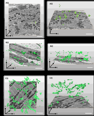 |
3D reconstruction of the tubular system of preterm G55/57, mid term G58/60 and term G66/68 of guinea pig left ventricles [318 multi-frame micrographs composed of 1 frames each in TIFF format] | Kashbour H, Taggart M | 2.9 GB | — |
| 2018-01-03 | 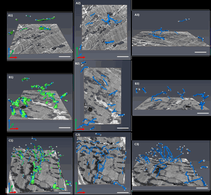 |
Segment /nodes network analysis of the tubular system of the prenatal stages of GP cardiomyocyte [254 multi-frame micrographs composed of 1 frames each in TIFF format] | Kashbour H, Taggart M | 1.9 GB | — |
| 2021-11-15 | 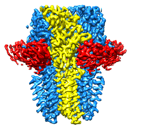 |
CryoEM structure of GABA(A)R-beta3 homopentamer at 3.4A from Tundra, 100kV microscope [4073 multi-frame micrographs composed of 18 frames each in MRC format] | Karia D, Koh AF, Yu L, Kotecha A | 2.2 TB | 3.4 Å |
| 2022-02-25 | 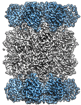 |
CryoEM structure of T20S proteasome at 3A from Tundra, 100kV microscope [5176 multi-frame micrographs composed of 18 frames each in MRC format] | Karia D, Koh AF, Yu L, Kotecha A | 1.2 TB | 3.0 Å |
| 2021-11-15 | 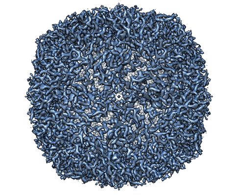 |
CryoEM structure of Apoferritin at 2.6A from Tundra, 100kV microscope [1896 multi-frame micrographs composed of 18 frames each in MRC format] | Karia D, Hlavenkova Z, Koh AF, Malinsky M, Yu L, Kotecha A | 1.0 TB | 2.6 Å |
| 2023-05-11 | 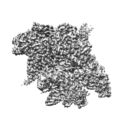 |
Cryo-EM structure of his-elemental paused elongation complex [6939 multi-frame micrographs composed of 50 frames each in TIFF format] | Kang JY, Mishanina TV, Bao Y, Chen J, Llewellyn E, Liu J, Darst SA, Landick R [Pubmed: 36795753] [DOI: 10.1073/pnas.2215945120] |
3.0 TB | 3.3 - 5.5 Å |
| 2023-04-25 | 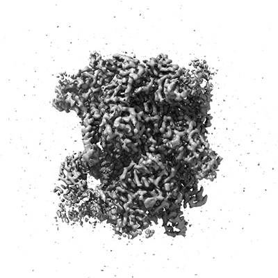 |
Cryo-EM structure of pre-consensus elemental paused elongation complex [4419 multi-frame micrographs composed of 50 frames each in TIFF format] | Kang JY, Mishanina TV, Bao Y, Chen J, Llewellyn E, Liu J, Darst SA, Landick R [Pubmed: 36795753] [DOI: 10.1073/pnas.2215945120] |
1.9 TB | 3.2 Å |
| 2023-05-11 | 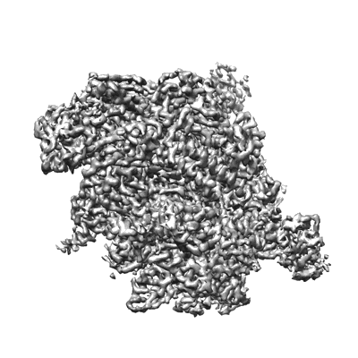 |
Cryo-EM structure of consensus elemental paused elongation complex [2525 multi-frame micrographs composed of 50 frames each in TIFF format] | Kang JY, Mishanina TV, Bao Y, Chen J, Llewellyn E, Liu J, Darst SA, Landick R [Pubmed: 36795753] [DOI: 10.1073/pnas.2215945120] |
1.1 TB | 3.3 - 3.8 Å |
| 2022-06-21 | 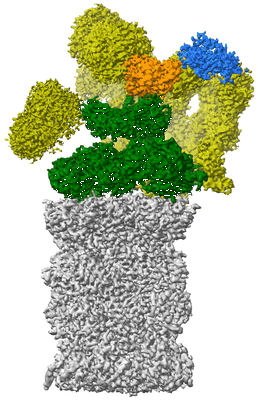 |
Cryo-EM structure of the plant 26S proteasome [multiple data sets in TIFF format] | Kandolf S, Grishkovskaya I, Meinhart A, Haselbach D [Pubmed: 35576154] [DOI: 10.1016/j.xplc.2022.100310] |
7.9 TB | 3.3 Å |
| 2023-06-30 | 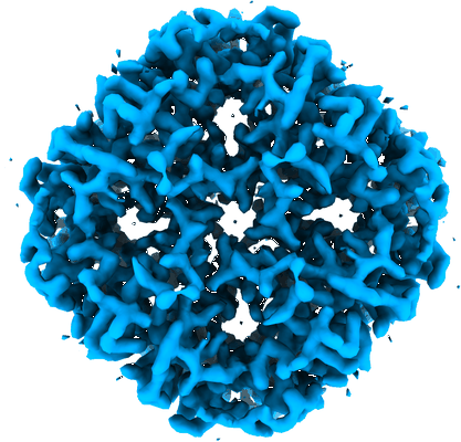 |
Cryo-electron micrographs of hAQP2 in DDM [6592 multi-frame micrographs composed of 40 frames each in TIFF format] | Kamegawa A, Suzuki H, Fujiyoshi Y [Pubmed: 37315821] [DOI: 10.1016/j.jsb.2023.107984] |
1.2 TB | 2.89 Å |
| 2023-11-14 | 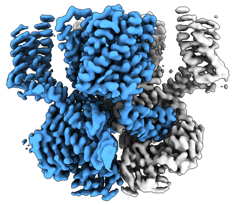 |
Ligand-free SpSLC9C1 in lipid nanodiscs [11299 multi-frame micrographs composed of 76 frames each in TIFF format] | Kalienkova V, Peter MF, Rheinberger J, Paulino C [Pubmed: 37880361] [DOI: 10.1038/s41586-023-06629-w] |
3.3 TB | 3.21 - 3.4 Å |
| 2023-11-14 | 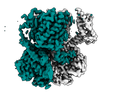 |
Ligand-free SpSLC9C1 in detergent [multiple data sets in TIFF format] | Kalienkova V, Peter MF, Rheinberger J, Paulino C [Pubmed: 37880361] [DOI: 10.1038/s41586-023-06629-w] |
2.1 TB | 3.05 - 3.3 Å |
| 2023-11-14 | 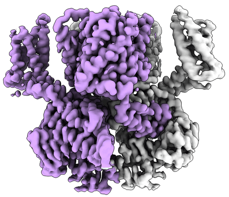 |
cGMP-bound SpSLC9C1 in lipid nanodiscs [multiple data sets in TIFF format] | Kalienkova V, Peter MF, Rheinberger J, Paulino C [Pubmed: 37880361] [DOI: 10.1038/s41586-023-06629-w] |
1.6 TB | 3.22 - 3.26 Å |
| 2023-12-01 | 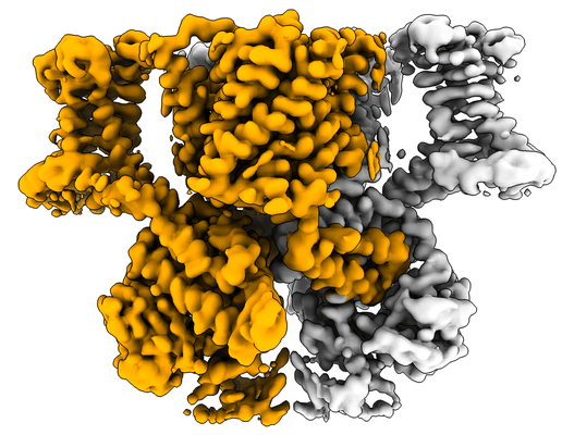 |
cAMP-bound SpSLC9C1 in lipid nanodiscs [multiple data sets in TIFF format] | Kalienkova V, Peter MF, Rheinberger J, Paulino C [Pubmed: 37880361] [DOI: 10.1038/s41586-023-06629-w] |
4.8 TB | 3.3 - 3.74 Å |
| 2024-02-28 | 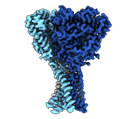 |
Apo Malacoceros FaNaC1 in lipid nanodiscs [multiple data sets in TIFF format] | Kalienkova V, Dandamudi M, Paulino C, Lynagh T [Pubmed: 38337033] [DOI: 10.1038/s41594-023-01198-y] |
1013.1 GB | 2.7 Å |
| 2024-02-28 | 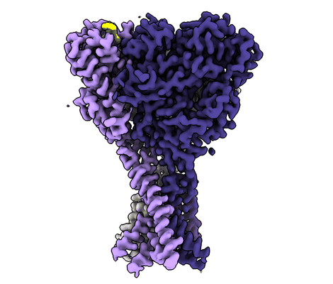 |
FMRFa-bound Malacoceros FaNaC1 in lipid nanodiscs [multiple data sets in TIFF format] | Kalienkova V, Dandamudi M, Paulino C, Lynagh T [Pubmed: 38337033] [DOI: 10.1038/s41594-023-01198-y] |
866.2 GB | 2.5 Å |
| 2024-02-28 | 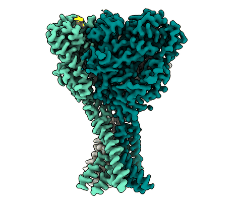 |
ASSFVRIa-bound Malacoceros FaNaC1 in lipid nanodiscs [multiple data sets in TIFF format] | Kalienkova V, Dandamudi M, Paulino C, Lynagh T [Pubmed: 38337033] [DOI: 10.1038/s41594-023-01198-y] |
1.3 TB | 2.4 Å |
| 2024-02-28 | 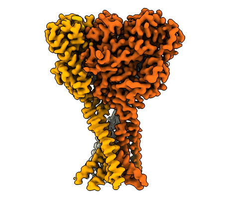 |
FMRFa-bound Malacoceros FaNaC1 in lipid nanodiscs in presence of diminazene [multiple data sets in TIFF format] | Kalienkova V, Dandamudi M, Paulino C, Lynagh T [Pubmed: 38337033] [DOI: 10.1038/s41594-023-01198-y] |
1.3 TB | 3.0 Å |
| 2020-11-27 | 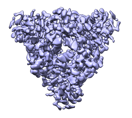 |
Cryo-EM structure of the prefusion state of canine distemper virus fusion protein ectodomain [1604 multi-frame micrographs composed of 48 frames each in TIFF format] | Kalbermatter DK, Shrestha NS, Gall FMG, Wyss MW, Riedl RR, Plattet PP, Fotiadis DF [Pubmed: 32647825] [DOI: 10.1016/j.yjsbx.2020.100021] |
474.1 GB | 4.3 Å |
| 2022-11-14 | 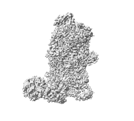 |
Cryo-electron microscopy of Dicer-1 and Its Partner Protein Loqs-PB complex with model pre-miRNA in presence of Ca2+ ions [3057 multi-frame micrographs composed of 40 frames each in TIFF format] | Jouravleva K, Golovenko D, Demo G, Dutcher RC, Hall TMT, Zamore PD, Korostelev AA [Pubmed: 36182693] [DOI: 10.1016/j.molcel.2022.09.002] |
1.4 TB | 3.06 Å |
| 2022-11-16 | 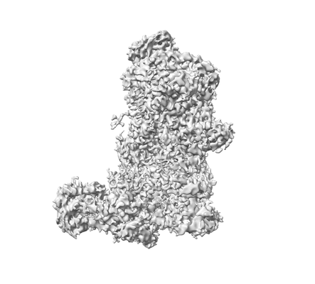 |
Cryo-electron microscopy of Dicer-1 and Its Partner Protein Loqs-PB complex with model pre-miRNA in presence of Mg2+ ions [8128 multi-frame micrographs composed of 38 frames each in TIFF format] | Jouravleva K, Golovenko D, Demo G, Dutcher RC, Hall TMT, Zamore PD, Korostelev AA [Pubmed: 36182693] [DOI: 10.1016/j.molcel.2022.09.002] |
2.9 TB | 3.26 - 3.73 Å |
