Electron Microscopy Public Image Archive
Electron Microscopy Public Image Archive
The EMPIAR-PDBj team at Osaka University assists Asian EM researchers with the transfer of big EM image data to EMPIAR. Instead of sending the data directly to the EBI (UK) via the internet, hard drives can also be sent to Osaka University by postal mail or via a courier service. As an alternative, internet transfer to our server in Osaka is also available. If you would like to take advantage of our submission services, please contact us first by e-mail before sending the data to us.
| Release date | Imageset | Title | Authors and references | Size | Resolution |
|---|---|---|---|---|---|
| 2023-04-12 |  |
Cryo-Electron Microscopy structure of OmcZ nanowires from Geobacter sulfurreducens [14005 multi-frame micrographs composed of 50 frames each in TIFF format] | Gu YG, Srikanth V, Salazar-Morales AI, Samatey FA, Malvankar NS, Guberman-Pfeffer MGP, Shen CS, Gupta KG, Londer YL, Giska FG, Batista VB [Pubmed: 36732469] [DOI: 10.1038/s41564-022-01315-5] |
6.5 TB | 3.4 Å |
| 2023-09-22 |  |
Structure of TDP-43 amyloid filaments from type A FTLD-TDP (individual 1) [91457 multi-frame micrographs composed of 40 frames each in TIFF format] | Arseni D, Ryskeldi-Falcon B [Pubmed: 37532939] [DOI: 10.1038/s41586-023-06405-w] |
14.5 TB | 2.39 Å |
| 2024-02-13 |  |
5-HT2AR bound to a novel agonist in complex with a mini-Gq protein and an active-state stabilizing single-chain variable fragment (scFv16) obtained by cryo-electron microscopy (cryoEM) [7573 multi-frame micrographs composed of 50 frames each in TIFF format] | Barros-Alvarez X, Kim K, Panova O, Roth BL, Skiniotis G [Pubmed: 36171289] [DOI: 10.1038/s41586-022-05258-z] |
3.4 TB | 3.45 Å |
| 2024-02-09 |  |
5-HT2B receptor bound to LSD obtained by cryo-electron microscopy (cryoEM) [12751 multi-frame micrographs composed of 50 frames each in TIFF format] | Barros-Alvarez X, Cao C, Panova O, Roth BL, Skiniotis G [Pubmed: 36087581] [DOI: 10.1016/j.neuron.2022.08.006] |
6.3 TB | 2.7 Å |
| 2023-05-17 | 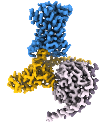 |
GPR56 (ADGRG1) 7TM domain bound to tethered agonist in complex with G protein heterotrimer [6773 multi-frame micrographs composed of 57 frames each in TIFF format] | Barros-Alvarez X, Panova O, Skiniotis G [Pubmed: 35418682] [DOI: 10.1038/s41586-022-04575-7] |
3.6 TB | 2.7 Å |
| 2023-05-17 | 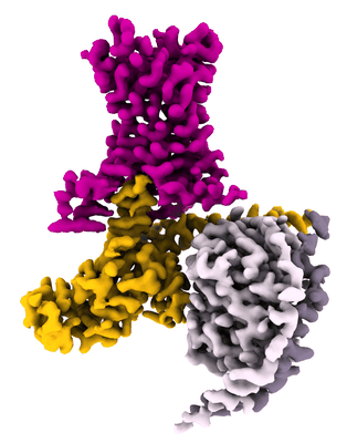 |
LPHN3 (ADGRL3) 7TM domain bound to tethered agonist in complex with G protein heterotrimer [8877 multi-frame micrographs composed of 63 frames each in TIFF format] | Barros-Alvarez X, Panova O, Skiniotis G [Pubmed: 35418682] [DOI: 10.1038/s41586-022-04575-7] |
5.3 TB | 2.9 Å |
| 2023-02-27 |  |
Cryo-EM micrographs of full-length human BIRC6 dimer with a bound DIABLO (SMAC) homodimer [multiple data sets in EER format] | Ehrmann JF, Grabarczyk DB, Clausen T [Pubmed: 36758105] [DOI: 10.1126/science.ade8873] |
24.2 TB | 7.2 Å |
| 2023-02-17 |  |
Cryo-EM micrographs of full-length human BIRC6 dimer [11578 multi-frame micrographs composed of 40 frames each in TIFF format] | Ehrmann JF, Grabarczyk DB, Clausen T [Pubmed: 36758105] [DOI: 10.1126/science.ade8873] |
3.0 TB | 7.0 Å |
| 2023-08-18 | 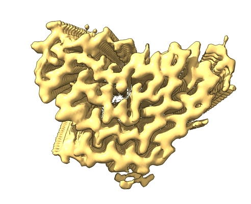 |
Structure of TDP-43 amyloid filaments from type A FTLD-TDP (individual 3) [36507 multi-frame micrographs composed of 40 frames each in TIFF format] | Arseni D, Ryskeldi-Falcon B [Pubmed: 37532939] [DOI: 10.1038/s41586-023-06405-w] |
5.7 TB | 2.39 Å |
| 2023-08-18 | 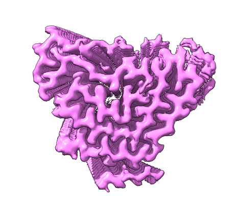 |
Structure of TDP-43 amyloid filaments from type A FTLD-TDP (individual 2) [33336 multi-frame micrographs composed of 40 frames each in TIFF format] | Arseni D, Ryskeldi-Falcon B [Pubmed: 37532939] [DOI: 10.1038/s41586-023-06405-w] |
5.3 TB | 2.39 Å |
| 2023-07-25 | 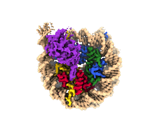 |
Cryo-EM structure of the human Sirtuin 6-nucleosome complex [11872 multi-frame micrographs composed of 40 frames each in TIFF format] | Chio US, Rechiche O, Bryll AR, Zhu J, Leith EM, Feldman JL, Peterson CL, Tan S, Armache JP [Pubmed: 37058572] [DOI: 10.1126/sciadv.adf7586] |
6.0 TB | 3.07 Å |
| 2023-08-18 | 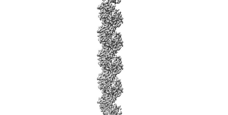 |
KpFtsZ single filament [6079 multi-frame micrographs composed of 60 frames each in TIFF format] | Fujita J, Amesaka H, Yoshizawa T, Hibino K, Kamimura N, Kuroda N, Konishi T, Kato Y, Hara M, Inoue T, Namba K, Tanaka SI, Matsumura H [Pubmed: 37429870] [DOI: 10.1038/s41467-023-39807-5] |
1.7 TB | 3.03 Å |
| 2023-08-18 |  |
KpFtsZ–Mb double helical tube [3096 multi-frame micrographs composed of 60 frames each in TIFF format] | Fujita J, Amesaka H, Yoshizawa T, Hibino K, Kamimura N, Kuroda N, Konishi T, Kato Y, Hara M, Inoue T, Namba K, Tanaka SI, Matsumura H [Pubmed: 37429870] [DOI: 10.1038/s41467-023-39807-5] |
794.4 GB | 2.67 Å |
| 2023-09-08 | 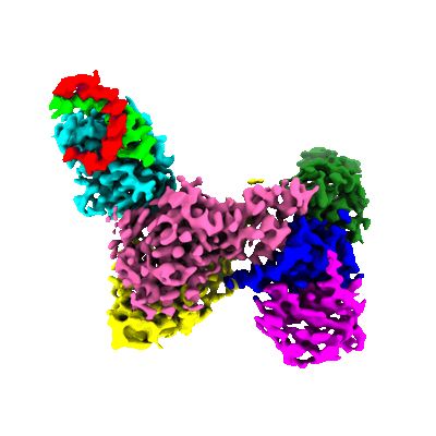 |
Cryo-EM micrographs of HIV-1 Vif bound to human APOBEC3H, CBF-beta, ELOB, ELOC, and N-terminal domain of CUL5 [14766 multi-frame micrographs composed of 49 frames each in TIFF format] | Ito F, Alvarez-Cabrera AL, Kim K, Zhou ZH, Chen XS [Pubmed: 37640699] [DOI: 10.1038/s41467-023-40955-x] |
2.2 TB | 3.24 - 3.54 Å |
| 2023-09-11 | 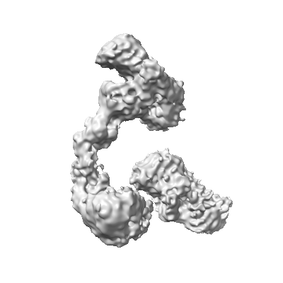 |
Cryo-EM micrographs of HIV-1 Vif bound to human APOBEC3H, CBF-beta, ELOB, ELOC, CUL5, and RBX2 [12546 multi-frame micrographs composed of 2130 frames each in TIFF format] | Ito F, Alvarez-Cabrera AL, Kim K, Zhou ZH, Chen XS [Pubmed: 37640699] [DOI: 10.1038/s41467-023-40955-x] |
11.9 TB | 5.14 Å |
| 2023-03-01 | 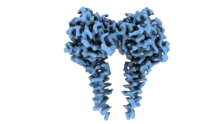 |
CryoEM structure of full-length dimeric ClbP [3888 multi-frame micrographs composed of 50 frames each in TIFF format] | Velilla JA, Walsh RM, Gaudet R [Pubmed: 36253550] [DOI: 10.1038/s41589-022-01142-z] |
1.1 TB | 3.73 Å |
| 2023-02-28 | 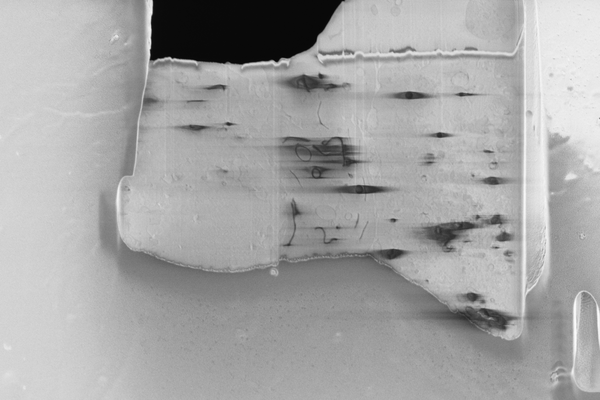 |
Cryo serial FIB/SEM of RPE-1 cells [18 micrographs in TIFF format] | Dumoux M, Glen T, Smith JLR, Ho EML, Perdigão LMA, Pennington A, Klumpe S, Yee NBY, Farmer DA, Lai PYA, Bowles W, Kelley R, Plitzko JM, Wu L, Basham M, Clare DK, Siebert CA, Darrow MC, Naismith JH, Grange M [Pubmed: 36805107] [DOI: 10.7554/elife.83623] |
826.6 MB | — |
| 2023-02-28 | 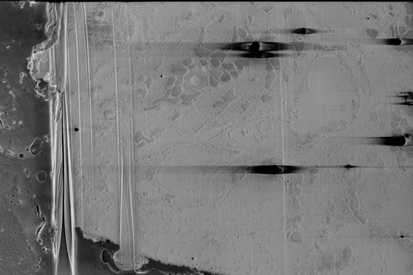 |
Cryo serial FIB/SEM of mouse heart tissue [136 micrographs in TIFF format] | Dumoux M, Glen T, Smith JLR, Ho EML, Perdigão LMA, Pennington A, Klumpe S, Yee NBY, Farmer DA, Lai PYA, Bowles W, Kelley R, Plitzko JM, Wu L, Basham M, Clare DK, Siebert CA, Darrow MC, Naismith JH, Grange M [Pubmed: 36805107] [DOI: 10.7554/elife.83623] |
18.0 GB | — |
| 2023-02-28 | 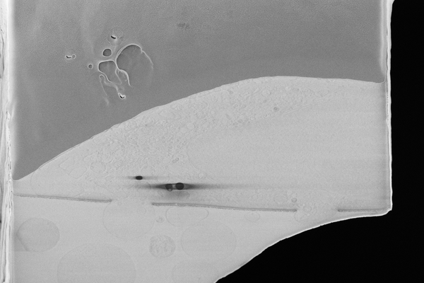 |
Cryo serial FIB/SEM of HeLa cells [46 micrographs in TIFF format] | Dumoux M, Glen T, Smith JLR, Ho EML, Perdigão LMA, Pennington A, Klumpe S, Yee NBY, Farmer DA, Lai PYA, Bowles W, Kelley R, Plitzko JM, Wu L, Basham M, Clare DK, Siebert CA, Darrow MC, Naismith JH, Grange M [Pubmed: 36805107] [DOI: 10.7554/elife.83623] |
6.7 GB | — |
| 2023-02-17 | 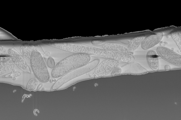 |
Cryo serial FIB/SEM of Rhodospirillum rubrum [43 micrographs in TIFF format] | Dumoux M, Glen T, Smith JLR, Ho EML, Perdigão LMA, Pennington A, Klumpe S, Yee NBY, Farmer DA, Lai PYA, Bowles W, Kelley R, Plitzko JM, Wu L, Basham M, Clare DK, Siebert CA, Darrow MC, Naismith JH, Grange M [Pubmed: 36805107] [DOI: 10.7554/elife.83623] |
5.7 GB | — |
| 2023-02-17 | 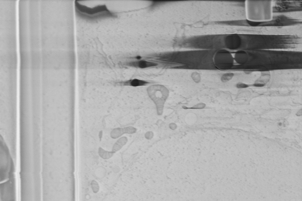 |
Cryo serial FIB/SEM of Vero cells [46 micrographs in TIFF format] | Dumoux M, Glen T, Smith JLR, Ho EML, Perdigão LMA, Pennington A, Klumpe S, Yee NBY, Farmer DA, Lai PYA, Bowles W, Kelley R, Plitzko JM, Wu L, Basham M, Clare DK, Siebert CA, Darrow MC, Naismith JH, Grange M [Pubmed: 36805107] [DOI: 10.7554/elife.83623] |
7.1 GB | — |
| 2023-02-17 | 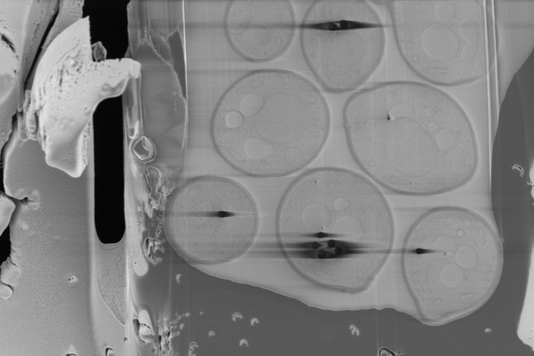 |
Cryo serial FIB/SEM of Saccharomyces cerevisiae [109 micrographs in TIFF format] | Dumoux M, Glen T, Smith JLR, Ho EML, Perdigão LMA, Pennington A, Klumpe S, Yee NBY, Farmer DA, Lai PYA, Bowles W, Kelley R, Plitzko JM, Wu L, Basham M, Clare DK, Siebert CA, Darrow MC, Naismith JH, Grange M [Pubmed: 36805107] [DOI: 10.7554/elife.83623] |
4.8 GB | — |
| 2023-02-28 | 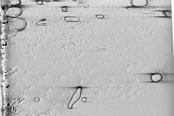 |
Cryo serial FIB SEM of mouse brain tissue [59 micrographs in TIFF format] | Dumoux M, Glen T, Smith JLR, Ho EML, Perdigão LMA, Pennington A, Klumpe S, Yee NBY, Farmer DA, Lai PYA, Bowles W, Kelley R, Plitzko JM, Wu L, Basham M, Clare DK, Siebert CA, Darrow MC, Naismith JH, Grange M [Pubmed: 36805107] [DOI: 10.7554/elife.83623] |
3.0 GB | — |
| 2023-02-28 | 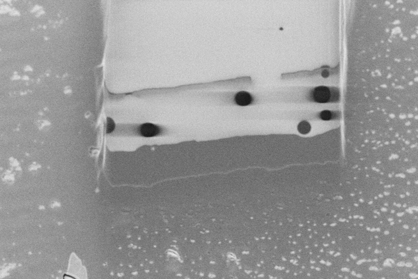 |
Cryo pFIB/SEM of PEG beads (test sample) [193 micrographs in TIFF format] | Dumoux M, Glen T, Smith JLR, Ho EML, Perdigão LMA, Pennington A, Klumpe S, Yee NBY, Farmer DA, Lai PYA, Bowles W, Kelley R, Plitzko JM, Wu L, Basham M, Clare DK, Siebert CA, Darrow MC, Naismith JH, Grange M [Pubmed: 36805107] [DOI: 10.7554/elife.83623] |
5.3 GB | — |
| 2023-06-27 | 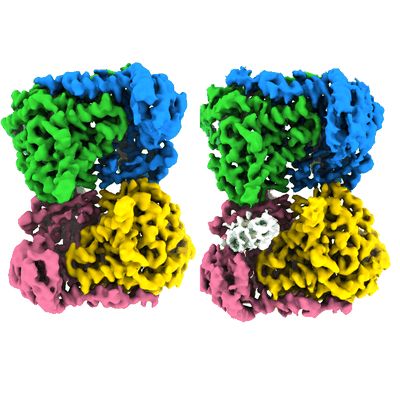 |
Unaligned cryo-EM micrographs of Human SHMT1 in complex with RNA [5450 multi-frame micrographs composed of 40 frames each in TIFF format] | Spizzichino S, Marabelli C, Chaves-Sanjuan A, Bolognesi M, Giardina G, Cutruzzola F | 3.9 TB | 3.52 Å |
