Electron Microscopy Public Image Archive
Electron Microscopy Public Image Archive
大阪大學的EMPIAR-PDBj團隊為亞洲EM研究人員向EMPIAR傳送大型EM圖像數據提供服務。 除了通過互聯網將數據直接傳送到EBI(UK),研究人員還可以通過郵政或快遞服務將數據硬盤發送到大阪大學,或者通過互聯網傳送到設置於大阪大學的服務器,然後由我們代為傳送至數據登錄網站。 如果您想使用此項服務,請先通過 電子郵件 與我們聯繫。
| Release date | Imageset | Title | Authors and references | Size | Resolution |
|---|---|---|---|---|---|
| 2020-10-14 | 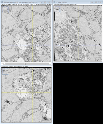 |
FIB SEM images of a Zebrafish hindbrain macrophage containing 2 Toxoplasma gondii tachizoites [multiple data sets in TIFF format] | Peddie CJ, Domart MC, Collinson L [Pubmed: 32461265] [DOI: 10.1242/dmm.043091] |
1.1 TB | — |
| 2020-12-09 | 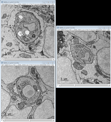 |
TEM images of a Zebrafish hindbrain cells containing Toxoplasma gondii tachizoites [multiple data sets in TIFF format] | Domart MC, Collinson L [Pubmed: 32461265] [DOI: 10.1242/dmm.043091] |
3.4 GB | — |
| 2022-03-28 |  |
Representative data from Near-native state imaging by cryo-soft-X-ray tomography reveals remodelling of cytoplasmic vesicles and mitochondria during HSV-1 infection [14 reconstructed volumes in MRC format] | Nahas KLN, Connor VC, Scherer KM, Kaminski CF, Harkiolaki M, Crump CM, Graham SC [DOI: 10.1101/2021.10.11.463900] |
9.9 GB | — |
| 2021-01-22 | 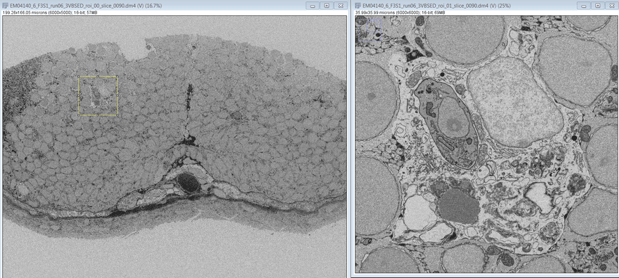 |
SBF SEM images of a Zebrafish hindbrain macrophage containing 2 Toxoplasma gondii tachizoites [multiple data sets in DM4 format] | Peddie CJ, Domart MC, Collinson L [Pubmed: 32461265] [DOI: 10.1242/dmm.043091] |
16.5 GB | — |
| 2020-12-09 | 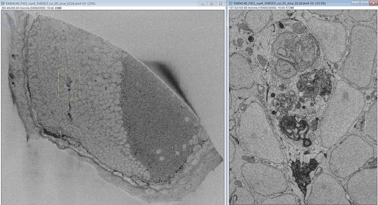 |
SBF SEM images of a Zebrafish hindbrain macrophage containing a replicating Toxoplasma gondii tachizoite [multiple data sets in DM4 format] | Peddie CJ, Domart MC, Collinson L [Pubmed: 32461265] [DOI: 10.1242/dmm.043091] |
338.4 GB | — |
| 2020-12-09 | 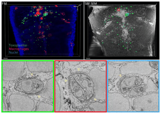 |
SBF SEM images of a Zebrafish hindbrain containing several Toxoplasma gondii tachizoites at different stages of replication [multiple data sets in DM4 and TIFF formats] | Peddie CJ, Domart MC, Collinson L [Pubmed: 32461265] [DOI: 10.1242/dmm.043091] |
175.2 GB | — |
| 2021-10-27 | 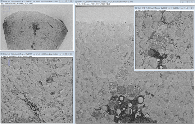 |
SBF SEM images of a Zebrafish hindbrain containing several Toxoplasma gondii tachizoites at different stages of replication [multiple data sets in DM4 format] | Peddie CJ, Domart MC, Collinson L [Pubmed: 32461265] [DOI: 10.1242/dmm.043091] |
553.9 GB | — |
| 2024-02-15 | 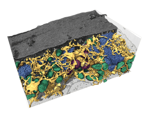 |
FIB-SEM dataset of a human bone osteosarcoma epithelial cell (U2-OS) [1168 micrographs in TIFF format] | Belevich I, Schertel A, Zaversek T, Szyrynska N, Saarnio S, Jokitalo E | 5.2 GB | — |
| 2022-03-21 |  |
CryoET of E. coli prepared with the Waffle Method [1504 multi-frame micrographs composed of 14 frames each in TIFF format] | Kelley K, Raczkowski AM, Klykov O, Jaroenlak P, Bobe D, Kopylov M, Eng ET, Bhabha G, Potter CS, Carragher B, Noble AJ [Pubmed: 35387991] [DOI: 10.1038/s41467-022-29501-3] |
94.0 GB | — |
| 2022-12-13 |  |
Focused ion beam-scanning electron microscopy links pathological myelin outfoldings to axonal changes in mice lacking Plp1 or Mag [1598 micrographs in TIFF format] | Steyer AM, Möbius W [Pubmed: 36354016] [DOI: 10.1002/glia.24290] |
31.3 GB | — |
| 2018-10-25 |  |
CryoET of bacterial RNA polymerase with several detergents [multiple data sets in MRC and RAW TEXT formats] | Chen J, Noble AJ, Kang JY, Darst SA [DOI: 10.1016/j.yjsbx.2019.100005] |
207.5 GB | — |
| 2022-03-15 | 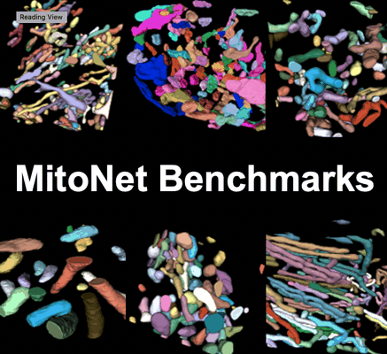 |
Seven benchmark datasets of instance segmentation of mitochondria: 6 diverse volume EM + 1 TEM (100 images) datasets [multiple data sets in TIFF format] | Narayan K, Conrad RW | 4.1 GB | — |
| 2022-12-13 |  |
Focused ion beam-scanning electron microscopy links pathological myelin outfoldings to axonal changes in mice lacking Plp1 or Mag [1553 micrographs in TIFF format] | Steyer AM, Möbius W [Pubmed: 36354016] [DOI: 10.1002/glia.24290] |
22.3 GB | — |
| 2023-11-06 |  |
Single particle cryo-EM structure of RIG-I:RNA:Riplet ternary complex [3420 multi-frame micrographs composed of 40 frames each in TIFF format] | Wang W, Pyle AM [DOI: 10.1038/s41467-023-42982-0] |
1.5 TB | — |
| 2022-12-13 |  |
Focused ion beam-scanning electron microscopy links pathological myelin outfoldings to axonal changes in mice lacking Plp1 or Mag [910 micrographs in TIFF format] | Steyer AM, Möbius W [Pubmed: 36354016] [DOI: 10.1002/glia.24290] |
13.7 GB | — |
| 2022-12-13 |  |
Focused ion beam-scanning electron microscopy links pathological myelin outfoldings to axonal changes in mice lacking Plp1 or Mag [1186 micrographs in TIFF format] | Steyer AM, Möbius W [Pubmed: 36354016] [DOI: 10.1002/glia.24290] |
28.2 GB | — |
| 2022-11-18 |  |
A high-throughput electron tomography workflow reveals over-elongated centrioles in relapsed-refractory multiple myeloma [multiple data sets in MRC format] | Köhrer S, Dittrich T, Schorb M, Weinhold N, Haberbosch I, Börmel M, Pajor G [Pubmed: 36452870] [DOI: 10.1016/j.crmeth.2022.100322] |
2.0 TB | — |
| 2023-11-13 |  |
Test subset: In situ cryo-ET dataset of Chlamydomonas reinhardtii prepared using cryo-plasmaFIB milling [18 tilt series in MRC format] | Kelley R, Zhang X, Obr M, Khavnekar S, Righetto R, Waltz F, Wietrzynski W, Michael A, Tagiltsev G, Beck F, Zhong E, Wan W, Briggs J, Plitzko J, Engel B, Kotecha A [Pubmed: 37613825] [DOI: 10.1093/micmic/ozad067.480] |
293.7 GB | — |
| 2022-11-23 | 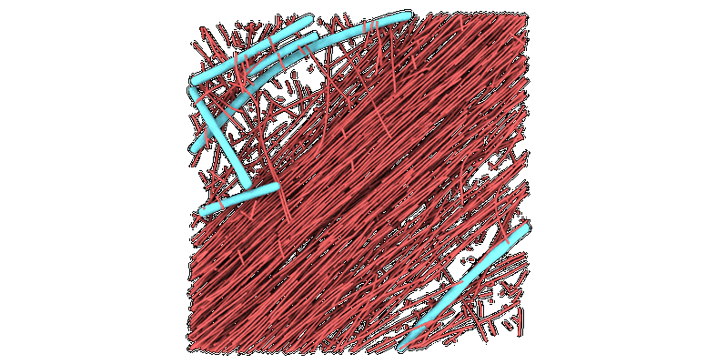 |
Cryo-electron tomograms of RPE1 cells with comprehensive annotation of actin filaments and microtubules [multiple data sets in TIFF and MRC formats] | Cheng DWC, Goetz SK, Mahamid J [Pubmed: 36690741] [DOI: 10.1038/s41592-022-01746-2] |
32.9 GB | — |
| 2020-08-06 | 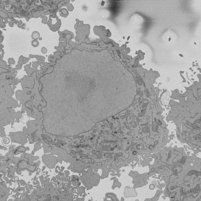 |
Cropped regions from Serial Block Face SEM of HeLa cell pellet with 10 nm pixels and 50 nm slices (benchmark dataset) [18 multi-frame micrographs composed of 300 frames each in TIFF format] | Peddie CP, Jones ML, Collinson LM | 15.6 GB | — |
| 2020-08-06 | 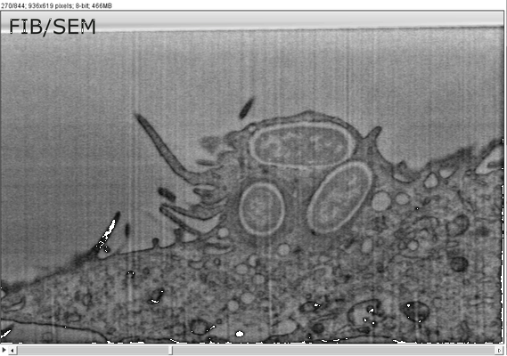 |
FIB/SEM sample dataset MCB-CLEM IV Weiner [1 multi-frame micrographs composed of 844 frames each in TIFF format] | Weiner A | 2.8 GB | — |
| 2024-01-15 | 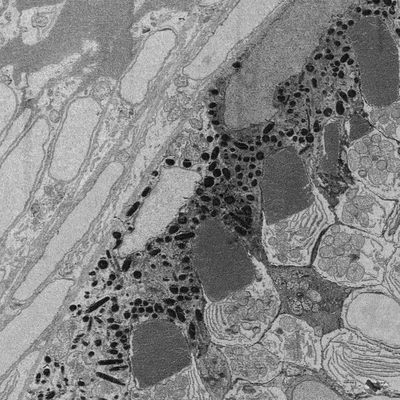 |
Developing retina in zebrafish 55 hpf larval eye. [16 reconstructed volumes in DM3 format] | Wilsch-Bräuninger M | 1.2 GB | — |
| 2019-02-01 |  |
Bdellovibrio electron cryotomography tilt-series acquired by continuous tilting [1 tilt series in MRC format] | Chreifi G, Chen S, Metskas LA, Kaplan M, Jensen GJ [Pubmed: 30639925] [DOI: 10.1016/j.jsb.2018.12.008] |
2.1 GB | — |
| 2023-10-23 |  |
High-throughput electron tomography identifies centriole over-elongation in plasma cell disorders [multiple data sets in MRC format] | Köhrer S, Dittrich T, Schorb M, Weinhold N, Haberbosch I, Börmel M, Pajor G, Goldschmidt H, Müller-Tidow C, Raab MS, John L, Seckinger A, Brobeil A, Dreger P, Tornóczky T, Pajor L, Hegenbart U, Schönland SO, Schwab Y, Krämer A [Pubmed: 37821581] [DOI: 10.1038/s41375-023-02056-y] |
7.7 TB | — |
| 2024-04-17 |  |
Spatial mapping of hepatic ER and mitochondria architecture reveals zonated remodeling in fasting and obesity [multiple data sets in TIFF format] | Parlakgul G | 2.9 TB | — |
