Electron Microscopy Public Image Archive
Electron Microscopy Public Image Archive
大阪大學的EMPIAR-PDBj團隊為亞洲EM研究人員向EMPIAR傳送大型EM圖像數據提供服務。 除了通過互聯網將數據直接傳送到EBI(UK),研究人員還可以通過郵政或快遞服務將數據硬盤發送到大阪大學,或者通過互聯網傳送到設置於大阪大學的服務器,然後由我們代為傳送至數據登錄網站。 如果您想使用此項服務,請先通過 電子郵件 與我們聯繫。
| Release date | Imageset | Title | Authors and references | Size | Resolution |
|---|---|---|---|---|---|
| 2021-02-10 |  |
Cryo-EM structure of K+-bound hERG channel [1496 multi-frame micrographs composed of 50 frames each in TIFF format] | Asai T, Adachi N, Moriya T, Kawasaki M, Suzuki K, Senda T, Murata T [Pubmed: 33450182] [DOI: 10.1016/j.str.2020.12.007] |
1.4 TB | 3.9 Å |
| 2021-02-10 | 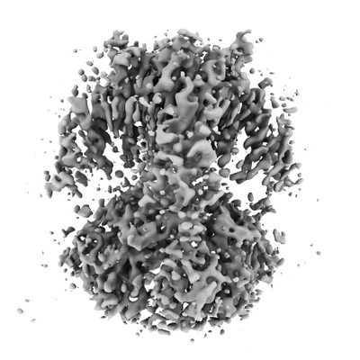 |
Cryo-EM structure of K+-bound hERG channel in the presence of astemizole [1865 multi-frame micrographs composed of 50 frames each in TIFF format] | Asai T, Adachi N, Moriya T, Kawasaki M, Suzuki K, Senda T, Murata T [Pubmed: 33450182] [DOI: 10.1016/j.str.2020.12.007] |
1.8 TB | 3.7 Å |
| 2023-08-18 | 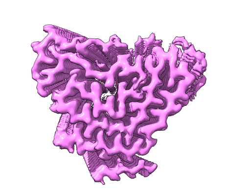 |
Structure of TDP-43 amyloid filaments from type A FTLD-TDP (individual 2) [33336 multi-frame micrographs composed of 40 frames each in TIFF format] | Arseni D, Ryskeldi-Falcon B [Pubmed: 37532939] [DOI: 10.1038/s41586-023-06405-w] |
5.3 TB | 2.39 Å |
| 2023-08-18 | 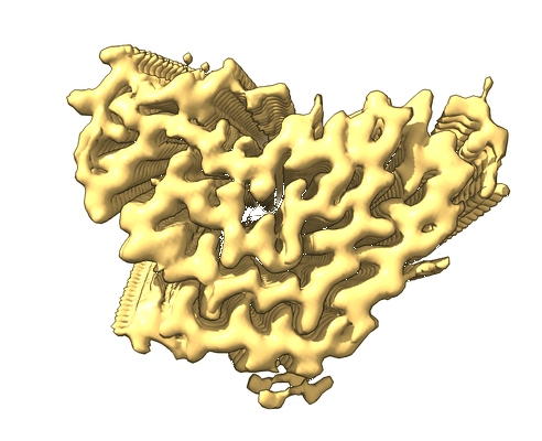 |
Structure of TDP-43 amyloid filaments from type A FTLD-TDP (individual 3) [36507 multi-frame micrographs composed of 40 frames each in TIFF format] | Arseni D, Ryskeldi-Falcon B [Pubmed: 37532939] [DOI: 10.1038/s41586-023-06405-w] |
5.7 TB | 2.39 Å |
| 2023-09-22 | 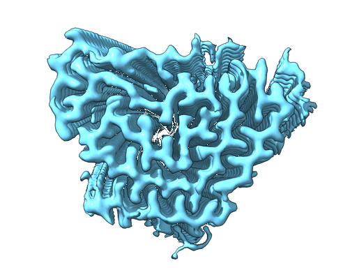 |
Structure of TDP-43 amyloid filaments from type A FTLD-TDP (individual 1) [91457 multi-frame micrographs composed of 40 frames each in TIFF format] | Arseni D, Ryskeldi-Falcon B [Pubmed: 37532939] [DOI: 10.1038/s41586-023-06405-w] |
14.5 TB | 2.39 Å |
| 2022-01-24 | 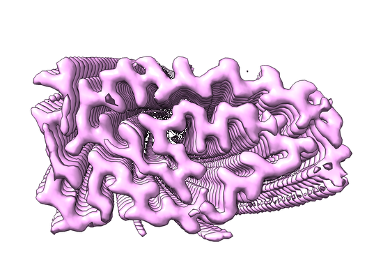 |
Structure of pathological TDP-43 filaments from ALS with FTLD (Individual 1, frontal cortex) [22896 multi-frame micrographs composed of 40 frames each in TIFF format] | Arseni D, Hasegawa M, Murzin AG, Kametani F, Arai M, Yoshida M, Ryskeldi-Falcon B [Pubmed: 34880495] [DOI: 10.1038/s41586-021-04199-3] |
3.9 TB | 2.59 Å |
| 2022-01-21 | 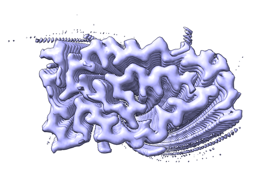 |
Structure of pathological TDP-43 filaments from ALS with FTLD (Individual 1, motor cortex) [12245 multi-frame micrographs composed of 40 frames each in TIFF format] | Arseni D, Hasegawa M, Murzin AG, Kametani F, Arai M, Yoshida M, Ryskeldi-Falcon B [Pubmed: 34880495] [DOI: 10.1038/s41586-021-04199-3] |
2.0 TB | 2.94 Å |
| 2022-01-24 | 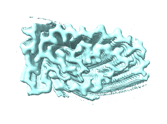 |
Structure of pathological TDP-43 filaments from ALS with FTLD (Individual 2, frontal cortex) [15991 multi-frame micrographs composed of 41 frames each in TIFF format] | Arseni D, Hasegawa M, Murzin AG, Kametani F, Arai M, Yoshida M, Ryskeldi-Falcon B [Pubmed: 34880495] [DOI: 10.1038/s41586-021-04199-3] |
2.9 TB | 2.94 Å |
| 2022-06-20 | 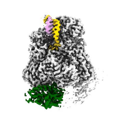 |
Cryo-EM data used for the determination of the structures of LACV-L in 3 different states: replication initiation state, transcription capped primer active site entry state and transcription initiation state [14341 multi-frame micrographs composed of 40 frames each in TIFF format] | Arragain B, Durieux Trouilleton Q, Baudin F, Provaznik J, Azevedo N, Cusack S, Schoehn G, Malet H [Pubmed: 35173159] [DOI: 10.1038/s41467-022-28428-z] |
2.2 TB | 2.8 - 3.6 Å |
| 2022-05-24 | 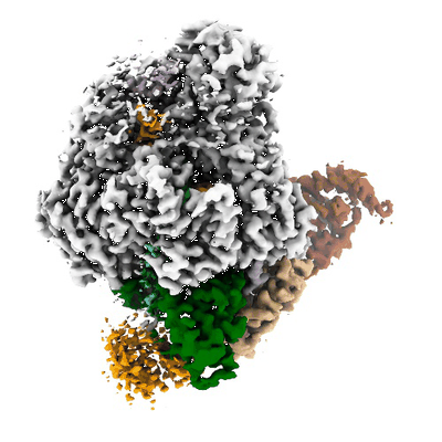 |
Cryo-EM data used for the determination of LACV-L structure in transcription early-elongation state [2510 multi-frame micrographs composed of 60 frames each in TIFF format] | Arragain B, Durieux Trouilleton Q, Baudin F, Provaznik J, Azevedo N, Cusack S, Schoehn G, Malet H [Pubmed: 35173159] [DOI: 10.1038/s41467-022-28428-z] |
721.4 GB | 3.3 Å |
| 2020-09-11 | 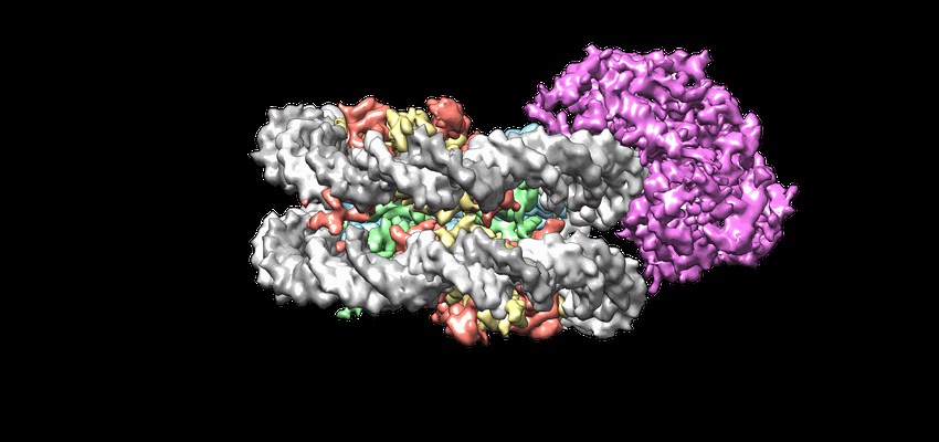 |
Cryo-EM structures of remodeler-nucleosome intermediates suggest allosteric control through the nucleosome [719 multi-frame micrographs composed of 30 frames each in MRCS format] | Armache J-P, Gamarra N, Johnson SL, Leonard JD, Wu S, Narlikar G, Cheng Y [Pubmed: 31210637] [DOI: 10.7554/eLife.46057] |
1.4 TB | 3.39 Å |
| 2021-11-16 |  |
The cryo-EM structure of the CENP-A nucleosome in complex with the phosphorylated CENP-C: CENP-A nucleosome in complex with phosphorylated CENP-C C-terminal domain (601-864) and CENP-N N-terminal domain (1-211) [8017 multi-frame micrographs composed of 50 frames each in TIFF format] | Ariyoshi M, Makino F, Watanabe R, Nakagawa R, Kato T, Namba K, Arimura Y, Fujita R, Kurumizaka H, Okumura EI, Hara M, Fukagawa T [Pubmed: 33463726] [DOI: 10.15252/embj.2020105671] |
1.6 TB | 4.5 - 7.8 Å |
| 2021-11-16 |  |
The cryo-EM structure of the CENP-A nucleosome in complex with the phosphorylated CENP-C:: CENP-A nucleosome in complex with phosphorylated CENP-C C-terminal domain(601-864) [6533 multi-frame micrographs composed of 50 frames each in TIFF format] | Ariyoshi M, Makino F, Watanabe R, Nakagawa R, Kato T, Namba K, Arimura Y, Fujita R, Kurumizaka H, Okumura EI, Hara M, Fukagawa T [Pubmed: 33463726] [DOI: 10.15252/embj.2020105671] |
1.4 TB | 6.8 Å |
| 2021-11-16 | 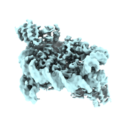 |
The cryo-EM structure of the CENP-A nucleosome in complex with the phosphorylated CENP-C:: CENP-A nucleosome in complex with CENP-C motif (655-675) and CENP-N N-terminal domain (1-211) [4630 multi-frame micrographs composed of 50 frames each in TIFF format] | Ariyoshi M, Makino F, Watanabe R, Nakagawa R, Kato T, Namba K, Arimura Y, Fujita R, Kurumizaka H, Okumura EI, Hara M, Fukagawa T [Pubmed: 33463726] [DOI: 10.15252/embj.2020105671] |
1.3 TB | 4.2 Å |
| 2021-09-24 |  |
Oligo nucleosome fraction from metaphase chromosome in Xenopus egg extract (lot1) [multiple data sets in TIFF format] | Arimura YA, Funabiki HF [Pubmed: 34478647] [DOI: 10.1016/j.molcel.2021.08.010] |
1.3 TB | 3.5 - 5.5 Å |
| 2021-09-24 |  |
Oligo nucleosome fraction from interphase chromosome in Xenopus egg extract lot 1 [1364 multi-frame micrographs composed of 50 frames each in TIFF format] | Arimura YA, Funabiki HF [Pubmed: 34478647] [DOI: 10.1016/j.molcel.2021.08.010] |
634.3 GB | 3.38 - 8.07 Å |
| 2021-09-24 | 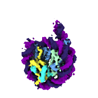 |
Mono nucleosome fraction from interphase chromosome in Xenopus egg extract lot1 [1656 multi-frame micrographs composed of 50 frames each in TIFF format] | Arimura YA, Funabiki HF [Pubmed: 34478647] [DOI: 10.1016/j.molcel.2021.08.010] |
749.0 GB | 5.12 Å |
| 2021-09-22 | 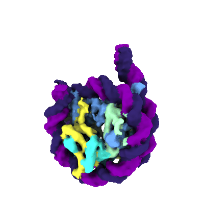 |
Mono nucleosome fraction from metaphase chromosome in Xenopus egg extract lot1 [1792 multi-frame micrographs composed of 50 frames each in TIFF format] | Arimura YA, Funabiki HF [Pubmed: 34478647] [DOI: 10.1016/j.molcel.2021.08.010] |
778.6 GB | 5.64 Å |
| 2021-09-24 | 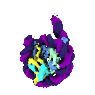 |
Di nucleosome fraction from interphase chromosome in Xenopus egg extract lot1 [1656 multi-frame micrographs composed of 50 frames each in TIFF format] | Arimura YA, Funabiki HF [Pubmed: 34478647] [DOI: 10.1016/j.molcel.2021.08.010] |
805.9 GB | 4.74 Å |
| 2021-09-22 | 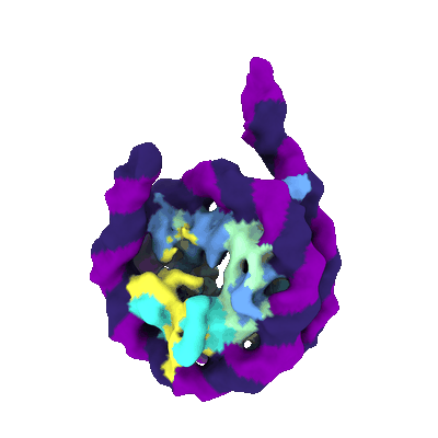 |
Di nucleosome fraction from metaphase chromosome in Xenopus egg extract lot1 [1386 multi-frame micrographs composed of 50 frames each in TIFF format] | Arimura YA, Funabiki HF [Pubmed: 34478647] [DOI: 10.1016/j.molcel.2021.08.010] |
825.4 GB | 8.1 Å |
| 2021-09-22 |  |
Oligo nucleosome fraction from metaphase chromosome in Xenopus egg extract lot2 [1456 multi-frame micrographs composed of 50 frames each in TIFF format] | Arimura YA, Funabiki HF [Pubmed: 34478647] [DOI: 10.1016/j.molcel.2021.08.010] |
696.5 GB | 3.77 - 4.32 Å |
| 2021-09-22 |  |
Oligo nucleosome fraction from interphase chromosome in Xenopus egg extract lot2 [1376 multi-frame micrographs composed of 50 frames each in TIFF format] | Arimura YA, Funabiki HF [Pubmed: 34478647] [DOI: 10.1016/j.molcel.2021.08.010] |
648.2 GB | 3.54 - 4.42 Å |
| 2021-09-22 |  |
Open linker DNA nucleosome reconstituted with GUB DNA [1576 multi-frame micrographs composed of 50 frames each in TIFF format] | Arimura YA, Funabiki HF [Pubmed: 34478647] [DOI: 10.1016/j.molcel.2021.08.010] |
777.0 GB | 4.52 Å |
| 2021-09-22 |  |
Closed linker DNA nucleosome reconstituted with GUB DNA [1320 multi-frame micrographs composed of 50 frames each in TIFF format] | Arimura YA, Funabiki HF [Pubmed: 34478647] [DOI: 10.1016/j.molcel.2021.08.010] |
624.4 GB | 3.77 Å |
| 2021-09-24 |  |
'Freed' nucleosome isolated from non-crosslinked interphase chromosome in Xenopus egg extract lot2 [multiple data sets in TIFF format] | Arimura YA, Funabiki HF [Pubmed: 34478647] [DOI: 10.1016/j.molcel.2021.08.010] |
1.1 TB | 5.44 Å |
