Electron Microscopy Public Image Archive
Electron Microscopy Public Image Archive
大阪大學的EMPIAR-PDBj團隊為亞洲EM研究人員向EMPIAR傳送大型EM圖像數據提供服務。 除了通過互聯網將數據直接傳送到EBI(UK),研究人員還可以通過郵政或快遞服務將數據硬盤發送到大阪大學,或者通過互聯網傳送到設置於大阪大學的服務器,然後由我們代為傳送至數據登錄網站。 如果您想使用此項服務,請先通過 電子郵件 與我們聯繫。
| Release date | Imageset | Title | Authors and references | Size | Resolution |
|---|---|---|---|---|---|
| 2020-10-09 |  |
Structure of two nucleosomes bridged by human PARP2 [multiple data sets in MRCS format] | Gaullier G, Morgan GP, Luger K [Pubmed: 33141820] [DOI: 10.1371/journal.pone.0240932] |
677.3 GB | 10.5 Å |
| 2022-04-29 | 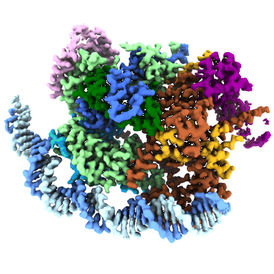 |
Structure of transcription factor UAF in complex with TBP and 35S rRNA promoter DNA [multiple data sets in TIFF format] | Baudin F, Murciano B, Fung HKH, Fromm SA, Mattei S, Mahamid J, Müller CW [Pubmed: 35442737] [DOI: 10.1126/sciadv.abn5725] |
3.1 TB | 2.8 Å |
| 2023-10-13 | 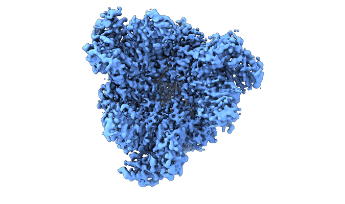 |
Structure of the peroxisomal Pex1/Pex6 ATPase complex bound to a substrate [multiple data sets in TIFF format] | Rüttermann MR, Koci MK, Lill PL, Geladas EDG, Kaschani FK, Klink BUK, Erdmann RE, Gatsogiannis CG [Pubmed: 37741838] [DOI: 10.1038/s41467-023-41640-9] |
4.9 TB | 4.1 - 4.7 Å |
| 2022-03-21 | 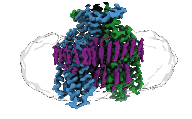 |
Structure of the ligand-free GPCR dimer Ste2 [9369 multi-frame micrographs composed of 53 frames each in EER format] | Velazhahan V, Tate CG [Pubmed: 35296853] [DOI: 10.1038/s41586-022-04498-3] |
8.2 TB | 3.1 Å |
| 2024-03-26 | 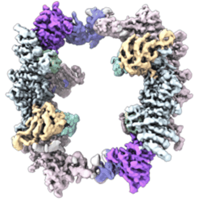 |
Structure of the human UBR5 HECT-type E3 ubiquitin ligase in a tetrameric form [17529 multi-frame micrographs composed of 75 frames each in TIFF format] | Wang FW [Pubmed: 37040767] [DOI: 10.1016/j.str.2023.03.010] |
7.6 TB | 3.5 Å |
| 2024-02-16 | 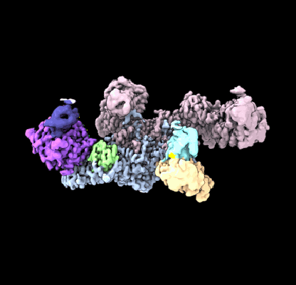 |
Structure of the human UBR5 HECT-type E3 ubiquitin ligase in a dimeric form [13094 multi-frame micrographs composed of 75 frames each in TIFF format] | Wang FW [Pubmed: 37040767] [DOI: 10.1016/j.str.2023.03.010] |
7.0 TB | 2.66 - 2.8 Å |
| 2021-03-05 | 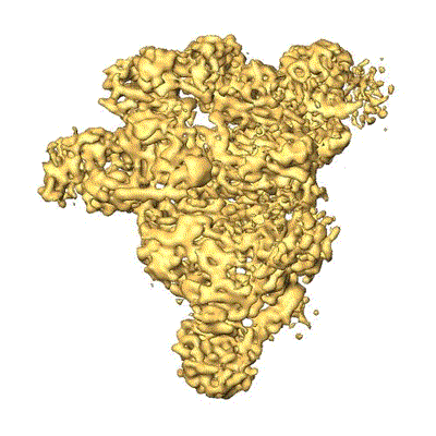 |
Structure of the human U4/U6.U5 tri-snRNP [4812 micrographs in MRC format] | Stark H [Pubmed: 26912367] [DOI: 10.1126/science.aad2085] |
300.9 GB | 7.0 Å |
| 2022-07-22 | 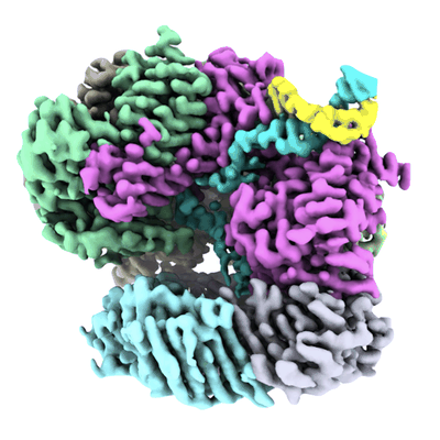 |
Structure of the human RAD17-RFC clamp loader and 9-1-1 checkpoint clamp bound to a dsDNA-ssDNA junction [8271 multi-frame micrographs composed of 882 frames each in EER format] | Day M, Oliver AW, Pearl LH [Pubmed: 35819203] [DOI: 10.1093/nar/gkac588] |
4.4 TB | 3.59 Å |
| 2019-05-02 | 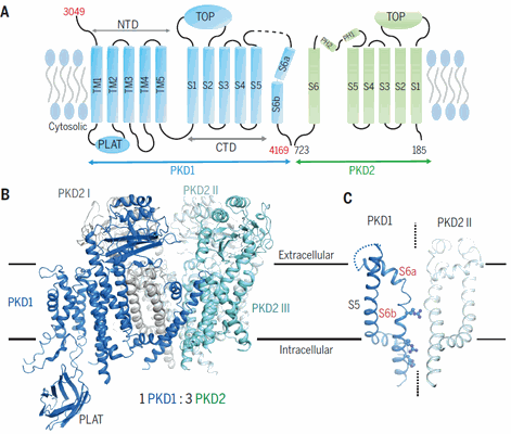 |
Structure of the human PKD1-PKD2 complex [stack of 3774 particles in MRCS format] | Su Q [Pubmed: 30093605] [DOI: 10.1126/science.aat9819] |
209.8 GB | 3.6 Å |
| 2017-02-09 | 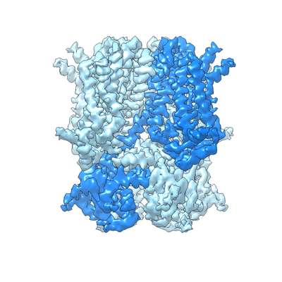 |
Structure of the human HCN1 hyperpolarization-activated cyclic nucleotide-gated ion channel [multiple data sets in MRC format] | Lee CH, MacKinnon R [Pubmed: 28086084] [DOI: 10.1016/j.cell.2016.12.023] |
66.5 GB | 3.5 Å |
| 2018-07-06 | 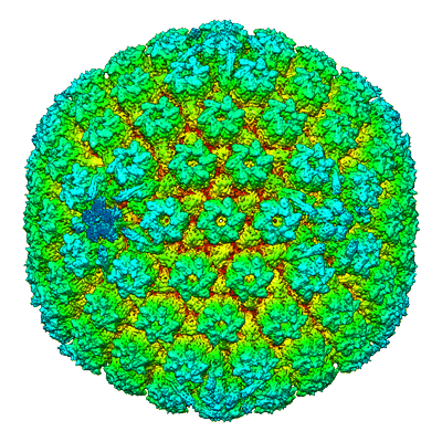 |
Structure of the herpes-simplex virus portal-vertex [3818 micrographs in MRC format] | McElwee M, Vijayakrishnan S, Rixon FJ, Bhella D [Pubmed: 29924793] [DOI: 10.1371/journal.pbio.2006191] |
238.6 GB | 7.7 Å |
| 2019-11-11 |  |
Structure of the green algal photosystem I supercomplex with a decameric light-harvesting complex I. [10989 multi-frame micrographs composed of 33 frames each in TIFF format] | Suga M, Ozawa S, Yoshida-Motomura K, Akita F, Miyazaki N, Takahashi Y [Pubmed: 31182847] [DOI: 10.1038/s41477-019-0438-4] |
8.0 TB | 2.8 - 2.9 Å |
| 2020-12-04 | 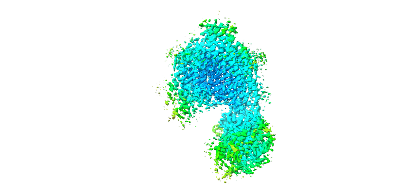 |
Structure of the class D GPCR Ste2 dimer coupled to two G proteins [multiple data sets in MRC and TIFF formats] | Velazhahan V, Ma N, Pándy-Szekeres G, Kooistra AJ, Lee Y, Gloriam DE, Vaidehi N, Tate CG [Pubmed: 33268889] [DOI: 10.1038/s41586-020-2994-1] |
1.3 TB | 3.3 Å |
| 2022-03-21 | 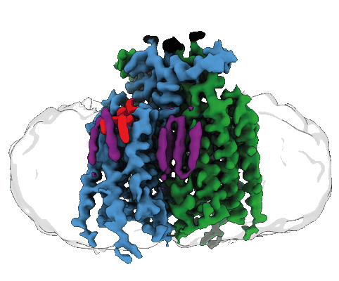 |
Structure of the agonist-bound GPCR dimer Ste2 [6944 multi-frame micrographs composed of 50 frames each in MRC format] | Velazhahan V, Tate CG [Pubmed: 35296853] [DOI: 10.1038/s41586-022-04498-3] |
1.3 TB | 3.46 - 3.53 Å |
| 2023-01-16 | 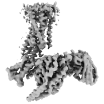 |
Structure of the active Gi-coupled human lysophosphatidic acid receptor 1 complexed with a potent agonist [6228 multi-frame micrographs composed of 48 frames each in TIFF format] | Akasaka H, Tanaka T, Sano FK, Matsuzaki Y, Shihoya W, Nureki O [Pubmed: 36109516] [DOI: 10.1038/s41467-022-33121-2] |
1.4 TB | 3.5 - 5.6 Å |
| 2018-01-03 | 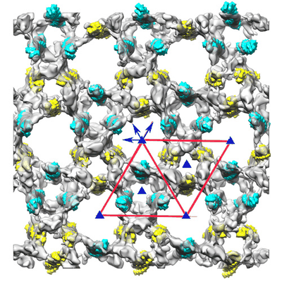 |
Structure of the Z-disk isolated from the indirect flight muscle of the honey bee [96 class averages in MRC format] | Rusu M, Hu Z, Taylor KA, Trinick J [Pubmed: 28733815] [DOI: 10.1007/s10974-017-9477-5] |
3.0 GB | 60.0 Å |
| 2024-04-09 | 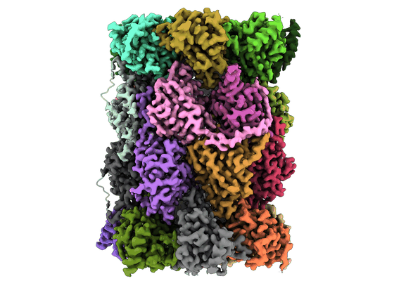 |
Structure of the Plasmodium falciparum 20S proteasome complexed with inhibitor TDI-8304 [29421 multi-frame micrographs composed of 75 frames each in TIFF format] | Hsu HC, Li H [Pubmed: 38097652] [DOI: 10.1038/s41467-023-44077-2] |
16.7 TB | 2.18 Å |
| 2024-04-03 | 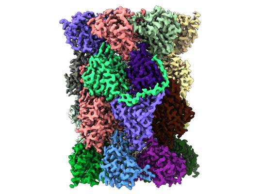 |
Structure of the Plasmodium falciparum 20S proteasome beta-6 A117D mutant complexed with inhibitor WLW-vs [24086 multi-frame micrographs composed of 65 frames each in TIFF format] | Hsu HC, Li H [Pubmed: 38097652] [DOI: 10.1038/s41467-023-44077-2] |
11.3 TB | 2.58 Å |
| 2023-07-14 | 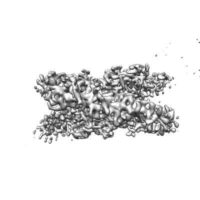 |
Structure of the Nucleosome Core Particle from Trypanosoma brucei [4913 multi-frame micrographs composed of 40 frames each in TIFF format] | Sandoval G, Deak G, Wapenaar H, Tuijtel MW, Wilson MD [Pubmed: 37427792] [DOI: 10.1093/nar/gkad577] |
1.4 TB | 3.3 Å |
| 2018-10-25 | 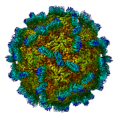 |
Structure of the Macrobrachium rosenbergii Nodavirus: A new genus within the Nodaviridae? [2459 micrographs in MRC format] | Ho KL, Gabrielsen M, Beh PL, Kueh CL, Thong QX, Streetley J, Tan WS, Bhella D [Pubmed: 30346944] [DOI: 10.1371/journal.pbio.3000038] |
130.4 GB | 3.28 Å |
| 2022-03-21 | 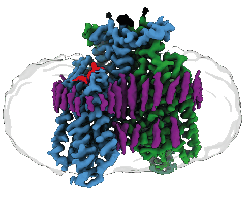 |
Structure of the GPCR dimer Ste2 bound to an antagonist [15751 multi-frame micrographs composed of 59 frames each in TIFF format] | Velazhahan V, Tate CG [Pubmed: 35296853] [DOI: 10.1038/s41586-022-04498-3] |
4.1 TB | 2.7 Å |
| 2022-07-18 | 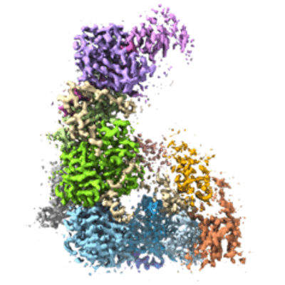 |
Structure of the Dicer-2-R2D2 heterodimer bound to a small RNA duplex [multiple data sets in TIFF format] | Yamaguchi S, Naganuma M, Nishizawa T, Kusakizako T, Tomari Y, Nishimasu H, Nureki O [Pubmed: 35768503] [DOI: 10.1038/s41586-022-04790-2] |
1.4 TB | 3.3 Å |
| 2020-09-30 | 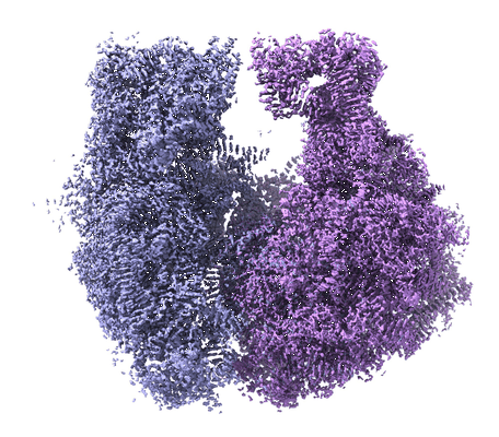 |
Structure of the Bacterial Ribosome at 2 Å Resolution [multiple data sets in TIFF format] | Watson ZL, Ward FR, Méheust R, Ad O, Schepartz A, Banfield JF, Cate JH [Pubmed: 32924932] [DOI: 10.7554/eLife.60482] |
2.1 TB | 1.98 Å |
| 2021-01-29 | 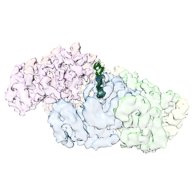 |
Structure of spastin bound to a glutamate-rich peptide implies a hand-over-hand mechanism of substrate translocation. [1200 multi-frame micrographs composed of 49 frames each in MRC format] | Han H, Schubert HL, Purdy MD, Yeager M, Sundquist WI, Hill CP [Pubmed: 31767681] [DOI: 10.1074/jbc.AC119.009890] |
1.8 TB | 4.2 Å |
| 2020-05-13 | 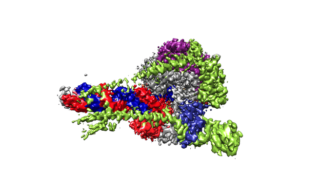 |
Structure of replicating SARS-CoV-2 polymerase [multiple data sets in TIFF and MRCS formats] | Hillen HS, Kokic G, Farnung L, Dienemann C, Tegunov D, Cramer P [Pubmed: 32438371] [DOI: 10.1038/s41586-020-2368-8] |
3.0 TB | 2.9 Å |
