Electron Microscopy Public Image Archive
Electron Microscopy Public Image Archive
大阪大學的EMPIAR-PDBj團隊為亞洲EM研究人員向EMPIAR傳送大型EM圖像數據提供服務。 除了通過互聯網將數據直接傳送到EBI(UK),研究人員還可以通過郵政或快遞服務將數據硬盤發送到大阪大學,或者通過互聯網傳送到設置於大阪大學的服務器,然後由我們代為傳送至數據登錄網站。 如果您想使用此項服務,請先通過 電子郵件 與我們聯繫。
| Release date | Imageset | Title | Authors and references | Size | Resolution |
|---|---|---|---|---|---|
| 2023-07-28 | 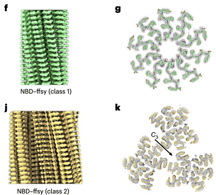 |
Cryo-EM micrographs of NBD–ffsy filaments [7680 multi-frame micrographs composed of 40 frames each in TIFF format] | Wang F [Pubmed: 37217766] [DOI: 10.1038/s41565-023-01401-7] |
1.7 TB | 3.1 - 3.2 Å |
| 2022-07-26 |  |
Cryo electron images of mitochondrial Hsp60 [8055 multi-frame micrographs composed of 22 frames each in TIFF format] | Walti MA [Pubmed: 34688687] [DOI: 10.1016/j.jmb.2021.167322] |
2.5 TB | 4.35 Å |
| 2018-06-18 |  |
RNA Polymerase III pre-initiation complex [multiple data sets in TIFF and MRC formats] | Vorländer MK, Khatter H, Wetzel R, Hagen WJH, Müller CW [Pubmed: 29345638] [DOI: 10.1038/nature25440] |
4.0 TB | 3.7 - 5.5 Å |
| 2018-06-11 |  |
RNA Polymerase III initially transcribing complex [multiple data sets in TIFF and MRC formats] | Vorländer MK, Khatter H, Wetzel R, Hagen WJH, Müller CW [Pubmed: 29345638] [DOI: 10.1038/nature25440] |
1.6 TB | 4.3 Å |
| 2020-11-11 | 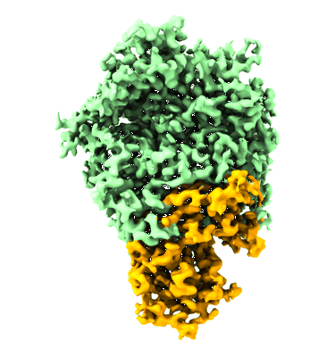 |
Cryo-EM structure of immunodominant protein P1 from human pathogen Mycoplasma pneumoniae on graphene oxide grid [multiple data sets in TIFF format] | Vizarraga D, Kawamoto A, Matsumoto U, Illanes R, Pérez-Luque R, Martín J, Mazzolini R, Bierge P, Pich OQ, Espasa M, Sanfeliu I, Esperalba J, Fernández-Huerta M, Scheffer MP, Pinyol J, Frangakis AS, Lluch-Senar M, Mori S, Shibayama K, Kenri T, Kato T, Namba K, Fita I, Miyata M, Aparicio D [Pubmed: 33057023] [DOI: 10.1038/s41467-020-18777-y] |
8.0 TB | 2.9 Å |
| 2024-02-08 |  |
Single particle Cryo EM of the LRRK2 I2020T mutant bound to GZD-824 [4102 multi-frame micrographs composed of 40 frames each in EER format] | Villagran Suarez A, Leschziner A [Pubmed: 38039358] [DOI: 10.1126/sciadv.adk6191] |
4.6 TB | 3.4 Å |
| 2024-02-09 |  |
Single particle Cryo EM of the C-terminal half LRRK2 G2019S mutant bound to GZD-824 [7988 multi-frame micrographs composed of 40 frames each in EER format] | Villagran Suarez A, Leschziner A [Pubmed: 38039358] [DOI: 10.1126/sciadv.adk6191] |
7.0 TB | 2.99 Å |
| 2024-03-21 |  |
Single particle Cryo EM of the C-terminal half LRRK2 I2020T mutant bound to GZD-824 [8386 multi-frame micrographs composed of 50 frames each in EER format] | Villagran Suarez A, Leschziner A [Pubmed: 38039358] [DOI: 10.1126/sciadv.adk6191] |
7.4 TB | 3.1 Å |
| 2022-04-25 |  |
Cryo EM structure of ΔRing6 LetB [10764 multi-frame micrographs composed of 30 frames each in TIFF format] | Vieni C, Coudray N, Bhabha G, Ekiert DC [Pubmed: 35077766] [DOI: 10.1016/j.jmb.2022.167463] |
3.0 TB | 3.2 Å |
| 2020-05-19 |  |
Whole-body integration of gene expression and single-cell morphology [11416 micrographs in TIFF format] | Vergara HM, Pape C, Meechan KI, Zinchenko V, Genoud C, Wanner AA, Mutemi KN, Titze B, Templin RM, Bertucci PY, Simakov O, Dürichen W, Machado P, Savage EL, Schermelleh L, Schwab Y, Friedrich RW, Kreshuk A, Tischer C, Arendt D [Pubmed: 34380046] [DOI: 10.1016/j.cell.2021.07.017] |
1.7 TB | — |
| 2020-07-14 |  |
Separating distinct macromolecular assemblies from cryo-EM images [2423 micrographs in MRC format] | Verbeke EJ, Zhou Y, Horton AP, Mallam AL, Taylor DW, Marcotte EM [Pubmed: 31726096] [DOI: 10.1016/j.jsb.2019.107416] |
128.5 GB | 4.0 - 19.0 Å |
| 2022-06-22 |  |
single particle cryo-EM of red blood cell lysate (hemolysate, hemoglobin reduced, filtered by SEC) [6608 multi-frame micrographs composed of 20 frames each in TIFF format] | Verbeke EJ [Pubmed: 35858567] [DOI: 10.1016/j.celrep.2022.111103] |
1.0 TB | 3.4 Å |
| 2021-11-02 | 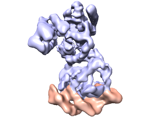 |
CryoEM single particle dataset for NanR dimer-DNA hetero-complex. [3465 multi-frame micrographs composed of 32 frames each in MRC format] | Venugopal H, Horne CR, Ramm G, Dobson RCJ [Pubmed: 33790291] [DOI: 10.1038/s41467-021-22253-6] |
364.8 GB | 3.9 Å |
| 2022-11-29 |  |
1.42 Angstrom Apoferritin structure determined using G1 Titan krios S-FEG operated at 300kV, zero loss imaging using Gatan BioQuantum energy filter operated at 10eV slit width and imaged using K2 camera. [multiple data sets in MRC format] | Venugopal H | 668.6 GB | 1.42 Å |
| 2023-07-10 |  |
Cryo electron tomography of Cytochalasin D-induced protrusions of Drosophila S2 cells - Datasets 1 - 4 [multiple data sets in TIFF and MRC formats] | Ventura Santos C, Carter AP, Rogers SL [Pubmed: 37034688] [DOI: 10.1101/2023.03.31.535077] |
446.0 GB | — |
| 2023-07-10 |  |
Cryo electron tomography of Cytochalasin D-induced protrusions of Drosophila S2 cells treated with DMSO or thapsigargin - Datasets 5 - 7 [multiple data sets in TIFF and MRC formats] | Ventura Santos C, Carter AP, Rogers SL [Pubmed: 37034688] [DOI: 10.1101/2023.03.31.535077] |
373.1 GB | — |
| 2023-07-10 |  |
Cryo electron tomography of Cytochalasin D-induced protrusions of Drosophila S2 alpha-tubulin acetyltransferase knock-out (dTAT KO) cells - Dataset 8 [multiple data sets in TIFF and MRC formats] | Ventura Santos C, Carter AP, Rogers SL [Pubmed: 37034688] [DOI: 10.1101/2023.03.31.535077] |
231.5 GB | — |
| 2023-07-10 |  |
Cryo electron tomography of induced protrusions of cofilin or control knock-down Drosophila S2 cells - Datasets 9 - 12 [multiple data sets in TIFF and MRC formats] | Ventura Santos C, Carter AP, Rogers SL [Pubmed: 37034688] [DOI: 10.1101/2023.03.31.535077] |
491.9 GB | — |
| 2023-10-03 |  |
Cryo electron tomography of Cytochalasin D-induced protrusions of Drosophila S2 cells treated with thapsigargin or MG132 [multiple data sets in TIFF and MRC formats] | Ventura Santos C, Carter AP [Pubmed: 37702953] [DOI: 10.15252/embr.202357264] |
107.6 GB | — |
| 2016-06-28 |  |
Designer nanoscale DNA assemblies programmed from the top down [50 micrographs in MRC format] | Veneziano R, Ratanalert S, Zhang K, Zhang F, Yan H, Chiu W, Bathe M [Pubmed: 27229143] [DOI: 10.1126/science.aaf4388] |
3.7 GB | 22.0 Å |
| 2016-06-28 | 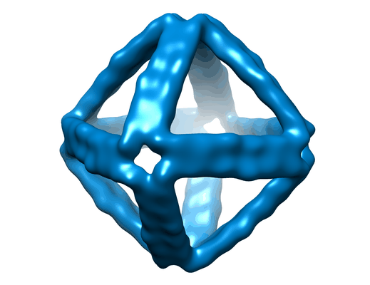 |
Designer nanoscale DNA assemblies programmed from the top down [100 micrographs in MRC format] | Veneziano R, Ratanalert S, Zhang K, Zhang F, Yan H, Chiu W, Bathe M [Pubmed: 27229143] [DOI: 10.1126/science.aaf4388] |
7.3 GB | 25.0 Å |
| 2016-06-28 | 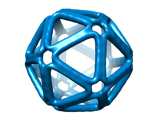 |
Designer nanoscale DNA assemblies programmed from the top down [177 micrographs in MRC format] | Veneziano R, Ratanalert S, Zhang K, Zhang F, Yan H, Chiu W, Bathe M [Pubmed: 27229143] [DOI: 10.1126/science.aaf4388] |
13.0 GB | 20.0 Å |
| 2016-08-18 | 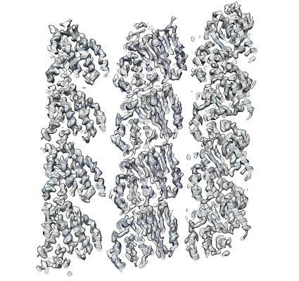 |
Structure and Dynamics of Single-isoform Recombinant Neuronal Human Tubulin [304 multi-frame micrographs composed of 23 frames each in MRC format] | Vemu A, Atherton J, Spector JO, Szyk A, Moores CA, Roll-Mecak A [Pubmed: 27129203] [DOI: 10.1074/jbc.C116.731133] |
487.7 GB | 4.0 Å |
| 2023-03-01 | 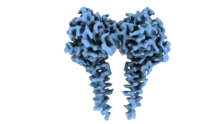 |
CryoEM structure of full-length dimeric ClbP [3888 multi-frame micrographs composed of 50 frames each in TIFF format] | Velilla JA, Walsh RM, Gaudet R [Pubmed: 36253550] [DOI: 10.1038/s41589-022-01142-z] |
1.1 TB | 3.73 Å |
| 2022-03-21 |  |
Structure of the GPCR dimer Ste2 bound to an antagonist [15751 multi-frame micrographs composed of 59 frames each in TIFF format] | Velazhahan V, Tate CG [Pubmed: 35296853] [DOI: 10.1038/s41586-022-04498-3] |
4.1 TB | 2.7 Å |
