Electron Microscopy Public Image Archive
Electron Microscopy Public Image Archive
大阪大學的EMPIAR-PDBj團隊為亞洲EM研究人員向EMPIAR傳送大型EM圖像數據提供服務。 除了通過互聯網將數據直接傳送到EBI(UK),研究人員還可以通過郵政或快遞服務將數據硬盤發送到大阪大學,或者通過互聯網傳送到設置於大阪大學的服務器,然後由我們代為傳送至數據登錄網站。 如果您想使用此項服務,請先通過 電子郵件 與我們聯繫。
| Release date | Imageset | Title | Authors and references | Size | Resolution |
|---|---|---|---|---|---|
| 2020-06-30 | 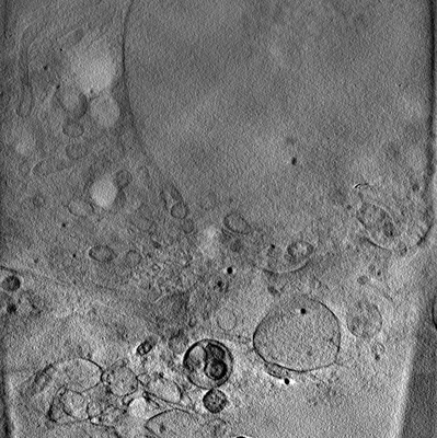 |
Soft X-ray Tomography of mock-infected U2OS cells [1 tilt series in MRC format] | Kounatidis I, Stanifer ML, Phillips MA, Paul-Gilloteaux P, Heiligenstein X, Wang H, Okolo CA, Fish TM, Spink MC, Stuart DI, Davis I, Boulant S, Grimes JM, Dobbie IM, Harkiolaki M [Pubmed: 32610083] [DOI: 10.1016/j.cell.2020.05.051] |
1.2 GB | — |
| 2020-06-30 | 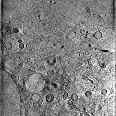 |
Soft X-ray Tomography of mock-infected U2OS cells [1 tilt series in MRC format] | Kounatidis I, Stanifer ML, Phillips MA, Paul-Gilloteaux P, Heiligenstein X, Wang H, Okolo CA, Fish TM, Spink MC, Stuart DI, Davis I, Boulant S, Grimes JM, Dobbie IM, Harkiolaki M [Pubmed: 32610083] [DOI: 10.1016/j.cell.2020.05.051] |
1.2 GB | — |
| 2020-06-30 | 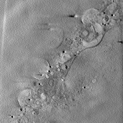 |
Soft X-ray Tomography of mock-infected U2OS cells [1 tilt series in MRC format] | Kounatidis I, Stanifer ML, Phillips MA, Paul-Gilloteaux P, Heiligenstein X, Wang H, Okolo CA, Fish TM, Spink MC, Stuart DI, Davis I, Boulant S, Grimes JM, Dobbie IM, Harkiolaki M [Pubmed: 32610083] [DOI: 10.1016/j.cell.2020.05.051] |
1.2 GB | — |
| 2020-06-30 | 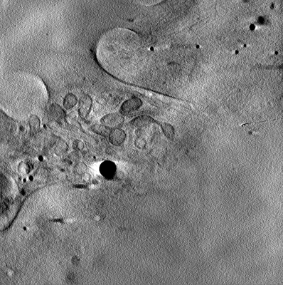 |
Soft X-ray Tomography of mock-infected U2OS cells [1 tilt series in MRC format] | Kounatidis I, Stanifer ML, Phillips MA, Paul-Gilloteaux P, Heiligenstein X, Wang H, Okolo CA, Fish TM, Spink MC, Stuart DI, Davis I, Boulant S, Grimes JM, Dobbie IM, Harkiolaki M [Pubmed: 32610083] [DOI: 10.1016/j.cell.2020.05.051] |
1.2 GB | — |
| 2024-01-15 | 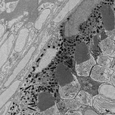 |
Developing retina in zebrafish 55 hpf larval eye. [16 reconstructed volumes in DM3 format] | Wilsch-Bräuninger M | 1.2 GB | — |
| 2024-02-15 | 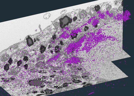 |
FIB-SEM dataset showing localization of a Golgi matrix protein GM130 in human hepatocellular carcinoma cell (Huh-7) [564 reconstructed volumes in TIFF format] | Belevich I, Jokitalo E | 1.1 GB | — |
| 2015-10-01 |  |
Tubulin Chaperone complexes TBC-DEG Q73L: alpha beta-tubulin complex [stack of 18361 particles in MRC format] | Nithianantham S, Le S, Seto E, Jia W, Leary J, Corbett KD, Moore JK, Al-Bassam J [Pubmed: 26208336] [DOI: 10.7554/eLife.08811] |
1.1 GB | 24.0 Å |
| 2023-01-18 | 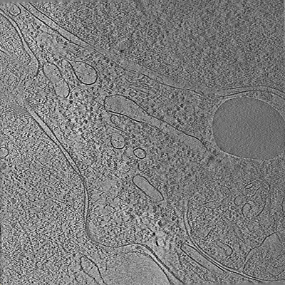 |
Tilt series of cell-cell contact of two PTK-1 cells [35 tilt series in MRC format] | Lemos M, Bezault A, Sauvanet C, Hanein D, Volkmann N [Pubmed: 36539423] [DOI: 10.1038/s41467-022-35409-9] |
1.1 GB | — |
| 2015-10-01 | 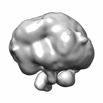 |
Tubulin Chaperone complexes TBC-DEG Q73L: alpha beta-tubulin:TBCC complex [stack of 16801 particles in MRC format] | Nithianantham S, Le S, Seto E, Jia W, Leary J, Corbett KD, Moore JK, Al-Bassam J [Pubmed: 26208336] [DOI: 10.7554/eLife.08811] |
1.0 GB | 24.0 Å |
| 2021-01-08 | 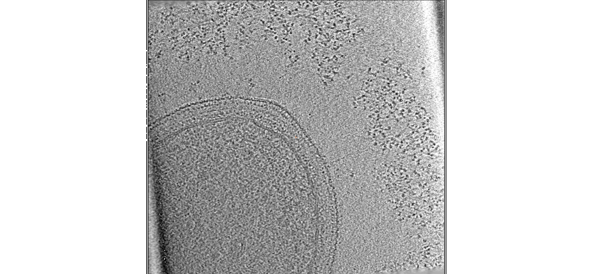 |
Nanoscale view of the Clostridium thermocellum cellulosome during cellulose degradation reveals an ecological strategy leading to phenotypic heterogeneity [37 tilt series in MRC format] | Tatli M, Moraïs S, Tovar-Herrera OE, Bomble Y, Bayer EA, Medalia O, Mizrahi I | 1004.3 MB | — |
| 2019-10-07 | 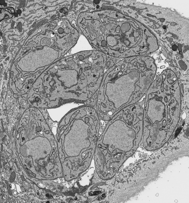 |
Processed FIB SEM images of a parasitophorous vacuole containing Toxoplasma gondii ∆CAP parasites. [1 multi-frame micrographs composed of 1 frames each in MRC format] | Hunt A, Russell MRG, Wagener J, Kent R, Carmeille R, Peddie CJ, Collinson L, Heaslip A, Ward GE, Treeck M [Pubmed: 31577230] [DOI: 10.7554/elife.50598] |
898.1 MB | — |
| 2022-01-11 | 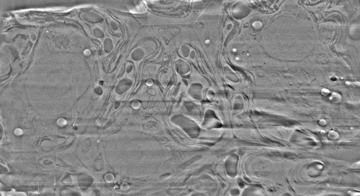 |
Cryo-FIB-SEM data on Chlamydomonas reinhardtii cells [37 micrographs in TIFF format] | Klumpe S [Pubmed: 34951584] [DOI: 10.7554/elife.70506] |
888.2 MB | — |
| 2023-02-28 | 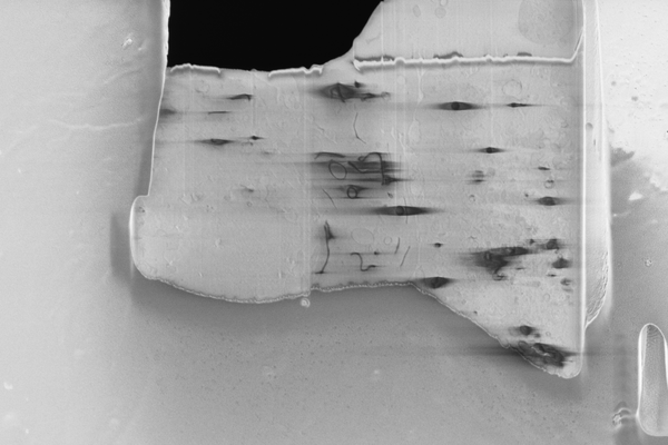 |
Cryo serial FIB/SEM of RPE-1 cells [18 micrographs in TIFF format] | Dumoux M, Glen T, Smith JLR, Ho EML, Perdigão LMA, Pennington A, Klumpe S, Yee NBY, Farmer DA, Lai PYA, Bowles W, Kelley R, Plitzko JM, Wu L, Basham M, Clare DK, Siebert CA, Darrow MC, Naismith JH, Grange M [Pubmed: 36805107] [DOI: 10.7554/elife.83623] |
826.6 MB | — |
| 2018-01-03 |  |
Segment /nodes network analysis of the tubular system of the Neonate and Adult GP cardiomyocyte [167 multi-frame micrographs composed of 1 frames each in TIFF format] | Kashbour H, Taggart M, White K | 787.5 MB | — |
| 2021-06-18 |  |
Subtomograms of nucleosomes extracted from cryo-tomograms of Drosophila melanogaster embryos [1 subtomograms in EM format] | Harastani M, Eltsov M, Leforestier A, Jonic S [Pubmed: 34095222] [DOI: 10.3389/fmolb.2021.663121] |
666.3 MB | — |
| 2019-10-07 |  |
Processed FIB SEM images of a parasitophorous vacuole containing Toxoplasma gondii ∆CAP parasites, complemented with CAP. [1 multi-frame micrographs composed of 1 frames each in MRC format] | Hunt A, Russell MRG, Wagener J, Kent R, Carmeille R, Peddie CJ, Collinson L, Heaslip A, Ward GE, Treeck M [Pubmed: 31577230] [DOI: 10.7554/elife.50598] |
583.8 MB | — |
| 2024-02-13 |  |
Plant SBF-SEM - Tobacco Leaf Chloroplast [130 micrographs in TIFF format] | Wickramanayake JS, Czymmek KJ [Pubmed: 37451777] [DOI: 10.1016/bs.mcb.2023.04.008] |
544.4 MB | — |
| 2018-02-08 |  |
Tilt-series of salmonella enterica wild-type bacterial flagellar motor [1 tilt series in MRC format] | Beeby M, Ribardo DA, Brennan CA, Ruby EG, Jensen GJ, Hendrixson DR [Pubmed: 26976588] [DOI: 10.1073/pnas.1518952113] |
328.0 MB | 69.4 Å |
| 2016-01-20 | 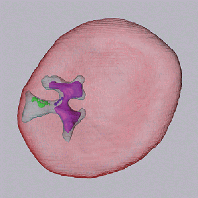 |
SBF-SEM of ring-stage malaria parasite infected red blood cell [120 micrographs in MRC format] | Sakaguchi M, Miyazaki N, Fujioka H, Kaneko O, Murata K [Pubmed: 26772147] [DOI: 10.1016/j.jsb.2016.01.003] |
293.0 MB | — |
| 2017-03-15 | 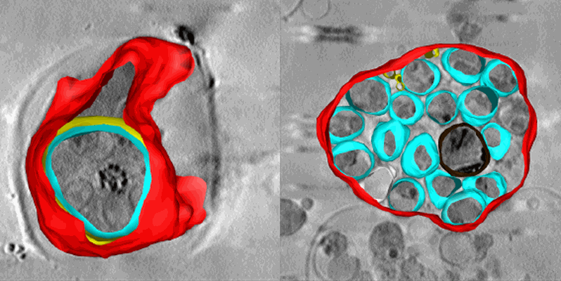 |
Soft X-ray tomography of Plasmodium falciparum infected human erythrocytes stalled in egress by the inhibitors Compound 2 and E64 [1 Soft X-ray tomograms in MRC format] | Hale VL, Saibil HR, Duke E, Fleck RA, Blackman MJ [Pubmed: 28292906] [DOI: 10.1073/pnas.1619441114] |
280.6 MB | — |
| 2016-01-20 | 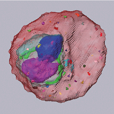 |
SBF-SEM of trophozoite-stage malaria parasite infected red blood cell [110 micrographs in MRC format] | Sakaguchi M, Miyazaki N, Fujioka H, Kaneko O, Murata K [Pubmed: 26772147] [DOI: 10.1016/j.jsb.2016.01.003] |
268.6 MB | — |
| 2018-01-17 |  |
Serial Block Face Scanning Electron Micrscopy dataset of fetal day 64 guinea pig psoas muscle in transverse [93 micrographs in TIFF format] | Cocks ET [DOI: 10.1111/jmi.12676] |
264.4 MB | — |
| 2023-06-20 |  |
Cryo-EPty SPA at CSA of 1.03 mrad [29 micrographs in MRC format] | Pei X, Zhou L, Huang C, Boyce M, Kim JS, Liberti E, Hu Y, Sasaki T, Nellist PD, Zhang P, Stuart DI, Kirkland AI, Wang P [Pubmed: 37230988] [DOI: 10.1038/s41467-023-38268-0] |
262.0 MB | 37.2 Å |
| 2021-11-08 |  |
Monitoring reversion of hepatitis C virus-induced cellular alterations by Direct-Acting Antivirals using cryo Soft X-ray Tomography and Infrared Microscopy [7 reconstructed volumes in TIFF format] | Perez-Berna AJ, Benseny-Cases N, Rodríguez MJ, Valcarcel R, Carrascosa JL, Gastaminzab P, Pereiroa E [Pubmed: 34726165] [DOI: 10.1107/S2059798321009955] |
260.2 MB | — |
