Electron Microscopy Public Image Archive
Electron Microscopy Public Image Archive
大阪大學的EMPIAR-PDBj團隊為亞洲EM研究人員向EMPIAR傳送大型EM圖像數據提供服務。 除了通過互聯網將數據直接傳送到EBI(UK),研究人員還可以通過郵政或快遞服務將數據硬盤發送到大阪大學,或者通過互聯網傳送到設置於大阪大學的服務器,然後由我們代為傳送至數據登錄網站。 如果您想使用此項服務,請先通過 電子郵件 與我們聯繫。
| Release date | Imageset | Title | Authors and references | Size | Resolution |
|---|---|---|---|---|---|
| 2020-12-11 | 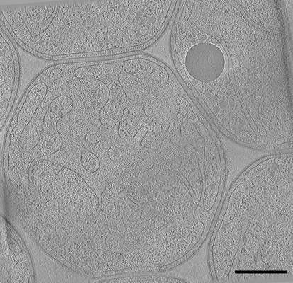 |
Cryo electron tomography after FIB-milling of Planctomycetes species Gemmata obscuriglobus (#1) [101 tilt series in MRC format] | Seeger C, Andersson SG [Pubmed: 32761170] [DOI: 10.1093/gbe/evaa159] |
2.7 GB | — |
| 2020-12-11 | 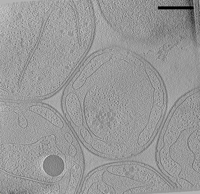 |
Cryo electron tomography after FIB-milling of Planctomycetes species Gemmata obscuriglobus (#2) [101 tilt series in MRC format] | Seeger C, Andersson SG [Pubmed: 32761170] [DOI: 10.1093/gbe/evaa159] |
2.7 GB | — |
| 2018-07-05 |  |
Cryo-EM structure of alpha-synuclein fibrils [118 multi-frame micrographs composed of 50 frames each in MRC format] | Guerrero-Ferreira R, Taylor NM, Mona D, Ringler P, Lauer ME, Riek R, Britschgi M, Stahlberg H [Pubmed: 29969391] [DOI: 10.7554/elife.36402] |
6.3 GB | — |
| 2020-12-11 | 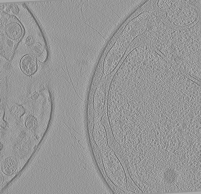 |
Cryo electron tomography after FIB-milling of Planctomycetes species Tuwongella immobilis (33k magnification) [multiple data sets in MRC format] | Seeger C, Andersson SG [Pubmed: 32761170] [DOI: 10.1093/gbe/evaa159] |
15.5 GB | — |
| 2022-12-13 | 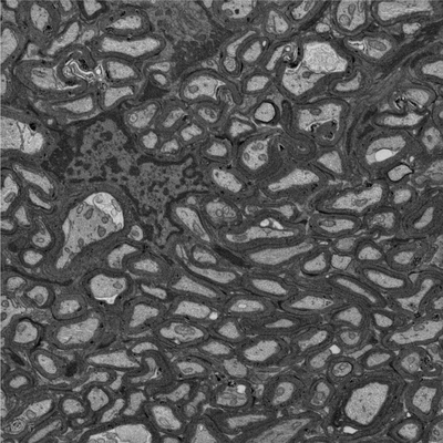 |
Focused ion beam-scanning electron microscopy links pathological myelin outfoldings to axonal changes in mice lacking Plp1 or Mag [940 micrographs in TIFF format] | Steyer AM, Möbius W [Pubmed: 36354016] [DOI: 10.1002/glia.24290] |
12.5 GB | — |
| 2022-12-13 | 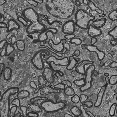 |
Focused ion beam-scanning electron microscopy links pathological myelin outfoldings to axonal changes in mice lacking Plp1 or Mag [1290 micrographs in TIFF format] | Steyer AM, Möbius W [Pubmed: 36354016] [DOI: 10.1002/glia.24290] |
22.4 GB | — |
| 2018-07-06 | 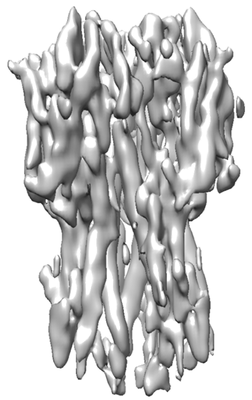 |
Single particle cryoEM of hemagglutinin with spot-to-plunge time of 500ms [multiple data sets in MRC format] | Noble AJ, Wei H, Dandey VP, Zhang Z, Potter CS, Carragher B [Pubmed: 30250056] [DOI: 10.1038/s41592-018-0139-3] |
51.1 GB | — |
| 2021-05-21 | 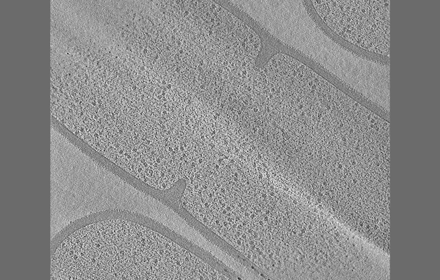 |
Tilt series of dividing vegetative and sporulating cells of Bacillus subtilis from the manuscript - Khanna et al., 2021 [multiple data sets in MRC format] | Khanna K, Lopez-Garrido J, Sugie J, Pogliano K, Villa E [Pubmed: 34018921] [DOI: 10.7554/eLife.62204] |
8.7 GB | — |
| 2022-03-14 | 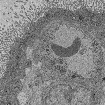 |
SBF SEM of Human term placental villi [multiple data sets in TIFF format] | Lewis RM [DOI: 10.1101/2022.01.26.477815] |
90.2 GB | — |
| 2018-08-09 |  |
Three-dimensional nanostructure of an intact microglia cell [multiple data sets in TIFF and IMOD formats] | Bolasco G, Weinhard L, Boissonnet T, Neujahr R, Gross CT [DOI: 10.3389/fnana.2018.00105] |
7.4 GB | — |
| 2020-10-14 | 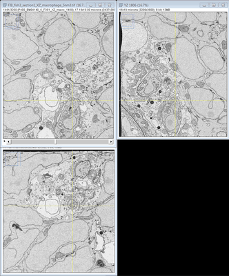 |
FIB SEM images of a Zebrafish hindbrain macrophage containing 2 Toxoplasma gondii tachizoites [multiple data sets in TIFF format] | Peddie CJ, Domart MC, Collinson L [Pubmed: 32461265] [DOI: 10.1242/dmm.043091] |
1.1 TB | — |
| 2020-12-09 | 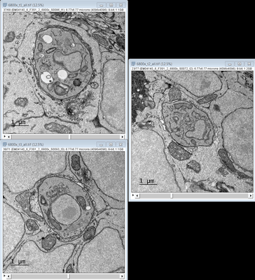 |
TEM images of a Zebrafish hindbrain cells containing Toxoplasma gondii tachizoites [multiple data sets in TIFF format] | Domart MC, Collinson L [Pubmed: 32461265] [DOI: 10.1242/dmm.043091] |
3.4 GB | — |
| 2022-03-28 | 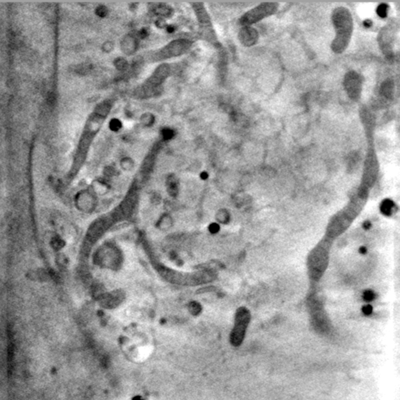 |
Representative data from Near-native state imaging by cryo-soft-X-ray tomography reveals remodelling of cytoplasmic vesicles and mitochondria during HSV-1 infection [14 reconstructed volumes in MRC format] | Nahas KLN, Connor VC, Scherer KM, Kaminski CF, Harkiolaki M, Crump CM, Graham SC [DOI: 10.1101/2021.10.11.463900] |
9.9 GB | — |
| 2021-01-22 | 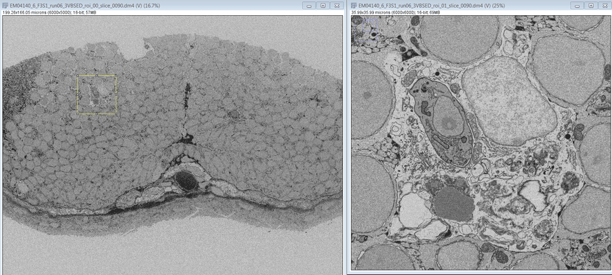 |
SBF SEM images of a Zebrafish hindbrain macrophage containing 2 Toxoplasma gondii tachizoites [multiple data sets in DM4 format] | Peddie CJ, Domart MC, Collinson L [Pubmed: 32461265] [DOI: 10.1242/dmm.043091] |
16.5 GB | — |
| 2020-12-09 | 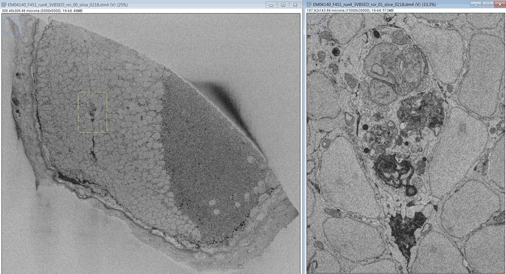 |
SBF SEM images of a Zebrafish hindbrain macrophage containing a replicating Toxoplasma gondii tachizoite [multiple data sets in DM4 format] | Peddie CJ, Domart MC, Collinson L [Pubmed: 32461265] [DOI: 10.1242/dmm.043091] |
338.4 GB | — |
| 2020-12-09 | 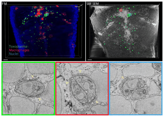 |
SBF SEM images of a Zebrafish hindbrain containing several Toxoplasma gondii tachizoites at different stages of replication [multiple data sets in DM4 and TIFF formats] | Peddie CJ, Domart MC, Collinson L [Pubmed: 32461265] [DOI: 10.1242/dmm.043091] |
175.2 GB | — |
| 2021-10-27 | 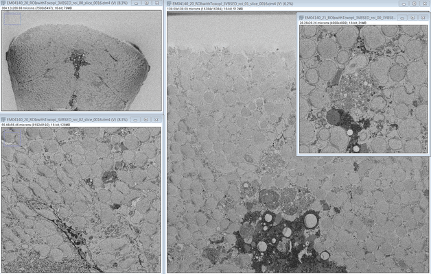 |
SBF SEM images of a Zebrafish hindbrain containing several Toxoplasma gondii tachizoites at different stages of replication [multiple data sets in DM4 format] | Peddie CJ, Domart MC, Collinson L [Pubmed: 32461265] [DOI: 10.1242/dmm.043091] |
553.9 GB | — |
| 2024-02-15 | 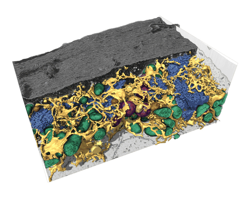 |
FIB-SEM dataset of a human bone osteosarcoma epithelial cell (U2-OS) [1168 micrographs in TIFF format] | Belevich I, Schertel A, Zaversek T, Szyrynska N, Saarnio S, Jokitalo E | 5.2 GB | — |
| 2022-03-21 | 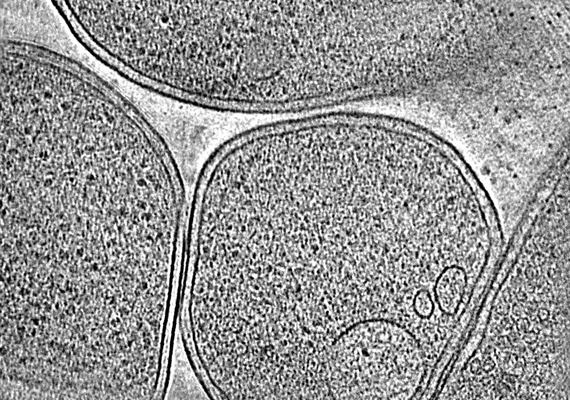 |
CryoET of E. coli prepared with the Waffle Method [1504 multi-frame micrographs composed of 14 frames each in TIFF format] | Kelley K, Raczkowski AM, Klykov O, Jaroenlak P, Bobe D, Kopylov M, Eng ET, Bhabha G, Potter CS, Carragher B, Noble AJ [Pubmed: 35387991] [DOI: 10.1038/s41467-022-29501-3] |
94.0 GB | — |
| 2022-12-13 | 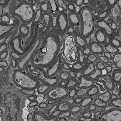 |
Focused ion beam-scanning electron microscopy links pathological myelin outfoldings to axonal changes in mice lacking Plp1 or Mag [1598 micrographs in TIFF format] | Steyer AM, Möbius W [Pubmed: 36354016] [DOI: 10.1002/glia.24290] |
31.3 GB | — |
| 2018-10-25 |  |
CryoET of bacterial RNA polymerase with several detergents [multiple data sets in MRC and RAW TEXT formats] | Chen J, Noble AJ, Kang JY, Darst SA [DOI: 10.1016/j.yjsbx.2019.100005] |
207.5 GB | — |
| 2022-03-15 | 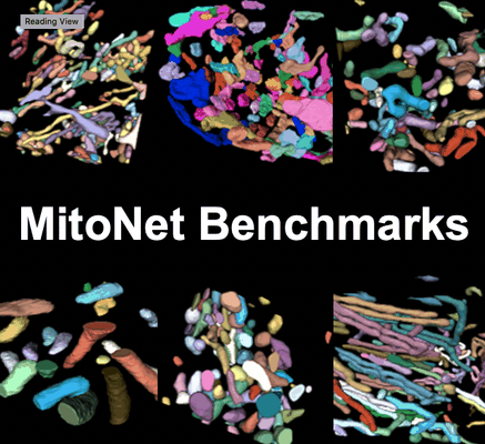 |
Seven benchmark datasets of instance segmentation of mitochondria: 6 diverse volume EM + 1 TEM (100 images) datasets [multiple data sets in TIFF format] | Narayan K, Conrad RW | 4.1 GB | — |
| 2022-12-13 |  |
Focused ion beam-scanning electron microscopy links pathological myelin outfoldings to axonal changes in mice lacking Plp1 or Mag [1553 micrographs in TIFF format] | Steyer AM, Möbius W [Pubmed: 36354016] [DOI: 10.1002/glia.24290] |
22.3 GB | — |
| 2023-11-06 |  |
Single particle cryo-EM structure of RIG-I:RNA:Riplet ternary complex [3420 multi-frame micrographs composed of 40 frames each in TIFF format] | Wang W, Pyle AM [DOI: 10.1038/s41467-023-42982-0] |
1.5 TB | — |
| 2022-12-13 |  |
Focused ion beam-scanning electron microscopy links pathological myelin outfoldings to axonal changes in mice lacking Plp1 or Mag [910 micrographs in TIFF format] | Steyer AM, Möbius W [Pubmed: 36354016] [DOI: 10.1002/glia.24290] |
13.7 GB | — |
