
Electron Microscopy Public Image Archive
Electron Microscopy Public Image Archive
大阪大学的EMPIAR-PDBj团队为亚洲EM研究人员向EMPIAR传送大型EM图像数据提供服务。 除了通过互联网将数据直接传送到EBI(UK),研究人员还可以通过邮政或快递服务将数据硬盘发送到大阪大学,或者通过互联网传送到设置于大阪大学的服务器,然后由我们代为传送至数据登录网站。 如果您想使用此项服务,请先通过 电子邮件 与我们联系。
| Release date | Imageset | Title | Authors and references | Size | Resolution |
|---|---|---|---|---|---|
| 2021-09-24 | 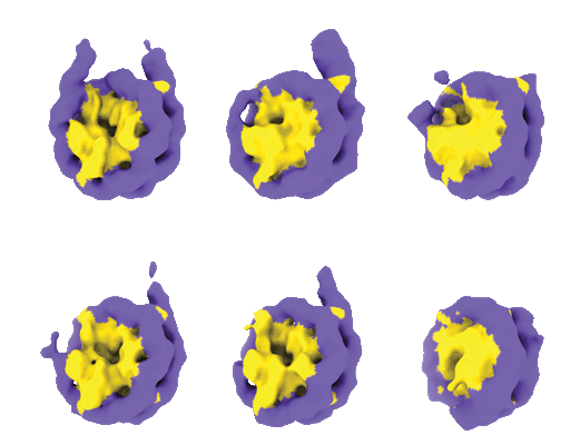 |
'Freed' nucleosome isolated from non-crosslinked interphase chromosome in Xenopus egg extract lot2 [multiple data sets in TIFF format] | Arimura YA, Funabiki HF [Pubmed: 34478647] [DOI: 10.1016/j.molcel.2021.08.010] |
1.1 TB | 5.44 Å |
| 2022-11-29 | 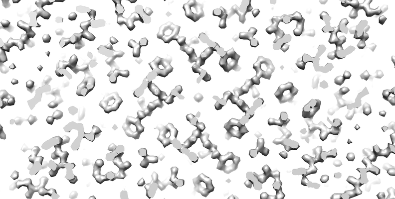 |
1.42 Angstrom Apoferritin structure determined using G1 Titan krios S-FEG operated at 300kV, zero loss imaging using Gatan BioQuantum energy filter operated at 10eV slit width and imaged using K2 camera. [multiple data sets in MRC format] | Venugopal H | 668.6 GB | 1.42 Å |
| 2020-06-26 | 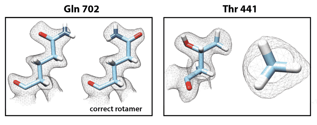 |
1.8 Å resolution structure of β-galactosidase with a 200 kV CRYO ARM electron microscope [4949 multi-frame micrographs composed of 40 frames each in TIFF format] | Merk A, Fukumura T, Zhu X, Darling JE, Grisshammer R, Ognjenovic J, Subramaniam S [Pubmed: 32695410] [DOI: 10.1107/S2052252520006855] |
1.1 TB | 1.8 Å |
| 2021-03-19 | 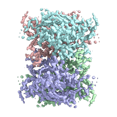 |
1.93 A cryo-EM structure of streptavidin [2277 multi-frame micrographs composed of 70 frames each in TIFF format] | Hiraizumi M, Yamashita K, Nisihzawa T, Kikkawa M, Nureki O | 373.1 GB | 1.93 Å |
| 2021-06-09 |  |
120kV MicroED structure of FUS (37-42) SYSGYS solved from merged datasets at 0.60 A [8 diffraction images in SMV format] | Zhou H, Luo F, Luo Z, Li D, Liu C, Li X [Pubmed: 31334636] [DOI: 10.1021/acs.analchem.9b01162] |
9.2 GB | 0.6 Å |
| 2020-10-23 |  |
170314, Five-day-old Col-0 Arabidopsis thaliana root, phloem pole unloading zone (339-381 um from the root tip) [multiple data sets in MRC format] | Paterlini A, Belevich I, Jokitalo E, Helariutta Y [Pubmed: 31182845] [DOI: 10.1038/s41477-019-0429-5] |
43.2 GB | — |
| 2020-11-18 |  |
2.05 angstrom resolution structure determination of sulfur oxygenase reductase using 200kV cryo-EM [2558 multi-frame micrographs composed of 50 frames each in MRC format] | Moriya T, Adachi N, Sato Y, Arakawa T, Kawasaki M, Yamada C, Fushinobu S, Senda T [Pubmed: 32775998] [DOI: 10.1016/j.yjsbx.2020.100030] |
1.7 TB | 2.05 - 2.24 Å |
| 2022-11-15 |  |
2.1 Å resolution structure of β-galactosidase obtained from Glacios equipped with Falcon 3 [multiple data sets in TIFF format] | Merk A, Darling JE, Grisshammer R, Ognjenović J | 4.8 TB | 2.1 Å |
| 2022-03-14 | 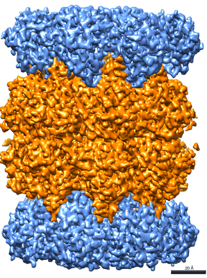 |
2.1Å T20S Proteosome from 200kV Glacios with Selectris Falcon 4 [4075 multi-frame micrographs composed of 854 frames each in EER format] | Koh FA, Khavnekar K, Kotecha A [Pubmed: 35377368] [DOI: 10.3791/63519] |
1.5 TB | 2.1 Å |
| 2016-04-15 | 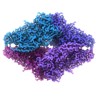 |
2.2 A resolution cryo-EM structure of beta-galactosidase in complex with a cell-permeant inhibitor [multiple data sets in MRC format] | Bartesaghi A, Merk A, Banerjee S, Matthies D, Wu X, Milne JL, Subramaniam S [Pubmed: 25953817] [DOI: 10.1126/science.aab1576] |
631.2 GB | 2.2 Å |
| 2022-04-01 | 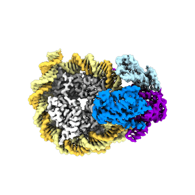 |
2.3 A structure of the ATP-dependent chromatin remodeler Chd1 bound to the nucleosome in a nucleotide-free state [multiple data sets in TIFF, MRC and MRCS formats] | Nodelman IM, Das S, Faustino AM, Fried SD, Bowman GD, Armache JP [Pubmed: 35173352] [DOI: 10.1038/s41594-021-00719-x] |
4.7 TB | 2.3 - 2.9 Å |
| 2020-05-26 | 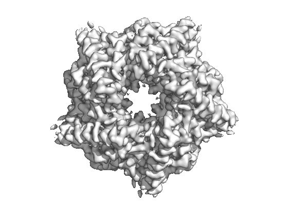 |
2.3 Angstrom cryo-EM reconstructions of HemQ from Geobacillus [258 multi-frame micrographs composed of 100 frames each in MRCS format] | Bromberg R, Guo Y, Borek D, Otwinowski Z [DOI: 10.1107/S2052252520002444] |
1.4 TB | 2.32 Å |
| 2020-05-27 | 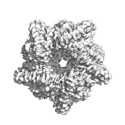 |
2.6 Angstrom cryo-EM reconstructions of HemQ from Geobacillus in the presence of substantial aberrations [257 multi-frame micrographs composed of 100 frames each in MRCS format] | Bromberg R, Guo Y, Borek D, Otwinowski Z [DOI: 10.1107/S2052252520002444] |
1.3 TB | 2.6 Å |
| 2020-05-27 | 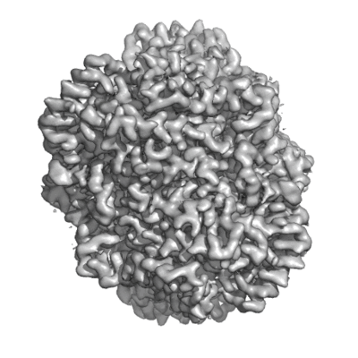 |
2.7 Angstrom cryo-EM reconstructions of glucose isomerase in the presence of substantial aberrations [202 multi-frame micrographs composed of 200 frames each in MRCS format] | Bromberg R, Guo Y, Borek D, Otwinowski Z [DOI: 10.1107/S2052252520002444] |
1.0 TB | 2.7 Å |
| 2021-06-09 | 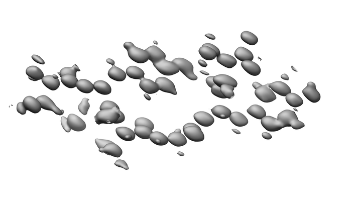 |
200kV MicroED structure of FUS (37-42) SYSGYS solved from merged datasets at 0.65 A [8 diffraction images in SMV format] | Zhou H, Luo F, Luo Z, Li D, Liu C, Li X [Pubmed: 31334636] [DOI: 10.1021/acs.analchem.9b01162] |
9.5 GB | 0.65 Å |
| 2014-01-03 | 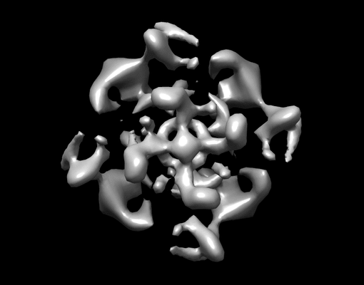 |
2D crystal images of the potassium channel MloK1 with and without cAMP ligand [multiple data sets in TIFF format] | Kowal J, Chami M, Baumgartner P, Arheit M, Chiu P-L, Rangl M, Scheuring S, Schroeder GF, Nimigean CM, Stahlberg H [Pubmed: 24469021] [DOI: 10.1038/ncomms4106] |
11.6 GB | 7.0 Å |
| 2020-07-14 | 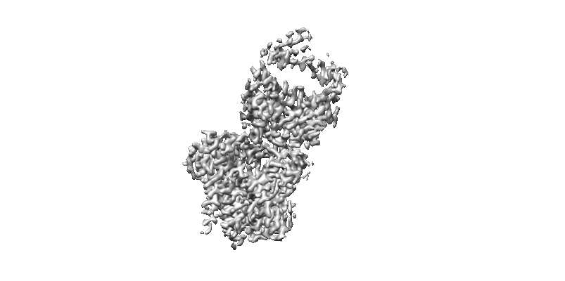 |
3 Å resolution single particle reconstruction of glucosyltransferase ALG6 in nanodisc [multiple data sets in MRC and TIFF formats] | Bloch JS, Pesciullesi G, Boilevin J, Nosol K, Irobalieva RN, Darbre T, Aebi M, Kossiakoff AA, Reymond JL, Locher KP [Pubmed: 32103179] [DOI: 10.1038/s41586-020-2044-z] |
2.8 TB | 3.0 Å |
| 2019-12-19 | 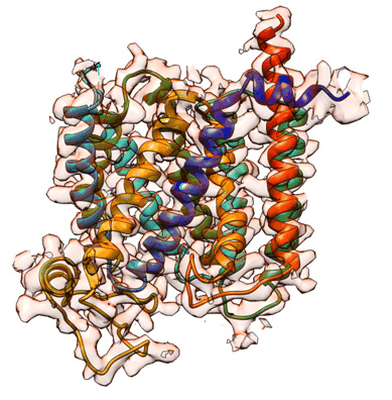 |
3.2 Å Single-Particle Cryo-EM Reconstruction of 49 kDa Membrane-Bound PfCRT Complexed with Fab [multiple data sets in MRCS and MRC formats] | Kim JK, Tan YZT, Wicht KJW, Erramilli SKE, Dhingra SKD, Okombo JO, Vendome JV, Hagenah LMH, Giacometti SIG, Warren ALW, Nosol KN, Roepe PDR, Potter CSP, Carragher BC, Kossiakoff AAK, Quick MQ, Fidock DAF, Mancia FM [Pubmed: 31776516] [DOI: 10.1038/s41586-019-1795-x] |
830.4 GB | 3.3 Å |
| 2021-03-05 | 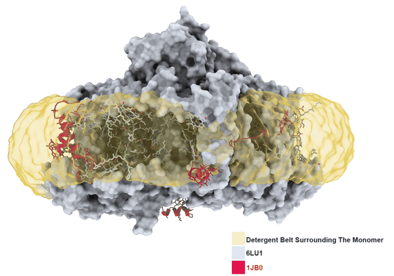 |
3.2 Å resolution structure of a functional monomeric Photosystem I from Thermosynechococcus elongatus BP-1 by single particle cryo-EM with a 200 kV CRYO ARM electron microscope [904 multi-frame micrographs composed of 60 frames each in TIFF format] | Coruh O, Frank A, Tanaka H, Kawamoto A, El-Mohsnawy E, Kato T, Namba K, Gerle C, Nowaczyk MM, Kurisu G [Pubmed: 33686186] [DOI: 10.1038/s42003-021-01808-9] |
226.1 GB | 3.2 Å |
| 2021-05-14 | 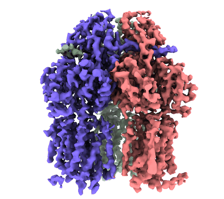 |
3.9 Angstrom reconstruction of E.coli AcrB embedded in the liposome [5757 multi-frame micrographs composed of 32 frames each in MRCS format] | Yao X, Fan X, Yan N [Pubmed: 32680969] [DOI: 10.1073/pnas.2009385117] |
2.4 TB | 3.9 Å |
| 2018-01-03 | 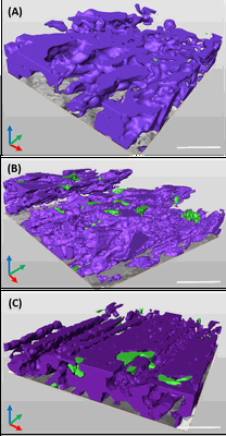 |
3D reconstruction of cardiac mitochondria in earlly prenatal (G55/57) and mid term(G58/60) and term (G66/68) GP cardiomyocyte [341 multi-frame micrographs composed of 1 frames each in TIFF format] | Kashbour H, Taggart M, White K | 1.7 GB | — |
| 2018-01-03 | 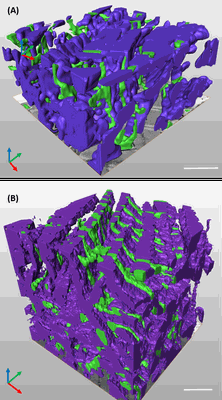 |
3D reconstruction of the cardiac mitochondria in Neaonate and Adult GP cardiomyocyte [262 multi-frame micrographs composed of 1 frames each in TIFF format] | Kashbour H, Taggart M, White K | 1.2 GB | — |
| 2018-01-03 | 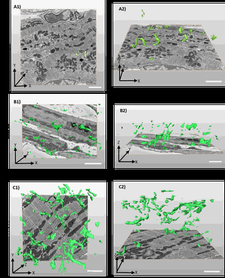 |
3D reconstruction of the tubular system of preterm G55/57, mid term G58/60 and term G66/68 of guinea pig left ventricles [318 multi-frame micrographs composed of 1 frames each in TIFF format] | Kashbour H, Taggart M | 2.9 GB | — |
| 2023-10-17 | 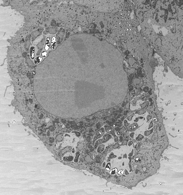 |
3D reconstructions of parasite development and the intracellular niche of the microsporidian pathogen E. intestinalis [multiple data sets in DM4 format] | Antao NVA, Lam CKL, Davydov AD, Riggi MR, Sall JS, Petzold CP, Liang FL, Iwasa JI, Ekiert DCE, Bhabha GB [Pubmed: 37425741] [DOI: 10.1101/2023.07.02.547383] |
537.9 GB | — |
| 2023-01-30 | 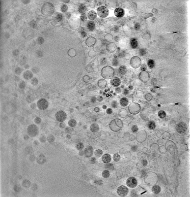 |
3D-surface reconstruction of cellular cryo-soft X-ray microscopy tomograms using semi-supervised deep learning [7 reconstructed volumes in MRC format] | Dyhr MCA, Sadeghi M, Moynova R, Knappe C, Kepsutlu Çakmak B, Werner S, Schneider G, McNally J, Noe F, Ewers H [DOI: 10.1101/2022.05.16.492055] |
23.5 GB | — |
