Electron Microscopy Public Image Archive
Electron Microscopy Public Image Archive
大阪大学的EMPIAR-PDBj团队为亚洲EM研究人员向EMPIAR传送大型EM图像数据提供服务。 除了通过互联网将数据直接传送到EBI(UK),研究人员还可以通过邮政或快递服务将数据硬盘发送到大阪大学,或者通过互联网传送到设置于大阪大学的服务器,然后由我们代为传送至数据登录网站。 如果您想使用此项服务,请先通过 电子邮件 与我们联系。
| Release date | Imageset | Title | Authors and references | Size | Resolution |
|---|---|---|---|---|---|
| 2023-12-01 | 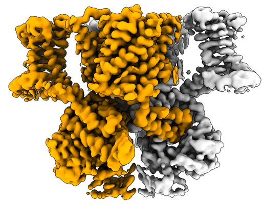 |
cAMP-bound SpSLC9C1 in lipid nanodiscs [multiple data sets in TIFF format] | Kalienkova V, Peter MF, Rheinberger J, Paulino C [Pubmed: 37880361] [DOI: 10.1038/s41586-023-06629-w] |
4.8 TB | 3.3 - 3.74 Å |
| 2023-12-18 | 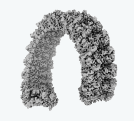 |
cA3-bound TIR-SAVED [3907 multi-frame micrographs composed of 50 frames each in MRC format] | Hogrel G, Guild A, Graham S, Rickman H, Grüschow S, Bertrand Q, Spagnolo L [Pubmed: 35948638] [DOI: 10.1038/s41586-022-05070-9] |
47.7 TB | 3.8 Å |
| 2018-10-23 | 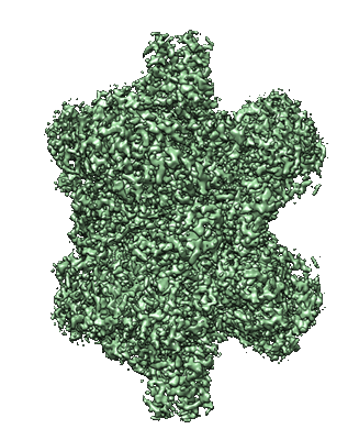 |
bovine liver glutamate dehydrogenase [4982 micrographs in MRC format] | Eng ET, Kelley K, Jordan KJ, Kopylov M, Carragher BO, Potter CS | 264.5 GB | 2.1 Å |
| 2019-05-09 | 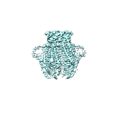 |
apo-LRRC8A in MSP2N2 nanodiscs [1779 multi-frame micrographs composed of 50 frames each in MRCS format] | Kern DM [Pubmed: 30775971] [DOI: 10.7554/eLife.42636] |
739.5 GB | 4.18 Å |
| 2019-01-29 |  |
afTMEM16/nanodisc complex in the presence of Ca2+ and 5mol% Ceramide 24:0 [2145 micrographs in MRC format] | Falzone M. E., Accardi A [Pubmed: 30648972] [DOI: 10.7554/eLife.43229] |
113.8 GB | 3.59 Å |
| 2019-01-29 |  |
afTMEM16/nanodisc complex in the presence of Ca2+ [2838 micrographs in MRC format] | Falzone M. E., Accardi A [Pubmed: 30648972] [DOI: 10.7554/eLife.43229] |
150.5 GB | 4.05 Å |
| 2019-01-30 |  |
afTMEM16/nanodisc complex in the absence of Ca2+ [3054 micrographs in MRC format] | Falzone ME, Rheinberger J, Di Lorenzo A, Accardi A [Pubmed: 31278385] [DOI: 10.1038/s41586-019-1377-y] |
162.0 GB | 4.2 Å |
| 2019-08-27 |  |
Yeast postcatalytic spliceosome, two cryoEM data sets at different magnifications [multiple data sets in MRC format] | Wilkinson ME, Nagai K [Pubmed: 31478901] [DOI: 10.1107/S2059798319010519] |
6.3 TB | 3.3 Å |
| 2020-08-18 | 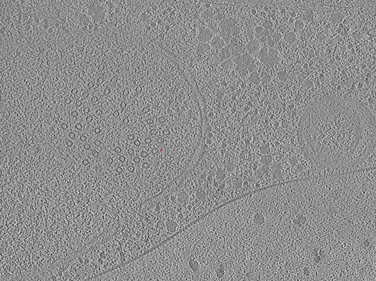 |
Yeast Tilt Series Collected on Lamella Generated by Fully Automated FIB Milling [1 tilt series in MRC format] | Zachs T, Schertel A, Medeiros J, Weiss GL, Hugener J, Matos J, Pilhofer M [Pubmed: 32149604] [DOI: 10.7554/eLife.52286] |
2.6 GB | 30.0 Å |
| 2021-05-10 |  |
Yeast C, Ci, C*, and P complex spliceosomes [multiple data sets in TIFF and MRCS formats] | Wilkinson ME, Fica SM, Galej WP, Nagai K [Pubmed: 27459055] [DOI: 10.1038/nature19316] |
8.9 TB | 2.8 - 10.0 Å |
| 2014-11-06 |  |
Yeast 80S Ribosome-Taura Syndrome Virus IRES complex, Frealign Input Particle Stack [stack of 416312 particles in MRC format] | Koh CS, Brilot AF, Grigorieff N, Korostelev AA [Pubmed: 24927574] [DOI: 10.1073/pnas.1406335111] |
273.6 GB | 6.1 Å |
| 2014-11-06 |  |
Yeast 80S Ribosome - tRNA- Kozak mRNA complexes, Frealign Input Particle Stack [stack of 86866 particles in MRC format] | Svidritskiy E, Brilot AF, Koh CS, Grigorieff N, Korostelev AA [Pubmed: 25043550] [DOI: 10.1016/j.str.2014.06.003] |
42.0 GB | 6.2 - 6.3 Å |
| 2020-05-19 |  |
Whole-body integration of gene expression and single-cell morphology [11416 micrographs in TIFF format] | Vergara HM, Pape C, Meechan KI, Zinchenko V, Genoud C, Wanner AA, Mutemi KN, Titze B, Templin RM, Bertucci PY, Simakov O, Dürichen W, Machado P, Savage EL, Schermelleh L, Schwab Y, Friedrich RW, Kreshuk A, Tischer C, Arendt D [Pubmed: 34380046] [DOI: 10.1016/j.cell.2021.07.017] |
1.7 TB | — |
| 2021-11-12 |  |
WT MDA5-dsRNA filaments in complex with ADP [4680 multi-frame micrographs composed of 40 frames each in TIFF format] | Yu Q, Modis Y [Pubmed: 34795277] [DOI: 10.1038/s41467-021-27062-5] |
791.8 GB | 3.4 - 3.9 Å |
| 2016-11-10 |  |
Volta phase plate with defocus cryo-EM dataset of Thermoplasma acidophilum 20S proteasome [427 multi-frame micrographs composed of 24 frames each in TIFF format] | Danev R, Tegunov D, Baumeister W [Pubmed: 28109158] [DOI: 10.1101/085530] |
92.5 GB | 2.2 - 2.4 Å |
| 2016-03-16 |  |
Volta phase plate in-focus dataset of T20S proteasome [158 multi-frame micrographs composed of 12 frames each in MRC format] | Danev R, Baumeister W [Pubmed: 26949259] [DOI: 10.7554/eLife.13046] |
50.3 GB | 3.2 Å |
| 2018-01-26 |  |
Volta phase plate data collection facilitates image processing and cryo-EM structure determination [435 multi-frame micrographs composed of 30 frames each in TIFF format] | von Loeffelholz O, Klaholz BP [Pubmed: 29337113] [DOI: 10.1016/j.jsb.2018.01.003] |
89.4 GB | 4.4 Å |
| 2018-01-25 |  |
Volta phase plate data collection facilitates image processing and cryo-EM structure determination [318 multi-frame micrographs composed of 30 frames each in TIFF format] | von Loeffelholz O, Klaholz BP [Pubmed: 29337113] [DOI: 10.1016/j.jsb.2018.01.003] |
64.0 GB | 3.9 Å |
| 2018-01-31 |  |
Volta phase plate data collection facilitates image processing and cryo-EM structure determination [219 multi-frame micrographs composed of 30 frames each in TIFF format] | von Loeffelholz O, Klaholz BP [Pubmed: 29337113] [DOI: 10.1016/j.jsb.2018.01.003] |
45.9 GB | 4.6 Å |
| 2016-02-04 |  |
Volta phase plate cryo-EM of the small protein complex Prx3 [multiple data sets in MRC and dat formats] | Khoshouei MK [Pubmed: 26817416] [DOI: 10.1038/ncomms10534] |
612.5 GB | 4.4 Å |
| 2015-02-26 |  |
VipA/VipB, sheath of the bacterial type IV secretion system, micrographs for helical reconstruction taken on a K2 detector [77 micrographs in MRC format] | Kudryashev M, Wang R, Brackmann M, Scherer S, Maier T, DiMaio F, Baker D, Stahlberg H, Egelman EH, Basler M [Pubmed: 25723169] [DOI: 10.1016/j.cell.2015.01.037] |
4.1 GB | 3.5 Å |
| 2023-05-22 | 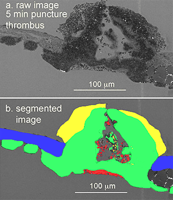 |
Venous puncture wound thrombi, 1, 5, 20 min post-puncture, 100 nm XY raw images, 20 nm XY pixels every 20 micons, 3 nm wide area TEM montages at selected depth [15 multi-frame micrographs composed of 1 frames each in DM4 format] | Storrie B, Leapman RD [Pubmed: 34531522] [DOI: 10.1038/s42003-021-02615-y] |
381.2 GB | — |
| 2016-08-15 |  |
VPP subtomogram averaging [11 class averages in MRC format] | Khoshouei M, Pfeffer S, Baumeister W, Foerster F, Danev R [Pubmed: 27235783] [DOI: 10.1016/j.jsb.2016.05.009] |
33.9 GB | 9.6 Å |
| 2021-11-08 |  |
VHUT-cryo-FIB, a method to fabricate frozen-hydrated lamella of tissue specimen for in situ cryo-electron tomography [13 multi-frame micrographs composed of 30 frames each in TIFF format] | Zhang J [Pubmed: 34174447] [DOI: 10.1016/j.jsb.2021.107763] |
112.1 GB | 18.0 Å |
| 2024-03-19 |  |
Unveiling the ultrastructural landscape of extracellular matrix via lift-out cryo-FIBSEM and cryo-ET [multiple data sets in TIFF and MRC formats] | Zens B., Fäßler F., Hansen J.M., Hauschild R., Datler J., Hodirnau V.V., Zheden V., Alanko J., Sixt M., Schur F.K.M. [DOI: 10.1083/jcb.202309125] |
183.0 GB | — |
