
Electron Microscopy Public Image Archive
Electron Microscopy Public Image Archive
Due to a storage failure, some data files are currently inaccessible (Markted: Incomplete dataset).
We are currently working to restore, but we are accepting priority requests.(email, inquiry).
We are restoring lost files from backups in the following order:
We apologize for the inconvenience and appreciate your understanding.
大阪大学的EMPIAR-PDBj团队为亚洲EM研究人员向EMPIAR传送大型EM图像数据提供服务。 除了通过互联网将数据直接传送到EBI(UK),研究人员还可以通过邮政或快递服务将数据硬盘发送到大阪大学,或者通过互联网传送到设置于大阪大学的服务器,然后由我们代为传送至数据登录网站。 如果您想使用此项服务,请先通过 电子邮件 与我们联系。
| Release date | Imageset | Title | Authors and references | Size | Resolution |
|---|---|---|---|---|---|
| 2023-04-13 | 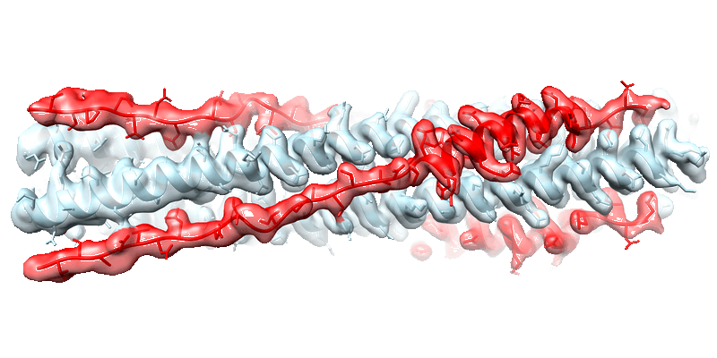 |
Cryo-EM structure of the SARS-CoV-2 HR1HR2 fusion core complex with extended HR2 [18846 multi-frame micrographs composed of 40 frames each in TIFF format] | Yang K, Brunger AT [Pubmed: 36122200] [DOI: 10.1073/pnas.2210990119] |
4.6 TB | 2.22 Å |
| 2023-04-13 | 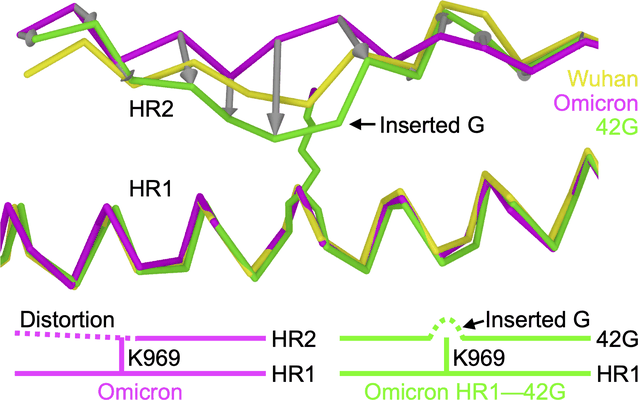 |
Structure-based design of a SARS-CoV-2 Omicron-specific inhibitor [multiple data sets in TIFF format] | Yang K, Brunger AT [Pubmed: 36940324] [DOI: 10.1073/pnas.2300360120] |
13.5 TB | 2.51 Å |
| 2022-07-22 | 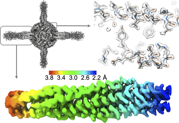 |
Structural conservation among variants of the SARS-CoV-2 spike postfusion bundle [multiple data sets in TIFF format] | Yang K, Brunger AT [Pubmed: 36940324] [DOI: 10.1073/pnas.2300360120] |
32.4 TB | 2.09 - 2.52 Å |
| 2020-09-02 | 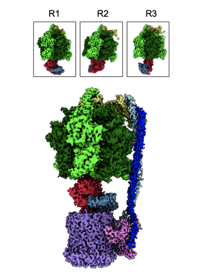 |
Structural basis of redox modulation on chloroplast ATP synthase (reduced form) [2063 micrographs in MRC format] | Yang JH, Williams D, Kandiah E, Fromme P, Chiu PL [Pubmed: 32879423] [DOI: 10.1038/s42003-020-01221-8] |
109.9 GB | 3.05 - 4.34 Å |
| 2022-02-28 | 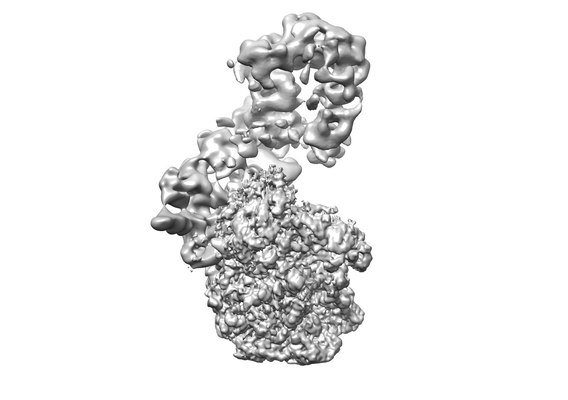 |
Structural visualization of de novo initiation of RNA polymerase II transcription [multiple data sets in TIFF format] | Yang C, Fujiwara R, Kim HJ, Basnet P, Zhu Y, Gorbea Colón JJ, Steimle S, Garcia BA, Kaplan CD, Murakami K [Pubmed: 35051353] [DOI: 10.1016/j.molcel.2021.12.020] |
14.3 TB | 3.0 - 7.6 Å |
| 2023-03-16 | 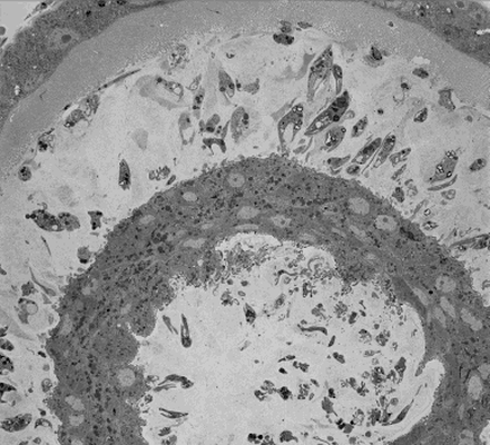 |
SBFSEM imaging of Leishmania haptomonads on the stomodeal valve in the sand fly [400 multi-frame micrographs composed of 1 frames each in MRC format] | Yanase R, Sunter JD [DOI: 10.1101/2022.10.28.514187] |
82.0 GB | — |
| 2023-03-17 | 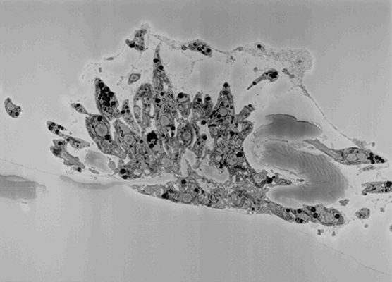 |
SBFSEM imaging of Leishmania haptomonad-like cells attached to plastic [252 multi-frame micrographs composed of 1 frames each in MRC format] | Yanase R, Sunter JD [DOI: 10.1101/2022.10.28.514187] |
33.6 GB | — |
| 2023-03-17 | 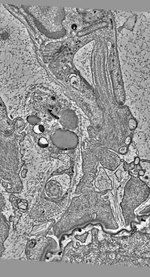 |
Serial section electron tomography of a Leishmania haptomonad on the stomodeal valve in the sand fly [670 multi-frame micrographs composed of 1 frames each in MRC format] | Yanase R, Sunter JD [DOI: 10.1101/2022.10.28.514187] |
6.7 GB | — |
| 2023-03-17 | 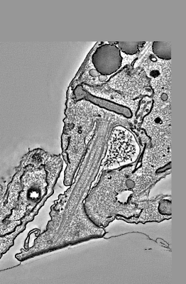 |
Serial section electron tomography of a Leishmania haptomonad-like cell attached to plastic [718 multi-frame micrographs composed of 1 frames each in MRC format] | Yanase R, Sunter JD [DOI: 10.1101/2022.10.28.514187] |
10.8 GB | — |
| 2020-09-03 |  |
ISWI-NCP complex in the ADPBeF-bound state [stack of 166165 particles in MRCS format] | Yan L, Wu H, Li X, Gao N, Chen Z [Pubmed: 30872815] [DOI: 10.1038/s41594-019-0199-9] |
41.7 GB | 3.37 Å |
| 2020-09-02 |  |
ISWI-NCP complex in the ADP-bound state [stack of 168430 particles in MRCS format] | Yan L, Wu H, Li X, Gao N, Chen Z [Pubmed: 32123390] [DOI: 10.1038/s41594-020-0388-6] |
36.2 GB | 2.9 Å |
| 2025-01-20 | 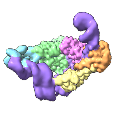 |
Cryo-EM structure of human TUT1 complexed with U6 snRNA [multiple data sets in TIFF format] | Yamashita S, Tomita K [DOI: 10.1093/nar/gkae1314] |
3.4 TB | 3.21 - 3.83 Å |
| 2022-07-18 | 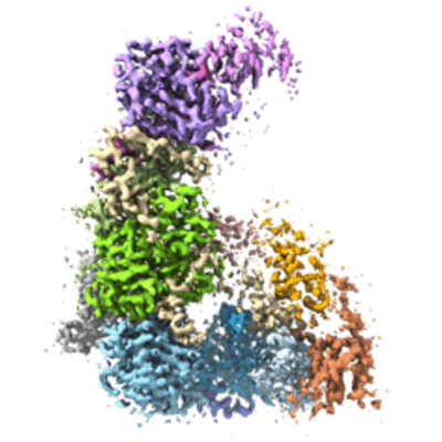 |
Structure of the Dicer-2-R2D2 heterodimer bound to a small RNA duplex [multiple data sets in TIFF format] | Yamaguchi S, Naganuma M, Nishizawa T, Kusakizako T, Tomari Y, Nishimasu H, Nureki O [Pubmed: 35768503] [DOI: 10.1038/s41586-022-04790-2] |
1.4 TB | 3.3 Å |
| 2026-06-17 |  |
Cryo-EM SPA dataset of arginine oxidase from Pseudomonas sp. TRU 7192 [5035 multi-frame micrographs composed of 40 frames each in TIFF format] | Yamaguchi H, Takahashi K, Numoto N, Suzuki H, Tatsumi M, Kamegawa A, Nishikawa K, Asano Y, Mizukoshi T, Miyano H, Fujiyoshi Y, Sugiki M [Pubmed: 39420599] [DOI: 10.1093/jb/mvae070] |
916.4 GB | 2.34 Å |
| 2025-07-06 |  |
Cryo-EM structure of the barley Yellow stripe 1 transporter [multiple data sets in TIFF format] | Yamagata A, Murata Y, Namba K, Terada T, Fukai S, Shirouzu M [Pubmed: 36424382] [DOI: 10.1038/s41467-022-34930-1] |
5.0 TB | 2.7 - 2.9 Å |
| 2025-06-26 |  |
Cryo-EM structures of SV2A in complex with antiepileptic drugs and botulinum neurotoxin A2 receptor-binding domain [multiple data sets in TIFF format] | Yamagata A, Ito K, Suzuki T, Dohmae N, Terada T, Shirouzu M [Pubmed: 38637505] [DOI: 10.1038/s41467-024-47322-4] |
6.6 TB | 2.82 - 3.38 Å |
| 2026-03-26 |  |
Clostridium perfringens iota toxin membrane-binding component Ib-prepore and Ib-pore [11549 multi-frame micrographs composed of 58 frames each in TIFF format] | Yamada TY, Sugita YS, Yoshida TY, Noda TN, Tsuge HT | 2.7 TB | 2.47 - 3.73 Å |
| 2022-11-14 | 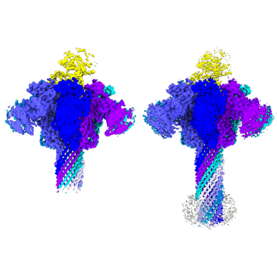 |
Cryo-EM structures of Ib-pore and Ia-bound Ib-pore [multiple data sets in TIFF format] | Yamada T, Yoshida T, Kawamoto A, Tsuge H [Pubmed: 32123390] [DOI: 10.1038/s41594-020-0388-6] |
8.0 TB | 2.8 - 2.9 Å |
| 2026-08-06 |  |
Iota toxin Ib D452A [4073 multi-frame micrographs composed of 52 frames each in TIFF format] | Yamada T, Nakanishi R, Sugita Y, Ninomiya Y, Noda T, Tsuge H | 933.7 GB | — |
| 2024-11-06 | 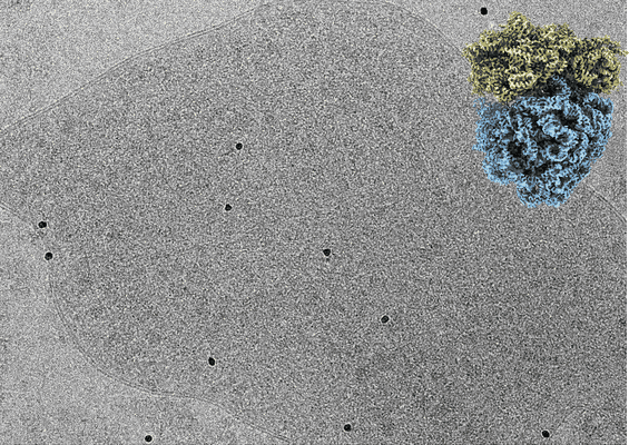 |
Tilt series of Mycoplasma pneumoniae cells treated with chloramphenicol collected on Gatan K3 camera [139 multi-frame micrographs composed of 10 frames each in TIFF format] | Xue L, Spahn CMT, Schacherl M, Mahamid J [Pubmed: 39668257] [DOI: 10.1038/s41594-024-01441-0] |
170.5 GB | 3.0 Å |
| 2021-06-04 | 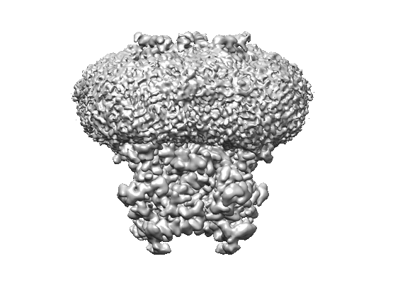 |
Cryo-EM structure of human rod CNGA1 channel in apo-state [stack of 274333 particles in MRCS format] | Xue J, Han Y, Zeng W, Wang Y, Jiang Y [Pubmed: 33651975] [DOI: 10.1016/j.neuron.2021.02.007] |
84.9 GB | 2.6 Å |
| 2024-08-24 | 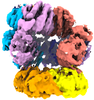 |
E. coli clamp loader with a closed clamp [multiple data sets in TIFF format] | Xu ZQ, Jergic S, Lo ATY, Pradhan AC, Brown HJ, Bouwer JC, Ghodke H, Lewis PJ, Tolun G, Oakley AJ, Dixon NE [Pubmed: 39333521] [DOI: 10.1038/s41467-024-52623-9] |
1.3 TB | 3.7 Å |
| 2025-06-07 |  |
E. coli clamp loader with primer-template DNA and an open clamp [8644 multi-frame micrographs composed of 50 frames each in MRC format] | Xu ZQ, Jergic S, Lo ATY, Pradhan AC, Brown HJ, Bouwer JC, Ghodke H, Lewis PJ, Tolun G, Oakley AJ, Dixon NE [Pubmed: 39333521] [DOI: 10.1038/s41467-024-52623-9] |
3.0 TB | 2.9 Å |
| 2025-01-24 | 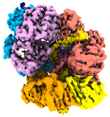 |
E. coli clamp loader with primer-template DNA and a closed clamp [7201 multi-frame micrographs composed of 60 frames each in MRC format] | Xu ZQ, Jergic S, Dixon NE, Lo ATY, Pradhan AC, Brown HJ, Bouwer JC, Ghodke H, Lewis PJ, Tolun G, Oakley AJ [Pubmed: 39333521] [DOI: 10.1038/s41467-024-52623-9] |
1.0 TB | 2.6 Å |
| 2025-06-07 |  |
E. coli clamp loader with primer-template DNA [3199 multi-frame micrographs composed of 50 frames each in MRC format] | Xu ZQ, Jergic S, Dixon NE, Lo ATY, Pradhan AC, Brown HJ, Bouwer JC, Ghodke H, Lewis PJ, Tolun G, Oakley AJ [Pubmed: 39333521] [DOI: 10.1038/s41467-024-52623-9] |
2.2 TB | 2.8 Å |
