
Electron Microscopy Public Image Archive
Electron Microscopy Public Image Archive
Due to a storage failure, some data files are currently inaccessible (Markted: Incomplete dataset).
We are currently working to restore, but we are accepting priority requests.(email, inquiry).
We are restoring lost files from backups in the following order:
We apologize for the inconvenience and appreciate your understanding.
大阪大学的EMPIAR-PDBj团队为亚洲EM研究人员向EMPIAR传送大型EM图像数据提供服务。 除了通过互联网将数据直接传送到EBI(UK),研究人员还可以通过邮政或快递服务将数据硬盘发送到大阪大学,或者通过互联网传送到设置于大阪大学的服务器,然后由我们代为传送至数据登录网站。 如果您想使用此项服务,请先通过 电子邮件 与我们联系。
| Release date | Imageset | Title | Authors and references | Size | Resolution |
|---|---|---|---|---|---|
| 2021-11-08 | 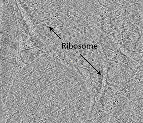 |
VHUT-cryo-FIB, a method to fabricate frozen-hydrated lamella of tissue specimen for in situ cryo-electron tomography [13 multi-frame micrographs composed of 30 frames each in TIFF format] | Zhang J [Pubmed: 34174447] [DOI: 10.1016/j.jsb.2021.107763] |
112.1 GB | 18.0 Å |
| 2026-10-07 |  |
Cryo-EM datasets of human Argonaute2 pre-RISC and mature RISC-target, PDB 9YM9, 9YMA, 9YMB, 9YMC, 9YMD, and 10BE [multiple data sets in TIFF and EER formats] | Zhang H, Nakanishi K [Pubmed: 42190652] [DOI: 10.1016/j.molcel.2026.04.029] |
11.9 TB | 3.02 - 3.87 Å |
| 2018-11-23 |  |
Cryo-EM Reconstruction of apo EsCas13d [multiple data sets in MRCS format] | Zhang C, Lyumkis D [Pubmed: 30241607] [DOI: 10.1016/j.cell.2018.09.001] |
66.9 GB | 6.5 Å |
| 2025-02-04 |  |
Structure of the IFN-lambda3/IFN-lambdaR1/IL-10Rbeta receptor complex with an engineered IL-10Rbeta [2928 multi-frame micrographs composed of 40 frames each in TIFF format] | Zhang B, Grubbe WS, Mendoza JL, Zhao M [Pubmed: 39827213] [DOI: 10.1038/s41467-025-56119-y] |
1.6 TB | 3.0 Å |
| 2026-05-20 |  |
Cryo-EM Structure of the IFN-lambda4/IFN-lambdaR1/IL-10Rbeta receptor complex with an engineered IL-10Rbeta [5130 multi-frame micrographs composed of 40 frames each in TIFF format] | Zhang B, Grubbe WS, Mendoza JL, Zhao M [Pubmed: 39827213] [DOI: 10.1038/s41467-025-56119-y] |
2.9 TB | 3.26 Å |
| 2024-03-19 | 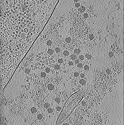 |
Unveiling the ultrastructural landscape of extracellular matrix via lift-out cryo-FIBSEM and cryo-ET [multiple data sets in TIFF and MRC formats] | Zens B., Fäßler F., Hansen J.M., Hauschild R., Datler J., Hodirnau V.V., Zheden V., Alanko J., Sixt M., Schur F.K.M. [Pubmed: 38506714] [DOI: 10.1083/jcb.202309125] |
183.0 GB | — |
| 2024-05-20 |  |
Cryo-EM micrographs of human α1B/βI+βIVb microtubules bound to GMPCPP decorated with TTLL6 [2770 micrographs in MRC format] | Zehr EA, Roll-Mecak A [Pubmed: 38658656] [DOI: 10.1038/s41589-024-01599-0] |
146.9 GB | 3.6 - 7.2 Å |
| 2026-03-13 |  |
Cryo-electron tomography of microtubules in axons from human cortical neurons derived from induced pluripotent stem cells (iPSCs). [5 reconstructed volumes in MRC format] | Zehr EA, Roll-Mecak A [Pubmed: N/A] [DOI: 10.1038/s41594-026-01787-7] |
544.4 GB | 2.69 Å |
| 2026-03-13 |  |
Cryo-electron tomography of microtubules in undifferentiated induced pluripotent stem cells (iPSCs) [5 reconstructed volumes in MRC format] | Zehr EA, Roll-Mecak A [DOI: 10.1038/s41594-026-01787-7] |
526.8 GB | 2.69 Å |
| 2026-03-13 |  |
Cryo-electron tomography of in vitro assembled, GDP-bound, porcine brain microtubules. [9 reconstructed volumes in MRC format] | Zehr EA, Roll-Mecak A [DOI: 10.1038/s41594-026-01787-7] |
722.7 GB | 2.69 Å |
| 2026-03-13 |  |
Cryo-electron tomography of in vitro assembled, GDP-bound, porcine brain microtubules. [10 reconstructed volumes in MRC format] | Zehr EA, Roll-Mecak A [Pubmed: N/A] [DOI: 10.1038/s41594-026-01787-7] |
553.1 GB | 2.69 Å |
| 2026-03-16 |  |
Cryo-electron tomography of in vitro assembled, GMPCPP-bound, porcine brain microtubules. [8 reconstructed volumes in MRC format] | Zehr EA, Roll-Mecak A [Pubmed: N/A] [DOI: 10.1038/s41594-026-01787-7] |
566.3 GB | 2.69 Å |
| 2026-03-13 |  |
Cryo-electron tomography of in vitro assembled, GMPCPP-bound, porcine brain microtubules. [16 reconstructed volumes in MRC format] | Zehr EA, Roll-Mecak A [Pubmed: N/A] [DOI: 10.1038/s41594-026-01787-7] |
1.4 TB | 2.69 Å |
| 2022-11-11 |  |
Structure of COPII coat - tilt series raw data [6601 multi-frame micrographs composed of 10 frames each in TIFF format] | Zanetti G, Hagen W, Hutchings J [Pubmed: 36468689] [DOI: 10.7554/eLife.83724] |
192.3 GB | 3.8 Å |
| 2026-04-20 |  |
Cryo-EM structure of the SN230G6 Fab - HLA-A*02:01 human alloantibody-HLA complex [12894 multi-frame micrographs composed of 40 frames each in TIFF format] | Zampieri V, Priddey A, Schneider S, Heidt S, Kosmoliaptsis V, Marquez JA | 2.0 TB | 2.78 Å |
| 2026-04-20 |  |
Cryo-EM structure of the WIM8E5 Fab - HLA-A*11:01 human alloantibody-HLA complex [18395 multi-frame micrographs composed of 40 frames each in TIFF format] | Zampieri V, Priddey A, Humm AS, Pellegrini E, Heidt S, Kosmoliaptsis V, Marquez JA | 2.6 TB | 3.23 Å |
| 2026-06-08 |  |
In extracto cryo-EM reveals eEF2 as a major hibernation factor on 60S and 80S particles [24552 micrographs in MRC format] | Zahra Seraj XZ, Ximena Zottig XZ, Chun-Ying Huang CYH, Anna B Loveland ABL, Stephen Diggs SD, Emily Sholi ES [Pubmed: 41394757] [DOI: 10.7554/eLife.110114.2] |
2.1 TB | — |
| 2020-08-18 | 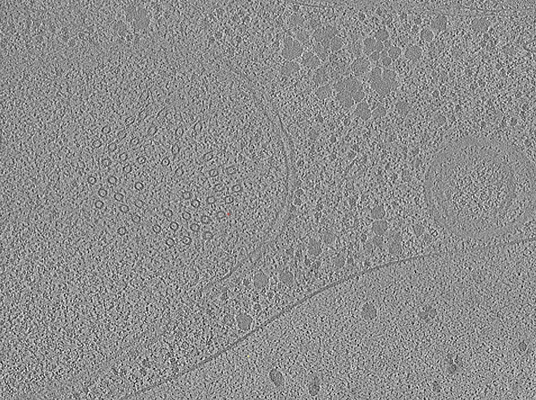 |
Yeast Tilt Series Collected on Lamella Generated by Fully Automated FIB Milling [1 tilt series in MRC format] | Zachs T, Schertel A, Medeiros J, Weiss GL, Hugener J, Matos J, Pilhofer M [Pubmed: 32149604] [DOI: 10.7554/eLife.52286] |
2.6 GB | 30.0 Å |
| 2026-02-03 |  |
Cryo-EM structure of the Chromera velia PSI supercomplex at 1.84 Angstrom resolution [multiple data sets in EER format] | Yuan X, Lukeš M, Qian P, Charras Q, Smrčka T, Man P, Kovářová L, Koník P, Heilmann E, Al-Amoudi A, Sobotka R, Naschberger A [Pubmed: 41423677] [DOI: 10.1038/s41467-025-67637-0] |
10.5 TB | 1.84 - 2.05 Å |
| 2026-06-29 |  |
Apoferritin with xLPP on [492 multi-frame micrographs composed of 34 frames each in TIFF format] | Yu Y, Olshin PK [DOI: 10.64898/2026.06.05.730245] |
112.6 GB | 1.87 Å |
| 2022-01-21 | 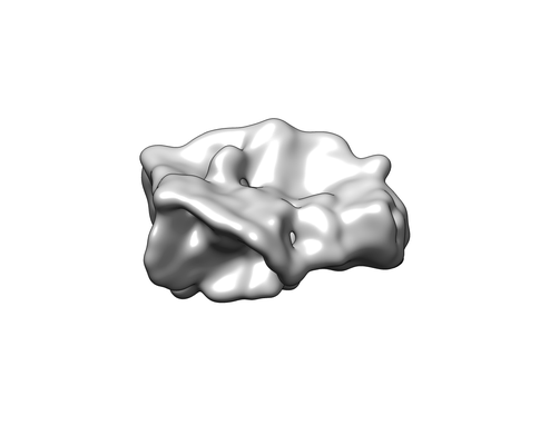 |
Structural Insights of Transcriptionally Active, Full-Length Androgen Receptor Coactivator Complexes [1958 multi-frame micrographs composed of 50 frames each in MRC format] | Yu X, Yi P [Pubmed: 32668201] [DOI: 10.1016/j.molcel.2020.06.031] |
5.1 TB | 13.0 Å |
| 2022-01-24 | 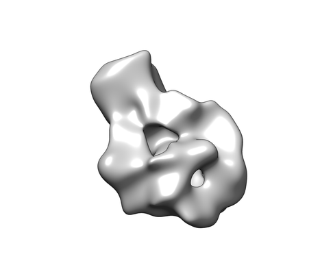 |
Structural Insights of Transcriptionally Active, Full-Length Androgen Receptor Coactivator Complexes [multiple data sets in MRC and TIFF formats] | Yu X, Yi P [Pubmed: 32668201] [DOI: 10.1016/j.molcel.2020.06.031] |
2.0 TB | 20.0 Å |
| 2019-04-05 |  |
mouse MDA5-dsRNA 10mM ATP Filamemt [2213 micrographs in MRC format] | Yu Q, Qu K, Modis YE [Pubmed: 30449722] [DOI: 10.1016/j.molcel.2018.10.012] |
138.9 GB | 3.87 Å |
| 2019-04-05 |  |
mouse MDA5-dsRNA 2.5-mM AMPPNP Filamemt [471 multi-frame micrographs composed of 75 frames each in MRC format] | Yu Q, Qu K, Modis YE [Pubmed: 30449722] [DOI: 10.1016/j.molcel.2018.10.012] |
1.1 TB | 4.06 Å |
| 2019-04-05 |  |
mouse MDA5-dsRNA filaments [592 multi-frame micrographs composed of 27 frames each in MRC format] | Yu Q, Qu K, Modis YE [Pubmed: 30449722] [DOI: 10.1016/j.molcel.2018.10.012] |
499.5 GB | 4.02 - 4.16 Å |
