Electron Microscopy Public Image Archive
Electron Microscopy Public Image Archive
大阪大学的EMPIAR-PDBj团队为亚洲EM研究人员向EMPIAR传送大型EM图像数据提供服务。 除了通过互联网将数据直接传送到EBI(UK),研究人员还可以通过邮政或快递服务将数据硬盘发送到大阪大学,或者通过互联网传送到设置于大阪大学的服务器,然后由我们代为传送至数据登录网站。 如果您想使用此项服务,请先通过 电子邮件 与我们联系。
| Release date | Imageset | Title | Authors and references | Size | Resolution |
|---|---|---|---|---|---|
| 2021-01-08 | 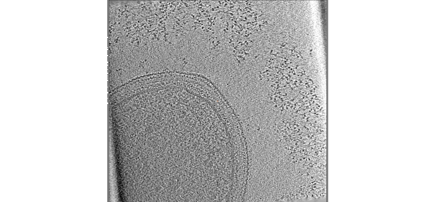 |
Nanoscale view of the Clostridium thermocellum cellulosome during cellulose degradation reveals an ecological strategy leading to phenotypic heterogeneity [37 tilt series in MRC format] | Tatli M, Moraïs S, Tovar-Herrera OE, Bomble Y, Bayer EA, Medalia O, Mizrahi I | 1004.3 MB | — |
| 2015-10-01 | 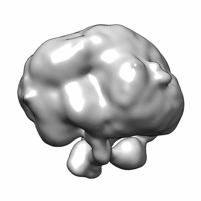 |
Tubulin Chaperone complexes TBC-DEG Q73L: alpha beta-tubulin:TBCC complex [stack of 16801 particles in MRC format] | Nithianantham S, Le S, Seto E, Jia W, Leary J, Corbett KD, Moore JK, Al-Bassam J [Pubmed: 26208336] [DOI: 10.7554/eLife.08811] |
1.0 GB | 24.0 Å |
| 2023-01-18 | 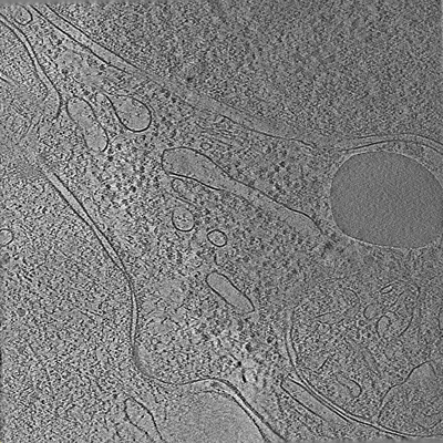 |
Tilt series of cell-cell contact of two PTK-1 cells [35 tilt series in MRC format] | Lemos M, Bezault A, Sauvanet C, Hanein D, Volkmann N [Pubmed: 36539423] [DOI: 10.1038/s41467-022-35409-9] |
1.1 GB | — |
| 2015-10-01 |  |
Tubulin Chaperone complexes TBC-DEG Q73L: alpha beta-tubulin complex [stack of 18361 particles in MRC format] | Nithianantham S, Le S, Seto E, Jia W, Leary J, Corbett KD, Moore JK, Al-Bassam J [Pubmed: 26208336] [DOI: 10.7554/eLife.08811] |
1.1 GB | 24.0 Å |
| 2024-02-15 | 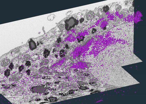 |
FIB-SEM dataset showing localization of a Golgi matrix protein GM130 in human hepatocellular carcinoma cell (Huh-7) [564 reconstructed volumes in TIFF format] | Belevich I, Jokitalo E | 1.1 GB | — |
| 2024-01-15 | 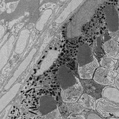 |
Developing retina in zebrafish 55 hpf larval eye. [16 reconstructed volumes in DM3 format] | Wilsch-Bräuninger M | 1.2 GB | — |
| 2020-06-30 |  |
Soft X-ray Tomography of mock-infected U2OS cells [1 tilt series in MRC format] | Kounatidis I, Stanifer ML, Phillips MA, Paul-Gilloteaux P, Heiligenstein X, Wang H, Okolo CA, Fish TM, Spink MC, Stuart DI, Davis I, Boulant S, Grimes JM, Dobbie IM, Harkiolaki M [Pubmed: 32610083] [DOI: 10.1016/j.cell.2020.05.051] |
1.2 GB | — |
| 2020-06-30 | 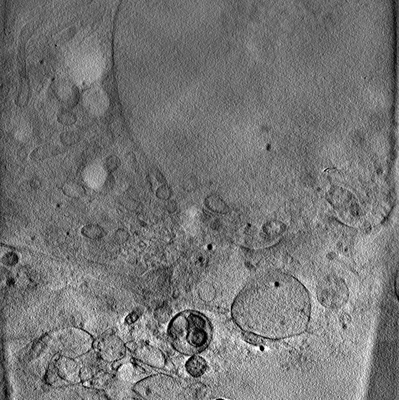 |
Soft X-ray Tomography of mock-infected U2OS cells [1 tilt series in MRC format] | Kounatidis I, Stanifer ML, Phillips MA, Paul-Gilloteaux P, Heiligenstein X, Wang H, Okolo CA, Fish TM, Spink MC, Stuart DI, Davis I, Boulant S, Grimes JM, Dobbie IM, Harkiolaki M [Pubmed: 32610083] [DOI: 10.1016/j.cell.2020.05.051] |
1.2 GB | — |
| 2023-03-03 | 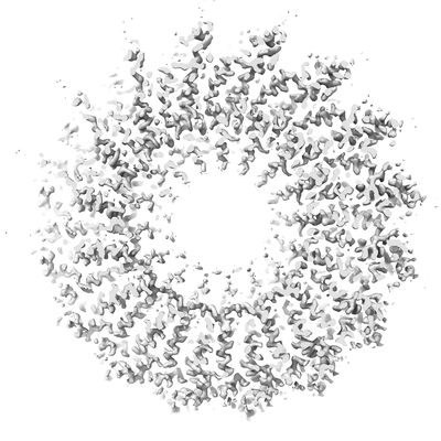 |
Cryo-iDPC-STEM structure of TMV - convergence semi-angle 4.0 mrad [multiple data sets in TIFF format] | Lazić I, Wirix M, Leidl ML, Sachse C [Pubmed: 36064775] [DOI: 10.1038/s41592-022-01586-0] |
1.2 GB | 3.5 Å |
| 2018-01-03 | 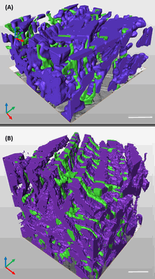 |
3D reconstruction of the cardiac mitochondria in Neaonate and Adult GP cardiomyocyte [262 multi-frame micrographs composed of 1 frames each in TIFF format] | Kashbour H, Taggart M, White K | 1.2 GB | — |
| 2021-03-05 | 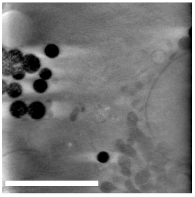 |
Dynabeads as Fiducials for SXT and SIM Correlation [700 tilt series in MRC format] | Okolo CA, Kounatidis I, Groen J, Nahas KL, Balint S, Fish T, Koronfel MA, López-Cortajarena A, Dobbie I, Pereiro E, Harkiolaki M | 1.2 GB | — |
| 2021-02-26 |  |
Cryo electron tomography of muscle tissue lamella from mice [1 tilt series in MRC format] | Zhang J, Zhang D, Sun L, Ji G, Huang X, Niu T, Xu J, Ma C, Zhu Y, Gao N, Xu W, Sun F [Pubmed: 34174447] [DOI: 10.1016/j.jsb.2021.107763] |
1.3 GB | — |
| 2016-11-18 | 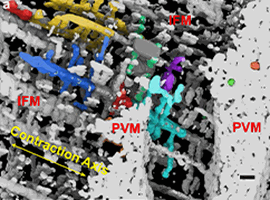 |
Focused Ion Beam-Scanning Electron Microscopy of mitochondrial reticulum in murine skeletal muscle [291 reconstructed volumes in MRC format] | Glancy B, Hartnell LM, Malide D, Yu ZX, Combs CA, Connelly PS, Subramaniam S, Balaban RS [Pubmed: 26223627] [DOI: 10.1038/nature14614] |
1.5 GB | — |
| 2023-08-18 |  |
Negative stain EM structure of the NF155 extracellular domain [95 micrographs in MRC format] | McKie SJ, Deane JE [Pubmed: 36996106] [DOI: 10.1073/pnas.2218823120] |
1.5 GB | 19.0 Å |
| 2015-06-18 |  |
A simulated cryoEM data set of GroEL particles [stack of 10000 particles in MRC format] | Deng Y, Sun F | 1.5 GB | — |
| 2023-10-17 | 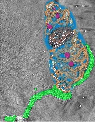 |
Soft X-ray Cryo Tomography of Trypanosoma [180 reconstructed volumes in MRC format] | Darrow MC [Pubmed: 28246039] [DOI: 10.1016/j.jsb.2017.02.007] |
1.5 GB | — |
| 2021-04-30 | 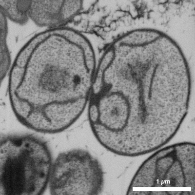 |
FIB-SEM of Gemmata obscuriglobus - a species of the Planctomycetes phylum [1 stitched maps in TIFF format] | Seeger C, Dyrhage K, Mahajan M, Odelgard A, Bergström Lind S, Andersson SGE [DOI: 10.3389/fmicb.2021.643045] |
1.6 GB | — |
| 2018-01-03 | 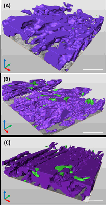 |
3D reconstruction of cardiac mitochondria in earlly prenatal (G55/57) and mid term(G58/60) and term (G66/68) GP cardiomyocyte [341 multi-frame micrographs composed of 1 frames each in TIFF format] | Kashbour H, Taggart M, White K | 1.7 GB | — |
| 2020-09-02 | 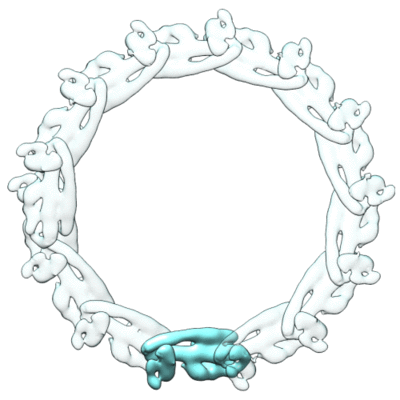 |
CryoEM reconstruction of ESCRT-III filament composed of IST1 NTD R16E K27E double mutant [stack of 4556 particles in MRCS format] | Nguyen HC, Talledge N, McCullough J, Sharma A, Moss FR, Iwasa JH, Vershinin MD, Sundquist WI, Frost A [Pubmed: 32251413] [DOI: 10.1038/s41594-020-0404-x] |
1.7 GB | 7.2 Å |
| 2018-01-03 | 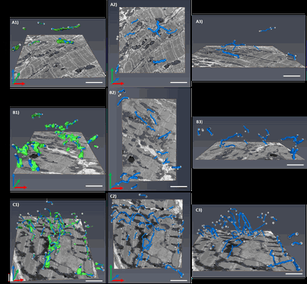 |
Segment /nodes network analysis of the tubular system of the prenatal stages of GP cardiomyocyte [254 multi-frame micrographs composed of 1 frames each in TIFF format] | Kashbour H, Taggart M | 1.9 GB | — |
| 2021-10-19 |  |
Multi-modal adaptor-clathrin contacts drive coated vesicle assembly [stack of 5720 particles in MRCS format] | Smith SM, Larocque G, Wood KM, Morris KL, Roseman AM, Sessions RB, Royle SJ, Smith CJ [Pubmed: 34487371] [DOI: 10.15252/embj.2021108795] |
1.9 GB | — |
| 2018-10-22 |  |
The in situ structures of mono-, di-, and trinucleosomes in human heterochromatin [59 tilt series in MRC format] | Cai S, Böck D, Pilhofer M, Gan L [Pubmed: 30091658] [DOI: 10.1091/mbc.E18-05-0331] |
1.9 GB | 21.0 - 24.0 Å |
