Electron Microscopy Public Image Archive
Electron Microscopy Public Image Archive
大阪大学的EMPIAR-PDBj团队为亚洲EM研究人员向EMPIAR传送大型EM图像数据提供服务。 除了通过互联网将数据直接传送到EBI(UK),研究人员还可以通过邮政或快递服务将数据硬盘发送到大阪大学,或者通过互联网传送到设置于大阪大学的服务器,然后由我们代为传送至数据登录网站。 如果您想使用此项服务,请先通过 电子邮件 与我们联系。
| Release date | Imageset | Title | Authors and references | Size | Resolution |
|---|---|---|---|---|---|
| 2018-05-04 |  |
Benchmarking cryo-EM single particle analysis workflow [699 multi-frame micrographs composed of 33 frames each in MRC format] | Kim LK, Rice WJ, Eng ET, Kopylov M, Cheng A, Raczkowski AR, Jordan KD, Bobe D, Potter CS, Carragher B [Pubmed: 29951483] [DOI: 10.3389/fmolb.2018.00050] |
37.3 GB | 2.5 - 2.8 Å |
| 2020-07-06 |  |
Subnanometer-resolution structure determination in situ by a hybrid subtomogram averaging - single particle cryoEM - workflow - on TMV [4 tilt series in MRC format] | Sanchez RM, Zhang Y, Chen W, Dietrich L, Kudryashev M [Pubmed: 32709843] [DOI: 10.1038/s41467-020-17466-0] |
37.5 GB | 5.24 Å |
| 2024-04-23 |  |
Cryo-Electron Tomography of Zebra Fish Muscle Tissue Biopsy [1 tilt series in MRC format] | Gervinskas G, Trepout S, Venugopal H, Ramm G | 37.6 GB | — |
| 2023-09-22 |  |
In-tissue cryo electron tomography of App^NL-G-F amyloid plaques [23 tilt series in MRC format] | Leistner C, Wilkinson M, Burgess A, Lovatt M, Goodbody S, Xu Y, Deuchars S, Radford SE, Ranson NA, Frank RAW [Pubmed: 37198197] [DOI: 10.1002/pro.3943] |
38.0 GB | 3.0 Å |
| 2014-02-21 | 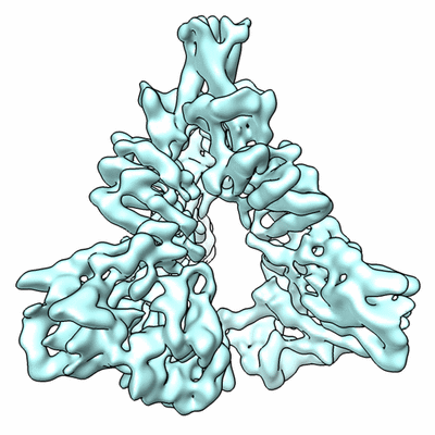 |
Low-contrast particle stack for HIV-1 Env gp160 precursor (MRC stack) [stack of 124478 particles in MRC format] | Mao Y, Wang L, Gu C, Herschhorn A, Desormeaux A, Finzi A, Xiang SH, Sodroski JG [Pubmed: 23757493] [DOI: 10.1073/pnas.1307382110] |
38.0 GB | 6.0 Å |
| 2024-04-23 |  |
Cryo-Electron Tomography of Mouse Brain Tissue Biopsy [1 tilt series in MRC format] | Gervinskas G, Trepout S, Venugopal H, Ramm G | 38.5 GB | — |
| 2023-09-05 |  |
Benchmark SBF SEM data of HeLa cells previously imaged by Zeiss LSM900 Airyscan microscopy [multiple data sets in DM4 and TIFF formats] | Domart MC, Collinson LM [DOI: 10.1101/2023.05.11.540445] |
39.2 GB | — |
| 2020-12-02 |  |
Apoferritin tilt series and frame series used to benchmark the resolution of both data types in M [multiple data sets in TIFF format] | Tegunov D, Xue L, Dienemann C, Cramer P, Mahamid J [Pubmed: 33542511] [DOI: 10.1038/s41592-020-01054-7] |
39.6 GB | 2.3 Å |
| 2018-05-30 |  |
Cryo-ET of natural chromatin from Ostreococcus tauri and Saccharyomyces cerevisiae [25 class averages in MRC format] | Cai S, Song Y, Chen C, Shi J [Pubmed: 29742050] [DOI: 10.1091/mbc.E17-07-0449] |
40.1 GB | — |
| 2018-04-05 |  |
CryoET of insulin-bound insulin receptor single particle with spot-to-plunge time of 200ms [multiple data sets in MRC format] | Noble AJ, Wei H, Dandey VP, Zhang Z, Potter CS, Carragher B [Pubmed: 30250056] [DOI: 10.1038/s41592-018-0139-3] |
40.4 GB | — |
| 2021-11-01 |  |
electron cryo-tomograms of axons from human cerebral organoids, expressing GFP-ESYT1 [multiple data sets in MRC and TIFF formats] | Hoffmann P, Giandomenico S, Ganeva I, Wozny M, Sutcliffe M, Lancaster M, Kukulski W [Pubmed: 34698018] [DOI: 10.7554/eLife.70269] |
40.9 GB | — |
| 2020-09-03 |  |
ISWI-NCP complex in the ADPBeF-bound state [stack of 166165 particles in MRCS format] | Yan L, Wu H, Li X, Gao N, Chen Z [Pubmed: 30872815] [DOI: 10.1038/s41594-019-0199-9] |
41.7 GB | 3.37 Å |
| 2014-11-06 |  |
Yeast 80S Ribosome - tRNA- Kozak mRNA complexes, Frealign Input Particle Stack [stack of 86866 particles in MRC format] | Svidritskiy E, Brilot AF, Koh CS, Grigorieff N, Korostelev AA [Pubmed: 25043550] [DOI: 10.1016/j.str.2014.06.003] |
42.0 GB | 6.2 - 6.3 Å |
| 2023-02-22 |  |
Electron cryo-tomography data of HeLa cells expressing untagged Cidec [multiple data sets in MRC format] | Ganeva I, Lim K, Boulanger J, Hoffmann PC, Muriel O, Borgeaud AC, Hagen WJH, Savage DB, Kukulski W [Pubmed: 36800289] [DOI: 10.1016/j.celrep.2023.112107] |
42.8 GB | — |
| 2023-01-18 |  |
Cryo iDPC-STEM single particle analysis of keyhole limpet hemocyanin [multiple data sets in TIFF format] | Mann DM, Lazic I, Wirix M, de Haas F [Pubmed: 36064775] [DOI: 10.1038/s41592-022-01586-0] |
42.9 GB | 6.51 Å |
| 2020-10-23 |  |
170314, Five-day-old Col-0 Arabidopsis thaliana root, phloem pole unloading zone (339-381 um from the root tip) [multiple data sets in MRC format] | Paterlini A, Belevich I, Jokitalo E, Helariutta Y [Pubmed: 31182845] [DOI: 10.1038/s41477-019-0429-5] |
43.2 GB | — |
| 2017-12-15 |  |
CryoET of rabbit muscle aldolase single particle [multiple data sets in MRC format] | Noble AJ, Dandey VP, Wei H, Brasch J, Chase J, Acharya P, Tan YZ, Zhang Z, Kim LY, Scapin G, Rapp M, Eng ET, Rice WJ, Cheng A, Negro CJ, Shapiro L, Kwong PD, Jeruzalmi D, des Georges A, Potter CS, Carragher B [Pubmed: 29809143] [DOI: 10.7554/eLife.34257] |
43.5 GB | — |
| 2019-06-07 |  |
AL amyloid fibril from a lambda 1 light chain [843 micrographs in MRC format] | Radamaker L, Schmidt M, Fändrich M, Fritz G [Pubmed: 30894526] [DOI: 10.1038/s41467-019-09032-0] |
44.7 GB | 3.3 Å |
| 2020-03-02 |  |
Tilt-schemes benchmarking for cryo electron tomography of HIV-1 CA-SP1 [multiple data sets in MRC format] | Turoňová B, Hagen WJH, Obr M, Mosalaganti S, Beugelink JW, Zimmerli CE, Kräusslich HG, Beck M [Pubmed: 32054835] [DOI: 10.1038/s41467-020-14535-2] |
45.6 GB | 4.2 Å |
| 2018-01-31 |  |
Volta phase plate data collection facilitates image processing and cryo-EM structure determination [219 multi-frame micrographs composed of 30 frames each in TIFF format] | von Loeffelholz O, Klaholz BP [Pubmed: 29337113] [DOI: 10.1016/j.jsb.2018.01.003] |
45.9 GB | 4.6 Å |
| 2017-12-18 | 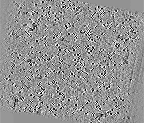 |
CryoET of apoferritin single particle [multiple data sets in MRC format] | Noble AJ, Dandey VP, Wei H, Brasch J, Chase J, Acharya P, Tan YZ, Zhang Z, Kim LY, Scapin G, Rapp M, Eng ET, Rice MJ, Cheng A, Negro CJ, Shapiro L, Kwong PD, Jeruzalmi D, des Georges A, Potter CS, Carragher B [Pubmed: 29809143] [DOI: 10.7554/eLife.34257] |
46.1 GB | — |
| 2021-10-25 | 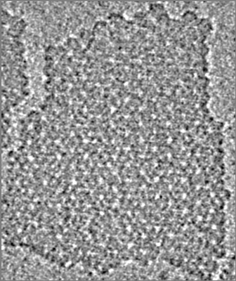 |
Cryo Electron Tomography of isolated S-layer patches from Ca.M.lanthanidiphila [multiple data sets in MRC format] | Gambelli L, Mesman R, Versantvoort W, Diebolder CA, Engel A, Evers W, Jetten MSM, Pabst M, Daum B, van Niftrik L [Pubmed: 34925275] [DOI: 10.3389/fmicb.2021.766527] |
46.3 GB | 21.0 Å |
| 2020-07-06 | 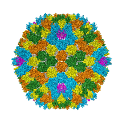 |
Single particle cryo-EM dataset of human adenovirus HAdV-D26 [stack of 8684 particles in MRCS format] | Abrishami V, Reddy VS, Huiskonen JT [Pubmed: 32470354] [DOI: 10.1016/j.pbiomolbio.2020.05.004] |
46.5 GB | 3.1 - 7.34 Å |
| 2023-10-10 | 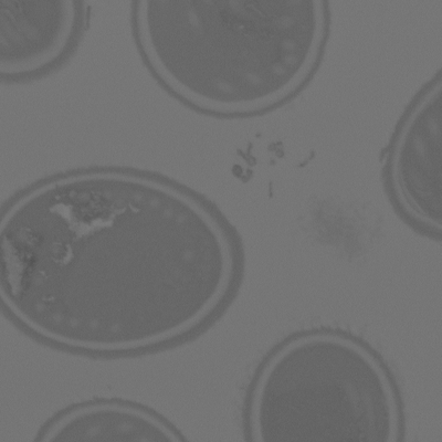 |
SBF-SEM micrographs of A. algerae spores, Ungerminated [250 micrographs in TIFF format] | Jaroenlak P, Cammer M, Davydov A, Sall J, Usmani M, Liang F, Ekiert D, Bhabha G [Pubmed: 32946515] [DOI: 10.1371/journal.ppat.1008738] |
46.6 GB | — |
| 2023-03-13 |  |
FIB-SEM images about Control/OPA1 KD NIH-3T3 cells [1984 multi-frame micrographs composed of 1 frames each in TIFF format] | Suga S, Nakamura K, Kawai H, Hirabayashi Y [DOI: 10.1101/2021.06.11.448083] |
46.8 GB | — |
