Electron Microscopy Public Image Archive
Electron Microscopy Public Image Archive
오사카 대학의 EMPIAR-PDBj 팀은, 아시아의 EM 연구자가 용량이 큰 EM 이미지를 EMPIAR 데이터베이스에 전송하는 것을 돕고 있습니다. 인터넷을 통하여 EBI (UK)에 직>접 데이터를 전송하는 대신, 이용자는 우편이나 택배를 통하여 하드 디스크를 오사카 대학으로 보내실 수 있습니다. 혹은 인터넷을 이용하여 오사카 대학의 서버로 전>송 하실 수 있습니다. 오사카 대학에 데이터 전송 서비스를 희망하시는 분은 데이터를 보내시기 전에 먼저 이메일 통하여 등록하시고 싶은 EM데이터에 관하여 상담하십시오.
| Release date | Imageset | Title | Authors and references | Size | Resolution |
|---|---|---|---|---|---|
| 2018-05-11 | 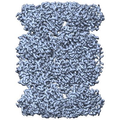 |
T. acidophilum 20S proteasome core movies obtained using Talos Arctica operating at 200 kV equipped with a K2 – image shift used for exposure target navigation [262 multi-frame micrographs composed of 68 frames each in MRC format] | Herzik Jr MA, Wu M, Lander GC [Pubmed: 28991891] [DOI: 10.1038/nmeth.4461] |
945.5 GB | 3.3 Å |
| 2018-05-11 | 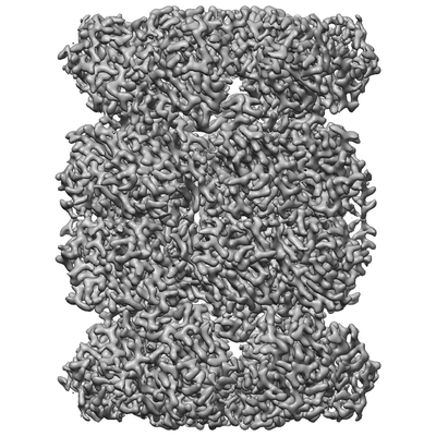 |
T. acidophilum 20S proteasome core movies obtained using Talos Arctica operating at 200 kV equipped with a K2 – stage position used for exposure target navigation [317 multi-frame micrographs composed of 68 frames each in MRC format] | Herzik Jr MA, Wu M, Lander GC [Pubmed: 28991891] [DOI: 10.1038/nmeth.4461] |
140.9 GB | 3.1 Å |
| 2015-03-06 | 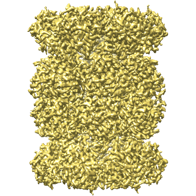 |
T20S Proteasome at 2.8 Å Resolution [multiple data sets in MRC format] | Campbell M, Veesler D, Cheng A, Potter CS, Carragher B [Pubmed: 25760083] [DOI: 10.7554/eLife.06380] |
2.0 TB | 2.8 Å |
| 2018-05-14 | 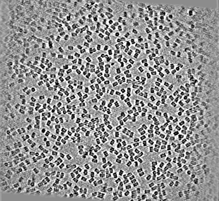 |
T20S proteasome single particle [586 micrographs in MRC format] | Noble AJ, Dandey VP, Wei H, Brasch J, Chase J, Acharya P, Tan YZ, Zhang Z, Kim LY, Scapin G, Rapp M, Eng ET, Rice MJ, Cheng A, Negro CJ, Shapiro L, Kwong PD, Jeruzalmi D, des Georges A, Potter CS, Carragher B [Pubmed: 29809143] [DOI: 10.7554/eLife.34257] |
116.1 GB | — |
| 2020-07-21 |  |
TASK2 in MSP1D1 lipid nanodisc at pH6.5 [3024 multi-frame micrographs composed of 50 frames each in TIFF format] | Li B, Brohawn SG [Pubmed: 32999458] [DOI: 10.1038/s41586-020-2770-2] |
2.0 TB | 3.45 Å |
| 2020-07-21 |  |
TASK2 in MSP1D1 lipid nanodisc at pH8.5 [3470 multi-frame micrographs composed of 50 frames each in TIFF format] | Li B, Brohawn SG [Pubmed: 32999458] [DOI: 10.1038/s41586-020-2770-2] |
2.3 TB | 3.52 Å |
| 2020-12-09 |  |
TEM images of a Zebrafish hindbrain cells containing Toxoplasma gondii tachizoites [multiple data sets in TIFF format] | Domart MC, Collinson L [Pubmed: 32461265] [DOI: 10.1242/dmm.043091] |
3.4 GB | — |
| 2020-08-12 |  |
TEM tomograms of Drosophila tracheal terminal cells during subcellular tube formation [multiple data sets in TIFF and MRC formats] | Mathew R, Rios-Barrera LD, Machado P, Schwab Y, Leptin M [Pubmed: 32657472] [DOI: 10.15252/embj.2020105332] |
366.7 GB | — |
| 2013-12-04 | 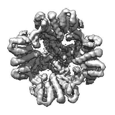 |
TRPV1 dataset taken on a K2 direct electron detector [multiple data sets in MRC format] | Liao M, Cao E, Julius D, Cheng Y [Pubmed: 24305160] [DOI: 10.1038/nature12822] |
6.3 TB | 3.275 Å |
| 2019-07-25 |  |
TRPV2 ion channel gating through allosteric domain coupling revealed by cryo-EM [1969 multi-frame micrographs composed of 35 frames each in MRCS format] | Serysheva II, Chiu W, Wensel TG [Pubmed: 30598551] [DOI: 10.1038/s41594-018-0168-8] |
3.6 TB | 4.0 Å |
| 2021-05-14 | 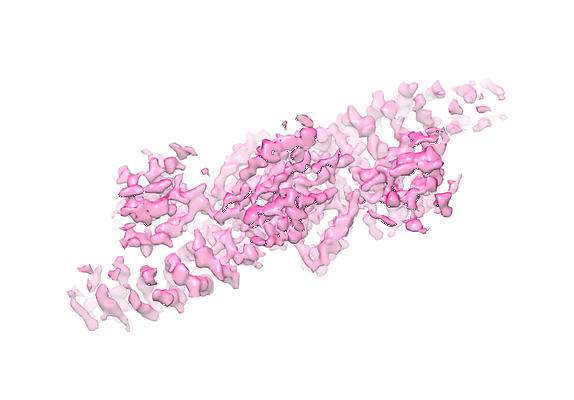 |
TSC complex particles [1767 micrographs in MRC format] | Ramlaul K, Fu W, Li H, de Martin Garrido N, He L, Trivedi M, Cui W, Aylett CHS, Wu G [Pubmed: 33307091] [DOI: 10.1016/j.jmb.2020.166743] |
93.7 GB | 4.2 Å |
| 2021-06-16 |  |
TSC complex particles [3507 multi-frame micrographs composed of 1 frames each in MRC format] | Ramlaul K, Fu W, Li H, de Martin Garrido N, He L, Trivedi M, Cui W, Aylett CHS, Wu G [Pubmed: 33307091] [DOI: 10.1016/j.jmb.2020.166743] |
13.1 TB | 4.2 Å |
| 2024-02-28 | 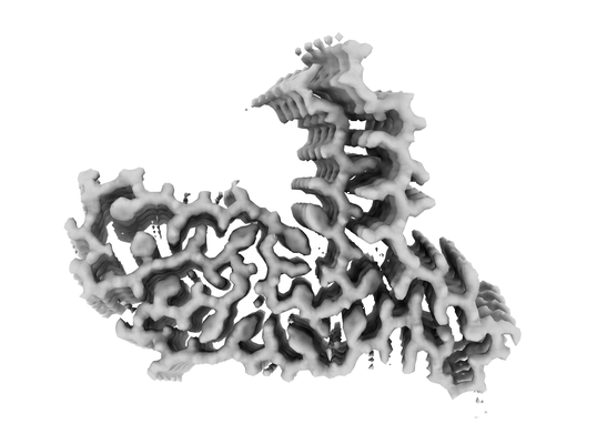 |
Taf15 amyloid filaments - Individual 1, frontal cortex [1980 multi-frame micrographs composed of 1407 frames each in EER format] | Tetter S, Arseni D, Murzin AG, Buhidma Y, Peak-Chew SYPC, Garringer HJ, Newell KL, Vidal R, Apostolova L, Lashley T, Ghetti B, Ryskeldi-Falcon B [Pubmed: 38057661] [DOI: 10.1038/s41586-023-06801-2] |
1.2 TB | 1.97 Å |
| 2024-03-21 | 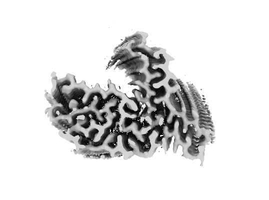 |
Taf15 amyloid filaments - Individual 3 [15621 multi-frame micrographs composed of 40 frames each in TIFF format] | Tetter S, Arseni D, Murzin AG, Buhidma Y, Peak-Chew SYPC, Garringer HJ, Newell KL, Vidal R, Apostolova L, Lashley T, Ghetti B, Ryskeldi-Falcon B [Pubmed: 38057661] [DOI: 10.1038/s41586-023-06801-2] |
2.5 TB | 2.63 Å |
| 2023-07-18 |  |
Targeted volume Correlative Light and Electron Microscopy of environmental marine microorganisms [1937 micrographs in TIFF format] | Mocaer K, Mizzon G, Gunkel M, Halavatyi A, Steyer AM, Oorschot V, Schorb M, Le Kieffre C, Yee DP, Chevalier F, Gallet B, Decelle J, Schwab Y, Ronchi P [DOI: 10.1101/2023.01.27.525698] |
14.5 GB | — |
| 2022-11-21 |  |
Tau Paired Helical Filament from Alzheimer's Disease incubated 1 hr. with EGCG [3890 multi-frame micrographs composed of 48 frames each in TIFF format] | Seidler PM, Murray KA, Boyer DR, Ge P, Sawaya MR, Eisenberg DS [Pubmed: 36114178] [DOI: 10.1038/s41467-022-32951-4] |
3.7 TB | 3.3 Å |
| 2022-11-21 |  |
Tau Paired Helical Filament from Alzheimer's Disease incubated with EGCG for 3 hours [6406 multi-frame micrographs composed of 30 frames each in MRCS format] | Seidler PM, Murray KA, Boyer DR, Ge P, Sawaya MR, Eisenberg DS [Pubmed: 36114178] [DOI: 10.1038/s41467-022-32951-4] |
1.7 TB | 3.8 Å |
| 2022-11-21 |  |
Tau Paired Helical Filament from Alzheimer's Disease not incubated with EGCG [2651 multi-frame micrographs composed of 48 frames each in TIFF format] | Seidler PM, Murray KA, Boyer DR, Ge P, Sawaya MR, Eisenberg DS [Pubmed: 36114178] [DOI: 10.1038/s41467-022-32951-4] |
2.5 TB | 3.4 Å |
| 2023-11-07 |  |
Tau filaments from the cellular fraction of Alzheimer's disease patient brain [12117 multi-frame micrographs composed of 40 frames each in TIFF format] | Ryskeldi-Falcon BR-F, Behr TSB [Pubmed: 37163117] [DOI: 10.1101/2023.04.30.537820] |
1.9 TB | 3.27 Å |
| 2023-11-13 |  |
Test subset: In situ cryo-ET dataset of Chlamydomonas reinhardtii prepared using cryo-plasmaFIB milling [18 tilt series in MRC format] | Kelley R, Zhang X, Obr M, Khavnekar S, Righetto R, Waltz F, Wietrzynski W, Michael A, Tagiltsev G, Beck F, Zhong E, Wan W, Briggs J, Plitzko J, Engel B, Kotecha A [Pubmed: 37613825] [DOI: 10.1093/micmic/ozad067.480] |
293.7 GB | — |
| 2020-12-02 |  |
Tetrameric Ptch1 complexed with ShhNp [5198 multi-frame micrographs composed of 32 frames each in MRC format] | Qian H, Yan N [Pubmed: 31127104] [DOI: 10.1038/s41467-019-10234-9] |
2.5 TB | 3.6 - 6.8 Å |
| 2020-06-19 |  |
Tetrameric SARS-CoV-2 ORF3a in a lipid nanodisc [7092 multi-frame micrographs composed of 50 frames each in TIFF format] | Kern DM, Sorum B, Mali SS, Hoel CM, Sridharan S, Remis JP, Toso DB, Kotecha A, Bautista DM, Brohawn SG [Pubmed: 34158638] [DOI: 10.1038/s41594-021-00619-0] |
4.8 TB | 6.5 Å |
| 2019-02-19 | 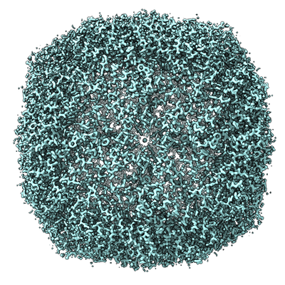 |
The 1.54 Å structure of Apoferritin by CRYOARM300 with cold-FEG [972 multi-frame micrographs composed of 50 frames each in TIFF format] | Kato T, Makino F, Nakane T, Terahara N, Kaneko T, Shimizu Y, Motoki S, Ishikawa I, Yonekura K, Namba K [DOI: 10.1017/S1431927619005725] |
145.9 GB | 1.54 Å |
| 2021-10-26 | 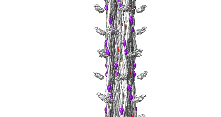 |
The CryoEM Structure of Drosophila Flight Muscle Thick Filaments at 7Å Resolution [1510 multi-frame micrographs composed of 44 frames each in MRC format] | Daneshparvar N, Taylor DW, O'Leary TS, Rahmani H, Abbasiyeganeh F, Previs MJ, Taylor KA [Pubmed: 32718994] [DOI: 10.1101/2020.06.05.136580] |
8.7 TB | 7.0 - 8.0 Å |
| 2023-08-20 | 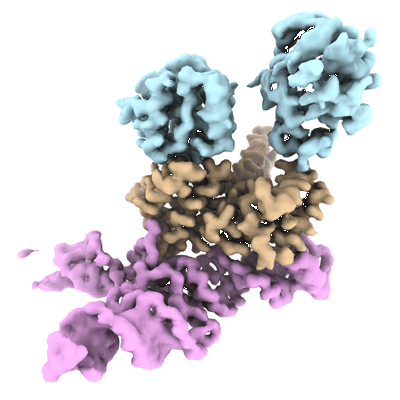 |
The J-K RNA fragment of encephalomyocarditis virus IRES in complex with eIF4G-HEAT1 and eIF4A [6194 multi-frame micrographs composed of 40 frames each in TIFF format] | Suzuki H, Fujiyoshi Y | 1018.5 GB | 3.76 Å |
