Electron Microscopy Public Image Archive
Electron Microscopy Public Image Archive
Our EMPIAR-PDBj will suspend the following operations from August 10, 2024 (Saturday) to August 18, 2024 (Sunday):
Web servers and data downloads will remain available during this period.
Additionally, please be aware that EMPIAR-EBI will also be closed for annotation operations from July 25, 2024 (Thursday) to August 18, 2024 (Sunday).
Thank you.
오사카 대학의 EMPIAR-PDBj 팀은, 아시아의 EM 연구자가 용량이 큰 EM 이미지를 EMPIAR 데이터베이스에 전송하는 것을 돕고 있습니다. 인터넷을 통하여 EBI (UK)에 직>접 데이터를 전송하는 대신, 이용자는 우편이나 택배를 통하여 하드 디스크를 오사카 대학으로 보내실 수 있습니다. 혹은 인터넷을 이용하여 오사카 대학의 서버로 전>송 하실 수 있습니다. 오사카 대학에 데이터 전송 서비스를 희망하시는 분은 데이터를 보내시기 전에 먼저 이메일 통하여 등록하시고 싶은 EM데이터에 관하여 상담하십시오.
| Release date | Imageset | Title | Authors and references | Size | Resolution |
|---|---|---|---|---|---|
| 2021-05-28 | 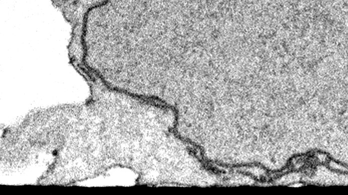 |
CLEM/FIB-SEM Imaging of T Cells after the Formation of Signaling Microclusters at the Immunological Synapse [160 micrographs in TIFF format] | Narayan K | 26.6 MB | — |
| 2021-04-14 | 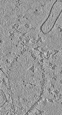 |
DNA origami signposts for identifying proteins on cell membranes by electron cryotomography [2 tilt series in MRC format] | Silvester E, Vollmer B, Pražák V, Vasishtan D, Machala EA, Whittle C, Black S, Bath J, Turberfield AJ, Grünewald K, Baker LA [Pubmed: 33606980] [DOI: 10.1016/j.cell.2021.01.033] |
38.8 MB | 36.44 Å |
| 2022-01-12 | 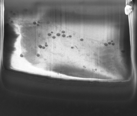 |
Cryo-FIB-SEM volume in a Sum159 human cell line [20 micrographs in TIFF format] | Klumpe S, Fung HKH, Goetz SK, Zagoriy I, Hampoelz B, Zhang X, Erdmann PS, Baumbach J, Müller CW, Beck M, Plitzko JM, Mahamid J [Pubmed: 34951584] [DOI: 10.7554/elife.70506] |
120.1 MB | — |
| 2021-02-12 | 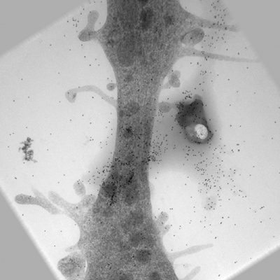 |
Ultra-high voltage electron microscope tomography tilt series of neurite section [1 tilt series in MRC format] | Nishida T, Yoshimura R, Nishi R, Imoto Y, Endo Y [Pubmed: 32133640] [DOI: 10.1111/jmi.12885] |
120.3 MB | — |
| 2021-02-12 | 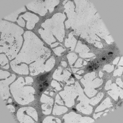 |
Ultra-high voltage electron microscope tomography tilt series of 0.7-μm-thick neurite section acquired at 6,000× magnification at an accelerating voltage of 1 MV [1 tilt series in MRC format] | Nishida T, Yoshimura R, Nishi R, Imoto Y, Endo Y [Pubmed: 32133640] [DOI: 10.1111/jmi.12885] |
120.3 MB | — |
| 2021-02-12 | 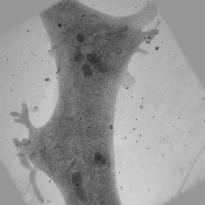 |
Ultra-high voltage electron microscope tomography tilt series of 0.7-μm-thick neurite section acquired at 15,000× magnification at an accelerating voltage of 1 MV [1 tilt series in MRC format] | Nishida T, Yoshimura R, Nishi R, Imoto Y, Endo Y [Pubmed: 32133640] [DOI: 10.1111/jmi.12885] |
120.3 MB | — |
| 2020-05-27 | 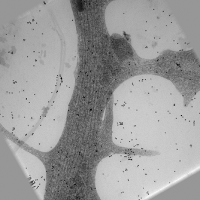 |
Ultra-high voltage electron microscope tomography tilt series of 0.7-μm-thick neurite section acquired at 20,000× magnification at an accelerating voltage of 1 MV [1 tilt series in MRC format] | Nishida T, Yoshimura R, Nishi R, Imoto Y, Endo Y [Pubmed: 32133640] [DOI: 10.1111/jmi.12885] |
120.3 MB | — |
| 2021-09-01 | 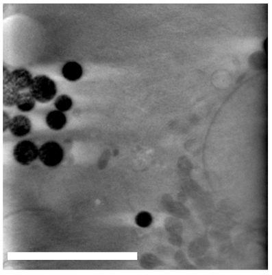 |
Lysosomes as Fiducials for SXT and SIM Correlation [166 tilt series in TIFF format] | Okolo CA, Kounatidis I, Groen J, Nahas KL, Balint S, Fish T, Koronfel MA, López-Cortajarena A, Dobbie I, Pereiro E, Harkiolaki M | 162.4 MB | — |
| 2016-01-20 | 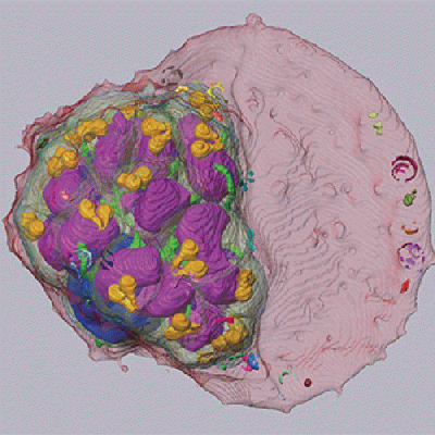 |
SBF-SEM of late schizont-stage malaria parasite infected red blood cell [90 micrographs in MRC format] | Sakaguchi M, Miyazaki N, Fujioka H, Kaneko O, Murata K [Pubmed: 26772147] [DOI: 10.1016/j.jsb.2016.01.003] |
219.7 MB | — |
| 2016-01-20 | 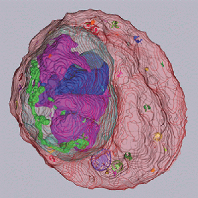 |
SBF-SEM of early schizont-stage malaria parasite infected red blood cell [80 micrographs in MRC format] | Sakaguchi M, Miyazaki N, Fujioka H, Kaneko O, Murata K [Pubmed: 26772147] [DOI: 10.1016/j.jsb.2016.01.003] |
222.1 MB | — |
| 2015-10-09 | 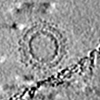 |
Cryo-electron tomography and subtomogram averaging of Rous-Sarcoma-Virus deltaMBD virus-like particles [1 class averages in MRC format] | Schur FKM, Dick RA, Hagen WJH, Vogt VM, Briggs JAG [Pubmed: 26223638] [DOI: 10.1128/JVI.01502-15] |
248.1 MB | — |
| 2021-11-08 | 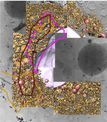 |
Monitoring reversion of hepatitis C virus-induced cellular alterations by Direct-Acting Antivirals using cryo Soft X-ray Tomography and Infrared Microscopy [7 reconstructed volumes in TIFF format] | Perez-Berna AJ, Benseny-Cases N, Rodríguez MJ, Valcarcel R, Carrascosa JL, Gastaminzab P, Pereiroa E [Pubmed: 34726165] [DOI: 10.1107/S2059798321009955] |
260.2 MB | — |
| 2023-06-20 | 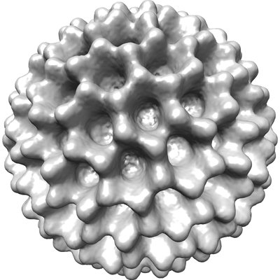 |
Cryo-EPty SPA at CSA of 1.03 mrad [29 micrographs in MRC format] | Pei X, Zhou L, Huang C, Boyce M, Kim JS, Liberti E, Hu Y, Sasaki T, Nellist PD, Zhang P, Stuart DI, Kirkland AI, Wang P [Pubmed: 37230988] [DOI: 10.1038/s41467-023-38268-0] |
262.0 MB | 37.2 Å |
| 2018-01-17 | 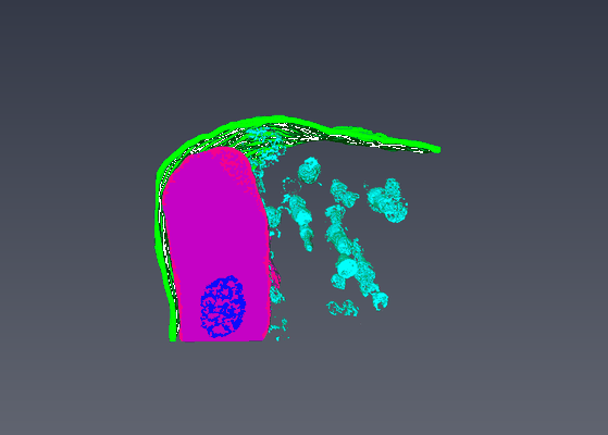 |
Serial Block Face Scanning Electron Micrscopy dataset of fetal day 64 guinea pig psoas muscle in transverse [93 micrographs in TIFF format] | Cocks ET [DOI: 10.1111/jmi.12676] |
264.4 MB | — |
| 2016-01-20 | 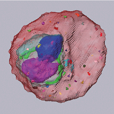 |
SBF-SEM of trophozoite-stage malaria parasite infected red blood cell [110 micrographs in MRC format] | Sakaguchi M, Miyazaki N, Fujioka H, Kaneko O, Murata K [Pubmed: 26772147] [DOI: 10.1016/j.jsb.2016.01.003] |
268.6 MB | — |
| 2017-03-15 | 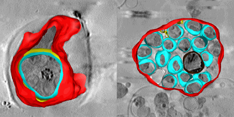 |
Soft X-ray tomography of Plasmodium falciparum infected human erythrocytes stalled in egress by the inhibitors Compound 2 and E64 [1 Soft X-ray tomograms in MRC format] | Hale VL, Saibil HR, Duke E, Fleck RA, Blackman MJ [Pubmed: 28292906] [DOI: 10.1073/pnas.1619441114] |
280.6 MB | — |
| 2016-01-20 | 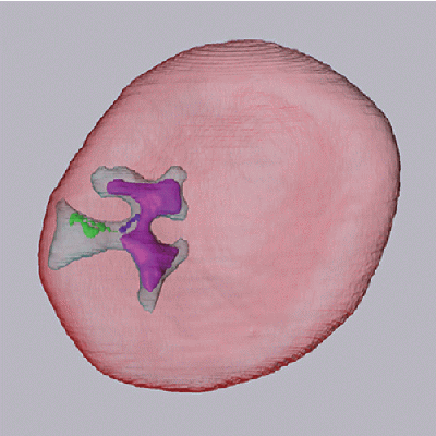 |
SBF-SEM of ring-stage malaria parasite infected red blood cell [120 micrographs in MRC format] | Sakaguchi M, Miyazaki N, Fujioka H, Kaneko O, Murata K [Pubmed: 26772147] [DOI: 10.1016/j.jsb.2016.01.003] |
293.0 MB | — |
| 2018-02-08 |  |
Tilt-series of salmonella enterica wild-type bacterial flagellar motor [1 tilt series in MRC format] | Beeby M, Ribardo DA, Brennan CA, Ruby EG, Jensen GJ, Hendrixson DR [Pubmed: 26976588] [DOI: 10.1073/pnas.1518952113] |
328.0 MB | 69.4 Å |
| 2024-02-13 |  |
Plant SBF-SEM - Tobacco Leaf Chloroplast [130 micrographs in TIFF format] | Wickramanayake JS, Czymmek KJ [Pubmed: 37451777] [DOI: 10.1016/bs.mcb.2023.04.008] |
544.4 MB | — |
| 2019-10-07 |  |
Processed FIB SEM images of a parasitophorous vacuole containing Toxoplasma gondii ∆CAP parasites, complemented with CAP. [1 multi-frame micrographs composed of 1 frames each in MRC format] | Hunt A, Russell MRG, Wagener J, Kent R, Carmeille R, Peddie CJ, Collinson L, Heaslip A, Ward GE, Treeck M [Pubmed: 31577230] [DOI: 10.7554/elife.50598] |
583.8 MB | — |
| 2021-06-18 |  |
Subtomograms of nucleosomes extracted from cryo-tomograms of Drosophila melanogaster embryos [1 subtomograms in EM format] | Harastani M, Eltsov M, Leforestier A, Jonic S [Pubmed: 34095222] [DOI: 10.3389/fmolb.2021.663121] |
666.3 MB | — |
| 2018-01-03 | 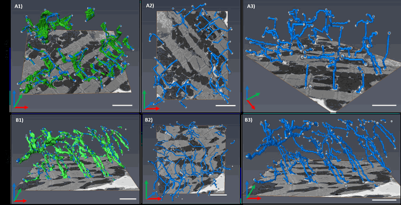 |
Segment /nodes network analysis of the tubular system of the Neonate and Adult GP cardiomyocyte [167 multi-frame micrographs composed of 1 frames each in TIFF format] | Kashbour H, Taggart M, White K | 787.5 MB | — |
| 2023-02-28 |  |
Cryo serial FIB/SEM of RPE-1 cells [18 micrographs in TIFF format] | Dumoux M, Glen T, Smith JLR, Ho EML, Perdigão LMA, Pennington A, Klumpe S, Yee NBY, Farmer DA, Lai PYA, Bowles W, Kelley R, Plitzko JM, Wu L, Basham M, Clare DK, Siebert CA, Darrow MC, Naismith JH, Grange M [Pubmed: 36805107] [DOI: 10.7554/elife.83623] |
826.6 MB | — |
| 2022-01-11 |  |
Cryo-FIB-SEM data on Chlamydomonas reinhardtii cells [37 micrographs in TIFF format] | Klumpe S [Pubmed: 34951584] [DOI: 10.7554/elife.70506] |
888.2 MB | — |
| 2019-10-07 |  |
Processed FIB SEM images of a parasitophorous vacuole containing Toxoplasma gondii ∆CAP parasites. [1 multi-frame micrographs composed of 1 frames each in MRC format] | Hunt A, Russell MRG, Wagener J, Kent R, Carmeille R, Peddie CJ, Collinson L, Heaslip A, Ward GE, Treeck M [Pubmed: 31577230] [DOI: 10.7554/elife.50598] |
898.1 MB | — |
