Electron Microscopy Public Image Archive
Electron Microscopy Public Image Archive
오사카 대학의 EMPIAR-PDBj 팀은, 아시아의 EM 연구자가 용량이 큰 EM 이미지를 EMPIAR 데이터베이스에 전송하는 것을 돕고 있습니다. 인터넷을 통하여 EBI (UK)에 직>접 데이터를 전송하는 대신, 이용자는 우편이나 택배를 통하여 하드 디스크를 오사카 대학으로 보내실 수 있습니다. 혹은 인터넷을 이용하여 오사카 대학의 서버로 전>송 하실 수 있습니다. 오사카 대학에 데이터 전송 서비스를 희망하시는 분은 데이터를 보내시기 전에 먼저 이메일 통하여 등록하시고 싶은 EM데이터에 관하여 상담하십시오.
| Release date | Imageset | Title | Authors and references | Size | Resolution |
|---|---|---|---|---|---|
| 2022-03-04 | 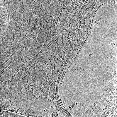 |
Cryo-electron tomographs of mouse hippocampal neurons [63 reconstructed volumes in MRC format] | Nedozralova H, Basnet N, Ibiricu I, Bodakuntla S, Biertümpfel C, Mizuno N [Pubmed: 35262630] [DOI: 10.1083/jcb.202106086] |
88.6 GB | — |
| 2020-06-30 | 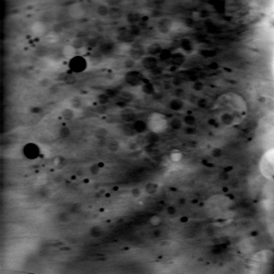 |
Soft X-ray Tomography of mock-infected U2OS cells [1 tilt series in MRC format] | Kounatidis I, Stanifer ML, Phillips MA, Paul-Gilloteaux P, Heiligenstein X, Wang H, Okolo CA, Fish TM, Spink MC, Stuart DI, Davis I, Boulant S, Grimes JM, Dobbie IM, Harkiolaki M [Pubmed: 32610083] [DOI: 10.1016/j.cell.2020.05.051] |
2.3 GB | — |
| 2020-06-30 | 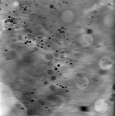 |
Soft X-ray Tomography of mock-infected U2OS cells [1 tilt series in MRC format] | Kounatidis I, Stanifer ML, Phillips MA, Paul-Gilloteaux P, Heiligenstein X, Wang H, Okolo CA, Fish TM, Spink MC, Stuart DI, Davis I, Boulant S, Grimes JM, Dobbie IM, Harkiolaki M [Pubmed: 32610083] [DOI: 10.1016/j.cell.2020.05.051] |
2.3 GB | — |
| 2020-06-30 |  |
Soft X-ray Tomography of mock-infected U2OS cells [1 tilt series in MRC format] | Kounatidis I, Stanifer ML, Phillips MA, Paul-Gilloteaux P, Heiligenstein X, Wang H, Okolo CA, Fish TM, Spink MC, Stuart DI, Davis I, Boulant S, Grimes JM, Dobbie IM, Harkiolaki M [Pubmed: 32610083] [DOI: 10.1016/j.cell.2020.05.051] |
1.2 GB | — |
| 2023-10-03 |  |
Adaptive traits of cysts of the snow alga Sanguina nivaloides unveiled by 3D subcellular imaging [multiple data sets in TIFF format] | Ezzedine J, Uwizeye C, Si Larbi G, Villain G, Louwagie M, Schilling M, Hagenmuller P, Gallet B, Stewart A, Petroutsos D, Devime F, Salze P, Liger L, Jouhet J, Dumont M, Ravanel S, Amato A, Valay JG, Jouneau PH, Falconet D, Marechal E [DOI: 10.21203/rs.3.rs-3038444/v1] |
104.7 GB | — |
| 2020-06-30 | 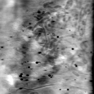 |
Soft X-ray Tomography of mock-infected U2OS cells [1 tilt series in MRC format] | Kounatidis I, Stanifer ML, Phillips MA, Paul-Gilloteaux P, Heiligenstein X, Wang H, Okolo CA, Fish TM, Spink MC, Stuart DI, Davis I, Boulant S, Grimes JM, Dobbie IM, Harkiolaki M [Pubmed: 32610083] [DOI: 10.1016/j.cell.2020.05.051] |
2.3 GB | — |
| 2023-10-09 | 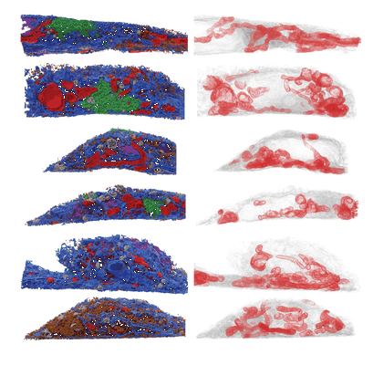 |
Quantitative subcellular reconstruction reveals a lipid mediated inter-organelle biogenesis network [multiple data sets in TIFF format] | Lee RG, Rudler DL, Raven SA, Peng L, Chopin A, Moh ESX, McCubbin T, Siira SJ, Fagan SV, DeBono NJ, Stentenbach M, Browne J, Rackham FF, Li J, Simpson KJ, Marcellin E, Packer NH, Reid GE, Padman BS, Rackham O, Filipovska A | 434.2 GB | — |
| 2020-06-30 | 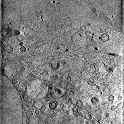 |
Soft X-ray Tomography of mock-infected U2OS cells [1 tilt series in MRC format] | Kounatidis I, Stanifer ML, Phillips MA, Paul-Gilloteaux P, Heiligenstein X, Wang H, Okolo CA, Fish TM, Spink MC, Stuart DI, Davis I, Boulant S, Grimes JM, Dobbie IM, Harkiolaki M [Pubmed: 32610083] [DOI: 10.1016/j.cell.2020.05.051] |
1.2 GB | — |
| 2021-04-14 | 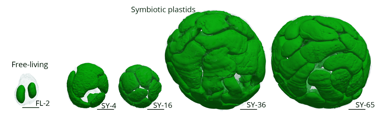 |
Cytoklepty in the plankton: a host strategy to optimize the bioenergetic machinery of endosymbiotic algae [500 multi-frame micrographs composed of 1 frames each in TIFF format] | Uwizeye C, Mars Brisbin M, Gallet B, LeKieffre C, Schieber NL, Wangpraseurt D, Decelle J [Pubmed: bioRxiv] [DOI: 10.1101/2020.12.08.416644] |
2.9 GB | — |
| 2018-05-02 | 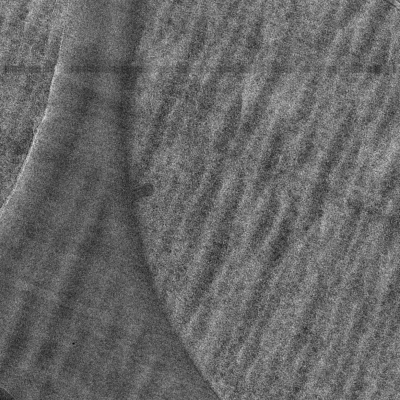 |
Correlative microscopy of vitreous sections provides insights into BAR-domain organisation in situ [9 tilt series in MRC format] | Bharat TAM, Hoffmann PC, Kukulski W [Pubmed: 29681471] [DOI: 10.1016/j.str.2018.03.015] |
27.9 GB | — |
| 2020-06-30 | 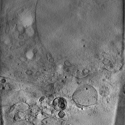 |
Soft X-ray Tomography of mock-infected U2OS cells [1 tilt series in MRC format] | Kounatidis I, Stanifer ML, Phillips MA, Paul-Gilloteaux P, Heiligenstein X, Wang H, Okolo CA, Fish TM, Spink MC, Stuart DI, Davis I, Boulant S, Grimes JM, Dobbie IM, Harkiolaki M [Pubmed: 32610083] [DOI: 10.1016/j.cell.2020.05.051] |
1.2 GB | — |
| 2022-01-28 |  |
CryoET of presequence protease single particle [multiple data sets in TIFF and MRC formats] | Liang WG, Wijaya J, Wei H, Noble AJ, Mancl JM, Mo S, Lee D, Lin King JV, Pan M, Liu C, Koehler CM, Zhao M, Potter CS, Carragher B, Li S, Tang WJ [Pubmed: 35383169] [DOI: 10.1038/s41467-022-29322-4] |
25.4 GB | — |
| 2020-06-30 | 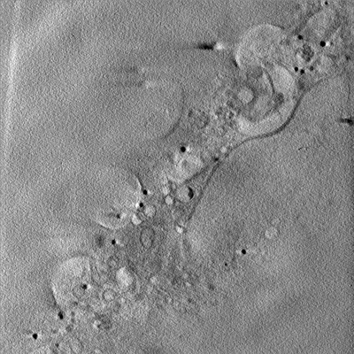 |
Soft X-ray Tomography of mock-infected U2OS cells [1 tilt series in MRC format] | Kounatidis I, Stanifer ML, Phillips MA, Paul-Gilloteaux P, Heiligenstein X, Wang H, Okolo CA, Fish TM, Spink MC, Stuart DI, Davis I, Boulant S, Grimes JM, Dobbie IM, Harkiolaki M [Pubmed: 32610083] [DOI: 10.1016/j.cell.2020.05.051] |
1.2 GB | — |
| 2020-06-30 | 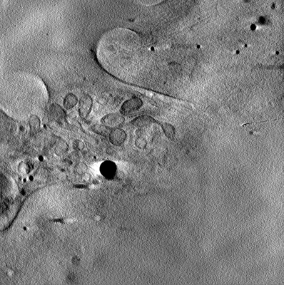 |
Soft X-ray Tomography of mock-infected U2OS cells [1 tilt series in MRC format] | Kounatidis I, Stanifer ML, Phillips MA, Paul-Gilloteaux P, Heiligenstein X, Wang H, Okolo CA, Fish TM, Spink MC, Stuart DI, Davis I, Boulant S, Grimes JM, Dobbie IM, Harkiolaki M [Pubmed: 32610083] [DOI: 10.1016/j.cell.2020.05.051] |
1.2 GB | — |
| 2023-05-10 | 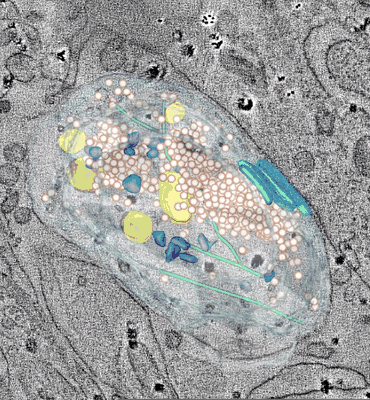 |
Microtubule depolymerization contributes to spontaneous neurotransmitter release [multiple data sets in MRC format] | Velasco C, Santarella-Mellwig R, Schorb M, Gao L, Thorn-Seshold O, Llobet A [Pubmed: 37147475] [DOI: 10.1038/s42003-023-04779-1] |
123.8 GB | — |
| 2024-05-14 |  |
Aligned tilt series of stationary phase Burkholderia thailandensis tomograms [40 tilt series in MRC format] | Welch MD, Khanna K [Pubmed: 38725941] [DOI: 10.17912/micropub.biology.001178] |
26.9 GB | — |
| 2021-06-18 | 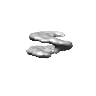 |
Subtomograms of nucleosomes extracted from cryo-tomograms of Drosophila melanogaster embryos [1 subtomograms in EM format] | Harastani M, Eltsov M, Leforestier A, Jonic S [Pubmed: 34095222] [DOI: 10.3389/fmolb.2021.663121] |
666.3 MB | — |
| 2022-05-11 | 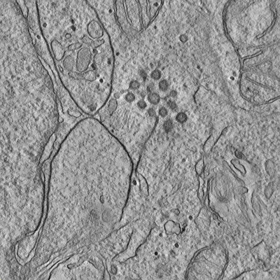 |
Tilt series and tomograms of cells expressing different non structural proteins (NSPs) of SARS-CoV-2 [multiple data sets in MRC format] | Polshchuk RS, Polishchuk E, De Matteis MA [Pubmed: 35551511] [DOI: 10.1038/s41586-022-04835-6] |
107.1 GB | — |
| 2018-04-05 | 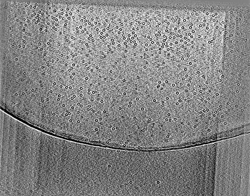 |
CryoET of apoferritin single particle with spot-to-plunge time of 100ms [multiple data sets in MRC and DYNAMO TBL AND OMD FILES formats] | Noble AJ, Wei H, Dandey VP, Zhang Z, Potter CS, Carragher B [Pubmed: 30250056] [DOI: 10.1038/s41592-018-0139-3] |
119.3 GB | — |
| 2023-03-13 | 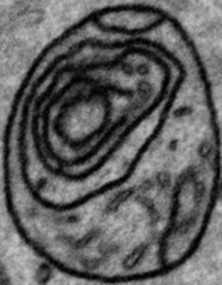 |
FIB-SEM images about Control/OPA1 KD NIH-3T3 cells [1984 multi-frame micrographs composed of 1 frames each in TIFF format] | Suga S, Nakamura K, Kawai H, Hirabayashi Y [DOI: 10.1101/2021.06.11.448083] |
46.8 GB | — |
| 2018-04-05 | 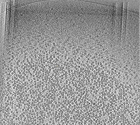 |
CryoET of apoferritin single particle with spot-to-plunge time of 400ms [multiple data sets in MRC and DYNAMO TBL AND OMD FILES formats] | Noble AJ, Wei H, Dandey VP, Zhang Z, Potter CS, Carragher B [Pubmed: 30250056] [DOI: 10.1038/s41592-018-0139-3] |
98.8 GB | — |
| 2023-07-10 | 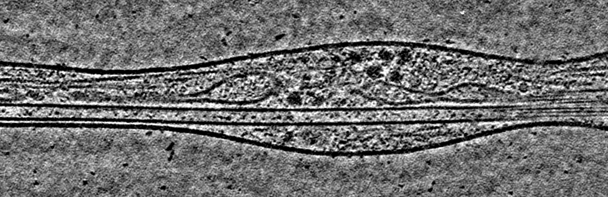 |
Cryo electron tomography of Cytochalasin D-induced protrusions of Drosophila S2 cells - Datasets 1 - 4 [multiple data sets in TIFF and MRC formats] | Ventura Santos C, Carter AP, Rogers SL [Pubmed: 37034688] [DOI: 10.1101/2023.03.31.535077] |
446.0 GB | — |
| 2018-04-05 | 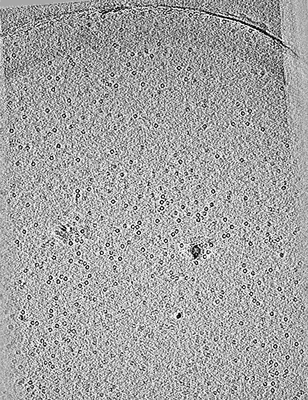 |
CryoET of apoferritin with 0.5 mM TCEP single particle with spot-to-plunge time of 170ms [multiple data sets in MRC and DYNAMO TBL AND OMD FILES formats] | Noble AJ, Wei H, Dandey VP, Zhang Z, Potter CS, Carragher B [Pubmed: 30250056] [DOI: 10.1038/s41592-018-0139-3] |
31.6 GB | — |
| 2023-07-10 | 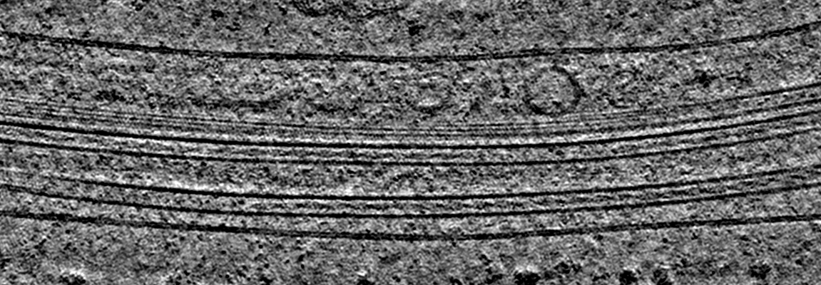 |
Cryo electron tomography of Cytochalasin D-induced protrusions of Drosophila S2 cells treated with DMSO or thapsigargin - Datasets 5 - 7 [multiple data sets in TIFF and MRC formats] | Ventura Santos C, Carter AP, Rogers SL [Pubmed: 37034688] [DOI: 10.1101/2023.03.31.535077] |
373.1 GB | — |
| 2018-04-05 |  |
CryoET of hemagglutinin single particle with spot-to-plunge time of 100ms [multiple data sets in MRC format] | Noble AJ, Wei H, Dandey VP, Zhang Z, Potter CS, Carragher B [Pubmed: 30250056] [DOI: 10.1038/s41592-018-0139-3] |
21.9 GB | — |
