Electron Microscopy Public Image Archive
Electron Microscopy Public Image Archive
오사카 대학의 EMPIAR-PDBj 팀은, 아시아의 EM 연구자가 용량이 큰 EM 이미지를 EMPIAR 데이터베이스에 전송하는 것을 돕고 있습니다. 인터넷을 통하여 EBI (UK)에 직>접 데이터를 전송하는 대신, 이용자는 우편이나 택배를 통하여 하드 디스크를 오사카 대학으로 보내실 수 있습니다. 혹은 인터넷을 이용하여 오사카 대학의 서버로 전>송 하실 수 있습니다. 오사카 대학에 데이터 전송 서비스를 희망하시는 분은 데이터를 보내시기 전에 먼저 이메일 통하여 등록하시고 싶은 EM데이터에 관하여 상담하십시오.
| Release date | Imageset | Title | Authors and references | Size | Resolution |
|---|---|---|---|---|---|
| 2023-12-04 |  |
Clostridium difficile binary toxin translocase CDTb tetradecamer in symmetric and asymmetric conformations [multiple data sets in MRC format] | Xu X, Ben-Hail D, des Georges A, Pozharski E [Pubmed: 31896582] [DOI: 10.1073/pnas.1919490117] |
704.4 GB | 2.8 - 3.1 Å |
| 2021-06-04 |  |
Cryo-EM structure of human rod CNGA1 channel in apo-state [stack of 274333 particles in MRCS format] | Xue J, Han Y, Zeng W, Wang Y, Jiang Y [Pubmed: 33651975] [DOI: 10.1016/j.neuron.2021.02.007] |
84.9 GB | 2.6 Å |
| 2022-11-14 |  |
Cryo-EM structures of Ib-pore and Ia-bound Ib-pore [multiple data sets in TIFF format] | Yamada T, Yoshida T, Kawamoto A, Tsuge H [Pubmed: 32123390] [DOI: 10.1038/s41594-020-0388-6] |
8.0 TB | 2.8 - 2.9 Å |
| 2022-07-18 |  |
Structure of the Dicer-2-R2D2 heterodimer bound to a small RNA duplex [multiple data sets in TIFF format] | Yamaguchi S, Naganuma M, Nishizawa T, Kusakizako T, Tomari Y, Nishimasu H, Nureki O [Pubmed: 35768503] [DOI: 10.1038/s41586-022-04790-2] |
1.4 TB | 3.3 Å |
| 2020-09-03 |  |
ISWI-NCP complex in the ADPBeF-bound state [stack of 166165 particles in MRCS format] | Yan L, Wu H, Li X, Gao N, Chen Z [Pubmed: 30872815] [DOI: 10.1038/s41594-019-0199-9] |
41.7 GB | 3.37 Å |
| 2020-09-02 |  |
ISWI-NCP complex in the ADP-bound state [stack of 168430 particles in MRCS format] | Yan L, Wu H, Li X, Gao N, Chen Z [Pubmed: 32123390] [DOI: 10.1038/s41594-020-0388-6] |
36.2 GB | 2.9 Å |
| 2023-03-16 |  |
SBFSEM imaging of Leishmania haptomonads on the stomodeal valve in the sand fly [400 multi-frame micrographs composed of 1 frames each in MRC format] | Yanase R, Sunter JD [DOI: 10.1101/2022.10.28.514187] |
82.0 GB | — |
| 2023-03-17 |  |
SBFSEM imaging of Leishmania haptomonad-like cells attached to plastic [252 multi-frame micrographs composed of 1 frames each in MRC format] | Yanase R, Sunter JD [DOI: 10.1101/2022.10.28.514187] |
33.6 GB | — |
| 2023-03-17 |  |
Serial section electron tomography of a Leishmania haptomonad on the stomodeal valve in the sand fly [670 multi-frame micrographs composed of 1 frames each in MRC format] | Yanase R, Sunter JD [DOI: 10.1101/2022.10.28.514187] |
6.7 GB | — |
| 2023-03-17 |  |
Serial section electron tomography of a Leishmania haptomonad-like cell attached to plastic [718 multi-frame micrographs composed of 1 frames each in MRC format] | Yanase R, Sunter JD [DOI: 10.1101/2022.10.28.514187] |
10.8 GB | — |
| 2022-02-28 |  |
Structural visualization of de novo initiation of RNA polymerase II transcription [multiple data sets in TIFF format] | Yang C, Fujiwara R, Kim HJ, Basnet P, Zhu Y, Gorbea Colón JJ, Steimle S, Garcia BA, Kaplan CD, Murakami K [Pubmed: 35051353] [DOI: 10.1016/j.molcel.2021.12.020] |
14.3 TB | 3.0 - 7.6 Å |
| 2020-09-02 |  |
Structural basis of redox modulation on chloroplast ATP synthase (reduced form) [2063 micrographs in MRC format] | Yang JH, Williams D, Kandiah E, Fromme P, Chiu PL [Pubmed: 32879423] [DOI: 10.1038/s42003-020-01221-8] |
109.9 GB | 3.05 - 4.34 Å |
| 2023-04-13 | 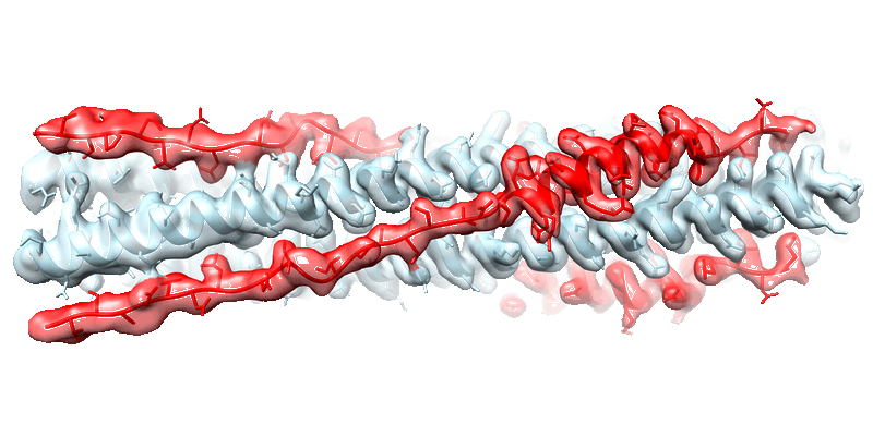 |
Cryo-EM structure of the SARS-CoV-2 HR1HR2 fusion core complex with extended HR2 [18846 multi-frame micrographs composed of 40 frames each in TIFF format] | Yang K, Brunger AT [Pubmed: 36122200] [DOI: 10.1073/pnas.2210990119] |
4.6 TB | 2.22 Å |
| 2023-04-13 | 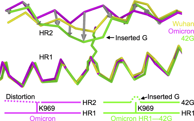 |
Structure-based design of a SARS-CoV-2 Omicron-specific inhibitor [multiple data sets in TIFF format] | Yang K, Brunger AT [Pubmed: 36940324] [DOI: 10.1073/pnas.2300360120] |
13.5 TB | 2.51 Å |
| 2022-07-22 | 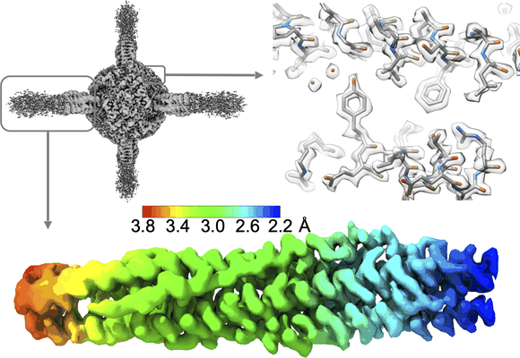 |
Structural conservation among variants of the SARS-CoV-2 spike postfusion bundle [multiple data sets in TIFF format] | Yang K, Brunger AT [Pubmed: 36940324] [DOI: 10.1073/pnas.2300360120] |
32.4 TB | 2.09 - 2.52 Å |
| 2023-12-04 | 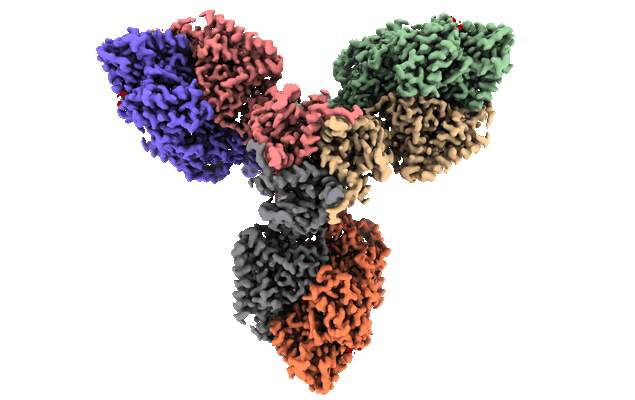 |
Single particle cryo-EM dataset of homohexameric 2-oxoglutarate dehydrogenase OdhA from Corynebacterium glutamicum in complex with the product succinyl-CoA [11827 multi-frame micrographs composed of 50 frames each in TIFF format] | Yang L, Mechaly A, Bellinzoni M [Pubmed: 37563123] [DOI: 10.1038/s41467-023-40253-6] |
4.0 TB | 2.07 Å |
| 2023-12-04 |  |
Single particle cryo-EM dataset of the homohexameric 2-oxoglutarate dehydrogenase OdhA from Corynebacterium glutamicum [13348 multi-frame micrographs composed of 40 frames each in TIFF format] | Yang L, Mechaly A, Bellinzoni M [Pubmed: 37563123] [DOI: 10.1038/s41467-023-40253-6] |
4.7 TB | 2.17 Å |
| 2023-12-04 |  |
Single particle cryo-EM dataset of the homohexameric 2-oxoglutarate dehydrogenase OdhA from Corynebacterium glutamicum with coenzyme A bound to the E2o domain [12202 multi-frame micrographs composed of 40 frames each in TIFF format] | Yang L, Mechaly A, Bellinzoni M [Pubmed: 37563123] [DOI: 10.1038/s41467-023-40253-6] |
4.2 TB | 2.17 Å |
| 2023-12-04 |  |
Single particle cryo-EM dataset of homohexameric 2-oxoglutarate dehydrogenase OdhA from Corynebacterium glutamicum following reaction with the 2-oxoglutarate analogue succinyl phosphonate [16647 multi-frame micrographs composed of 60 frames each in TIFF format] | Yang L, Mechaly A, Bellinzoni M [Pubmed: 37563123] [DOI: 10.1038/s41467-023-40253-6] |
6.2 TB | 2.26 Å |
| 2023-12-04 |  |
Single particle cryo-EM dataset of the complex between Corynebacterium glutamicum homohexameric 2-oxoglutarate dehydrogenase OdhA and the FHA-protein inhibitor OdhI [19443 multi-frame micrographs composed of 40 frames each in TIFF format] | Yang L, Mechaly A, Bellinzoni M [Pubmed: 37563123] [DOI: 10.1038/s41467-023-40253-6] |
6.6 TB | 2.29 Å |
| 2020-02-28 |  |
Three-Dimensional Reconstructions of Mouse Circumvallate Taste Buds Using Serial Blockface Scanning Electron Microscopy: I. Cell Types and the Apical Region of the Taste Bud [1194 multi-frame micrographs composed of 1 frames each in TIFF format] | Yang R, Dzowo YK, Wilson CE, Russell RL, Kidd GJ, Salcedo E, Lasher RS, Kinnamon JC, Finger TE [Pubmed: 31587284] [DOI: 10.1002/cne.24779] |
184.9 GB | — |
| 2022-11-14 |  |
The structure of PldA-PA3488 complex [3732 micrographs in MRC format] | Yang X, Li Z, Zhao L, She Z, Gao Z, Sui SF, Dong Y, Li Y [Pubmed: 36216841] [DOI: 10.1038/s41467-022-33690-2] |
327.7 GB | 3.05 Å |
| 2022-01-24 |  |
Single particle cryo-EM dataset of sarkosyl-insoluble fraction from the frontal cortex of an individual with pathological aging of amyloid-β 42 filaments [1189 multi-frame micrographs composed of 48 frames each in TIFF format] | Yang Y, Arseni D, Zhang W, Huang M, Lovestam SKA, Schweighauser M, Kotecha A, Murzin AG, Peak-Chew SY, Macdonald J, Lavenir I, Garringer HJ, Gelpi E, Newell KL, Kovacs GG, Vidal R, Ghetti B, Falcon B, Scheres SHW, Goedert M [Pubmed: 35025654] [DOI: 10.1126/science.abm7285] |
1.0 TB | 2.8 Å |
| 2022-01-25 |  |
Single particle cryo-EM dataset of sarkosyl-insoluble fraction from the frontal cortex of an individual with Alzheimer’s disease of amyloid-β 42 filaments [1762 multi-frame micrographs composed of 40 frames each in TIFF format] | Yang Y, Arseni D, Zhang WJ, Huang M, Lovestam SKA, Schweighauser M, Kotecha A, Murzin AG, Peak-Chew S, Macdonald J, Lavenir I, Garringer HJ, Gelpi E, Newell KL, Kovacs GG, Vidal R, Ghetti B, Falcon B, Scheres SHW, Goedert M [Pubmed: 35025654] [DOI: 10.1126/science.abm7285] |
531.9 GB | 2.5 Å |
| 2022-11-11 |  |
Single particle cryo-EM dataset of sarkosyl-insoluble fraction from the cingulate cortex of an individual with Parkinson's disease dementia -synuclein filaments [10874 multi-frame micrographs composed of 42 frames each in TIFF format] | Yang Y, Shi Y, Schweighauser M, Zhang X, Kotecha A, Murzin AG, Garringer HJ, Cullinane PW, Saito Y, Foroud T, Warner TT, Hasegawa K, Vidal R, Murayama S, Revesz T, Ghetti B, Hasegawa M, Lashley T, Scheres SHW, Goedert M [Pubmed: 36108674] [DOI: 10.1038/s41586-022-05319-3] |
1.2 TB | 2.2 Å |
