Electron Microscopy Public Image Archive
Electron Microscopy Public Image Archive
오사카 대학의 EMPIAR-PDBj 팀은, 아시아의 EM 연구자가 용량이 큰 EM 이미지를 EMPIAR 데이터베이스에 전송하는 것을 돕고 있습니다. 인터넷을 통하여 EBI (UK)에 직>접 데이터를 전송하는 대신, 이용자는 우편이나 택배를 통하여 하드 디스크를 오사카 대학으로 보내실 수 있습니다. 혹은 인터넷을 이용하여 오사카 대학의 서버로 전>송 하실 수 있습니다. 오사카 대학에 데이터 전송 서비스를 희망하시는 분은 데이터를 보내시기 전에 먼저 이메일 통하여 등록하시고 싶은 EM데이터에 관하여 상담하십시오.
| Release date | Imageset | Title | Authors and references | Size | Resolution |
|---|---|---|---|---|---|
| 2022-12-13 | 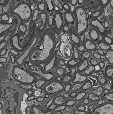 |
Focused ion beam-scanning electron microscopy links pathological myelin outfoldings to axonal changes in mice lacking Plp1 or Mag [1598 micrographs in TIFF format] | Steyer AM, Möbius W [Pubmed: 36354016] [DOI: 10.1002/glia.24290] |
31.3 GB | — |
| 2018-10-25 | 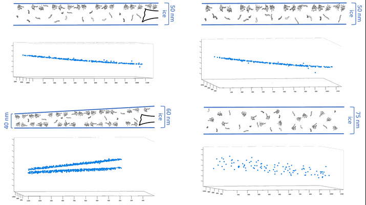 |
CryoET of bacterial RNA polymerase with several detergents [multiple data sets in MRC and RAW TEXT formats] | Chen J, Noble AJ, Kang JY, Darst SA [DOI: 10.1016/j.yjsbx.2019.100005] |
207.5 GB | — |
| 2022-03-15 | 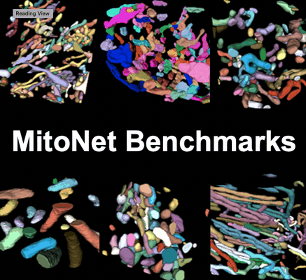 |
Seven benchmark datasets of instance segmentation of mitochondria: 6 diverse volume EM + 1 TEM (100 images) datasets [multiple data sets in TIFF format] | Narayan K, Conrad RW | 4.1 GB | — |
| 2022-12-13 | 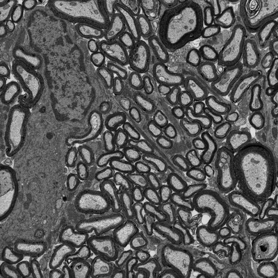 |
Focused ion beam-scanning electron microscopy links pathological myelin outfoldings to axonal changes in mice lacking Plp1 or Mag [1553 micrographs in TIFF format] | Steyer AM, Möbius W [Pubmed: 36354016] [DOI: 10.1002/glia.24290] |
22.3 GB | — |
| 2023-11-06 |  |
Single particle cryo-EM structure of RIG-I:RNA:Riplet ternary complex [3420 multi-frame micrographs composed of 40 frames each in TIFF format] | Wang W, Pyle AM [DOI: 10.1038/s41467-023-42982-0] |
1.5 TB | — |
| 2022-12-13 |  |
Focused ion beam-scanning electron microscopy links pathological myelin outfoldings to axonal changes in mice lacking Plp1 or Mag [910 micrographs in TIFF format] | Steyer AM, Möbius W [Pubmed: 36354016] [DOI: 10.1002/glia.24290] |
13.7 GB | — |
| 2022-12-13 |  |
Focused ion beam-scanning electron microscopy links pathological myelin outfoldings to axonal changes in mice lacking Plp1 or Mag [1186 micrographs in TIFF format] | Steyer AM, Möbius W [Pubmed: 36354016] [DOI: 10.1002/glia.24290] |
28.2 GB | — |
| 2022-11-18 |  |
A high-throughput electron tomography workflow reveals over-elongated centrioles in relapsed-refractory multiple myeloma [multiple data sets in MRC format] | Köhrer S, Dittrich T, Schorb M, Weinhold N, Haberbosch I, Börmel M, Pajor G [Pubmed: 36452870] [DOI: 10.1016/j.crmeth.2022.100322] |
2.0 TB | — |
| 2023-11-13 |  |
Test subset: In situ cryo-ET dataset of Chlamydomonas reinhardtii prepared using cryo-plasmaFIB milling [18 tilt series in MRC format] | Kelley R, Zhang X, Obr M, Khavnekar S, Righetto R, Waltz F, Wietrzynski W, Michael A, Tagiltsev G, Beck F, Zhong E, Wan W, Briggs J, Plitzko J, Engel B, Kotecha A [Pubmed: 37613825] [DOI: 10.1093/micmic/ozad067.480] |
293.7 GB | — |
| 2022-11-23 |  |
Cryo-electron tomograms of RPE1 cells with comprehensive annotation of actin filaments and microtubules [multiple data sets in TIFF and MRC formats] | Cheng DWC, Goetz SK, Mahamid J [Pubmed: 36690741] [DOI: 10.1038/s41592-022-01746-2] |
32.9 GB | — |
| 2020-08-06 | 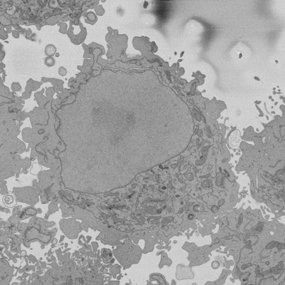 |
Cropped regions from Serial Block Face SEM of HeLa cell pellet with 10 nm pixels and 50 nm slices (benchmark dataset) [18 multi-frame micrographs composed of 300 frames each in TIFF format] | Peddie CP, Jones ML, Collinson LM | 15.6 GB | — |
| 2020-08-06 | 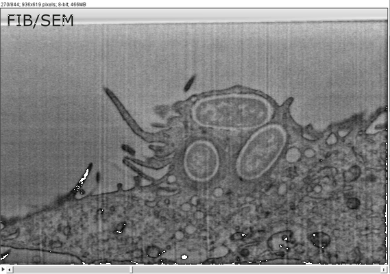 |
FIB/SEM sample dataset MCB-CLEM IV Weiner [1 multi-frame micrographs composed of 844 frames each in TIFF format] | Weiner A | 2.8 GB | — |
| 2024-01-15 | 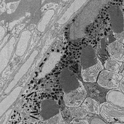 |
Developing retina in zebrafish 55 hpf larval eye. [16 reconstructed volumes in DM3 format] | Wilsch-Bräuninger M | 1.2 GB | — |
| 2019-02-01 | 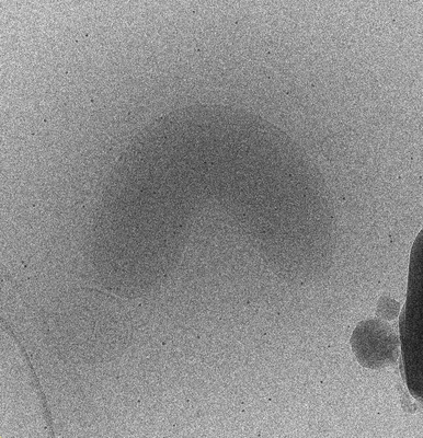 |
Bdellovibrio electron cryotomography tilt-series acquired by continuous tilting [1 tilt series in MRC format] | Chreifi G, Chen S, Metskas LA, Kaplan M, Jensen GJ [Pubmed: 30639925] [DOI: 10.1016/j.jsb.2018.12.008] |
2.1 GB | — |
| 2023-10-23 |  |
High-throughput electron tomography identifies centriole over-elongation in plasma cell disorders [multiple data sets in MRC format] | Köhrer S, Dittrich T, Schorb M, Weinhold N, Haberbosch I, Börmel M, Pajor G, Goldschmidt H, Müller-Tidow C, Raab MS, John L, Seckinger A, Brobeil A, Dreger P, Tornóczky T, Pajor L, Hegenbart U, Schönland SO, Schwab Y, Krämer A [Pubmed: 37821581] [DOI: 10.1038/s41375-023-02056-y] |
7.7 TB | — |
| 2024-04-17 |  |
Spatial mapping of hepatic ER and mitochondria architecture reveals zonated remodeling in fasting and obesity [multiple data sets in TIFF format] | Parlakgul G | 2.9 TB | — |
| 2019-02-01 |  |
Bdellovibrio bacteriovorus electron cryotomography tilt-series acquired by fast-incremental method [1 tilt series in MRC format] | Chreifi G, Chen S, Metskas LA, Kaplan M, Jensen GJ [Pubmed: 30639925] [DOI: 10.1016/j.jsb.2018.12.008] |
5.2 GB | — |
| 2021-11-16 |  |
Subcellular architecture collodaria photosymbiosis [7 multi-frame micrographs composed of 1000 frames each in TIFF format] | Decelle JD [Pubmed: 34499794] [DOI: 10.1111/1462-2920.15766] |
10.3 GB | — |
| 2024-01-16 | 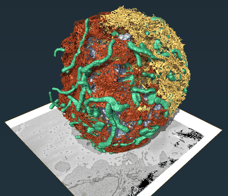 |
REEP3 and REEP4 determine the tubular morphology of the endoplasmic reticulum during mitosis [multiple data sets in DM4 and TIFF formats] | Belevich I, Jokitalo E [Pubmed: 30995177] [DOI: 10.1091/mbc.e18-11-0698] |
8.7 GB | — |
| 2019-05-07 |  |
A collection of yeast cell cryo-ET data [1488 tilt series in MRC format] | Gan L, Ng CT, Chen C, Cai S [Pubmed: 27605704] [DOI: 10.1091/mbc.E16-07-0506] |
2.5 TB | — |
| 2023-10-17 | 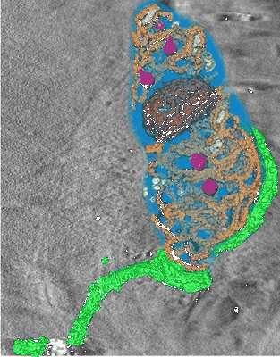 |
Soft X-ray Cryo Tomography of Trypanosoma [180 reconstructed volumes in MRC format] | Darrow MC [Pubmed: 28246039] [DOI: 10.1016/j.jsb.2017.02.007] |
1.5 GB | — |
| 2022-05-03 | 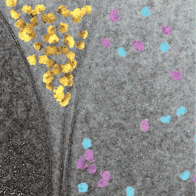 |
In situ single particle classification reveals distinct 60S maturation intermediates in cells [multiple data sets in MRC format] | Lucas BA, Zhang K, Loerch S, Grigorieff N [DOI: 10.1101/2022.04.10.487797] |
10.5 GB | — |
| 2023-01-03 |  |
Movies of apoferritin collected at different dose rates on the Direct Electron Apollo direct detector - 15 eps [972 multi-frame micrographs composed of 76 frames each in TIFF format] | Peng R, Fu X, Mendez JH, Randolph PS, Bammes BE, Stagg SM [Pubmed: 36578473] [DOI: 10.1016/j.yjsbx.2022.100080] |
297.2 GB | — |
| 2019-06-07 |  |
Scanning electron diffraction data collected from peptide microcrystals [30000 micrographs in DM4 format] | Gallagher-Jones M, Rodriguez JA, Bustillo K, Ophus C [Pubmed: 30675524] [DOI: 10.1038/s42003-018-0263-8] |
144.3 GB | — |
| 2020-12-11 |  |
Integrative imaging reveals SARS-CoV-2 induced reshaping of subcellular morphology [multiple data sets in MRC, BIG DATA VIEWER HDF5 and TIFF formats] | Cortese M, Lee JY, Cerikan B, Neufeldt CJ, Oorschot VMJ, Köhrer S, Hennies J, Schieber NL, Ronchi P, Mizzon G, Romero-Brey I, Santarella-Mellwig R, Schorb M, Boermel M, Mocaer K, Beckwith MS, Templin RM, Gross V, Pape C, Tischer C, Frankish J, Horvat NK, Laketa V, Stanifer M, Boulant S, Ruggieri A, Chatel-Chaix L, Schwab Y, Bartenschlager R [Pubmed: 33245857] [DOI: 10.1016/j.chom.2020.11.003] |
1.1 TB | — |
