Electron Microscopy Public Image Archive
Electron Microscopy Public Image Archive
오사카 대학의 EMPIAR-PDBj 팀은, 아시아의 EM 연구자가 용량이 큰 EM 이미지를 EMPIAR 데이터베이스에 전송하는 것을 돕고 있습니다. 인터넷을 통하여 EBI (UK)에 직>접 데이터를 전송하는 대신, 이용자는 우편이나 택배를 통하여 하드 디스크를 오사카 대학으로 보내실 수 있습니다. 혹은 인터넷을 이용하여 오사카 대학의 서버로 전>송 하실 수 있습니다. 오사카 대학에 데이터 전송 서비스를 희망하시는 분은 데이터를 보내시기 전에 먼저 이메일 통하여 등록하시고 싶은 EM데이터에 관하여 상담하십시오.
| Release date | Imageset | Title | Authors and references | Size | Resolution |
|---|---|---|---|---|---|
| 2021-03-12 | 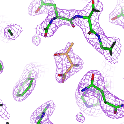 |
PKM2 in complex with L-threonine [476 multi-frame micrographs composed of 75 frames each in TIFF format] | Saur M, Hartshorn MJ, Dong J, Reeks J, Bunkoczi G, Jhoti H, Williams PA [Pubmed: 31877353] [DOI: 10.1016/j.drudis.2019.12.006] |
620.8 GB | 2.6 Å |
| 2022-07-27 |  |
PMCA-amplified α-synuclein fibrils, Multiple System Atrophy patient-derived seeds [4474 micrographs in MRC format] | Frieg B, Geraets JA, Schröder GF | 392.8 GB | 3.02 Å |
| 2022-07-27 |  |
PMCA-amplified α-synuclein fibrils, Parkinson's Disease patient-derived seeds [6780 micrographs in MRC format] | Frieg B, Geraets JA, Schröder GF | 595.3 GB | 3.3 Å |
| 2023-01-31 | 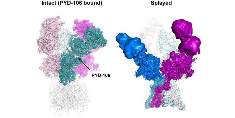 |
PYD-106 bound human GluN1a-GluN2C NMDA receptor in the presence of D-cycloserine and glutamate [11714 multi-frame micrographs composed of 30 frames each in TIFF format] | Chou TH [Pubmed: 36309015] [DOI: 10.1016/j.molcel.2022.10.008] |
2.1 TB | 3.72 - 4.19 Å |
| 2018-10-05 | 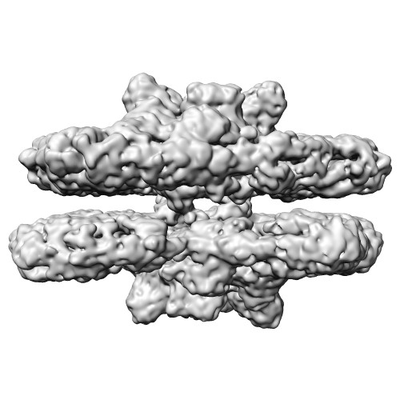 |
Paired C2S2M PSII-LHCII supercomplexes from thylakoid membranes of Pisum sativum [6929 micrographs in MRC format] | Melero R [Pubmed: 28855679] [DOI: 10.1038/s41598-017-10700-8] |
433.1 GB | 14.0 Å |
| 2022-11-29 |  |
Parallel cryo electron tomography (PACE-tomo) of 70S ribosomes (121 tilt series) [multiple data sets in TIFF and MRC formats] | Eisenstein F, Danev R [Pubmed: 36456783] [DOI: 10.1038/s41592-022-01690-1] |
490.1 GB | 3.1 Å |
| 2022-05-20 | 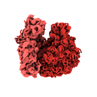 |
Parallel cryo electron tomography (PACE-tomo) of 70S ribosomes (200 kV, side-entry holder) [multiple data sets in MRC format] | Eisenstein F, Danev R [Pubmed: 36456783] [DOI: 10.1038/s41592-022-01690-1] |
222.8 GB | 5.8 - 6.5 Å |
| 2022-04-26 | 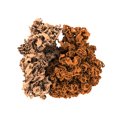 |
Parallel cryo electron tomography (PACE-tomo) of 70S ribosomes [multiple data sets in TIFF and MRC formats] | Eisenstein F, Danev R [Pubmed: 36456783] [DOI: 10.1038/s41592-022-01690-1] |
282.6 GB | 3.1 Å |
| 2022-06-14 | 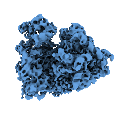 |
Parallel cryo electron tomography (PACE-tomo) of 80S ribosomes in situ [multiple data sets in MRC format] | Eisenstein F, Danev R [Pubmed: 36456783] [DOI: 10.1038/s41592-022-01690-1] |
132.3 GB | 8.2 Å |
| 2022-10-04 | 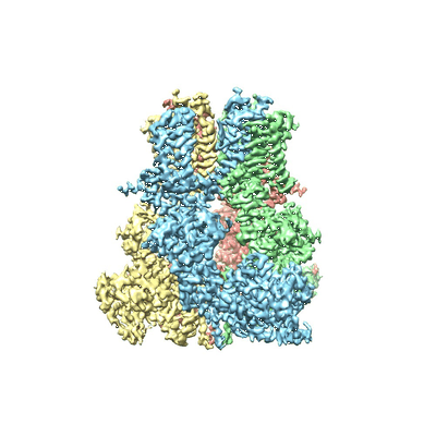 |
Particle stack from TRPM8 bound to calcium dataset [multiple data sets in MRC format] | Diver MM, Cheng Y, Julius D [Pubmed: 31488702] [DOI: 10.1126/science.aax6672] |
129.7 GB | 3.2 Å |
| 2022-06-13 | 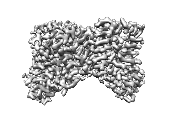 |
Particle stacks for VcINDY in 300mM NaCl [stack of 144865 particles in MRC format] | Sauer DB, Marden JJ, Song JM, Wang DN [Pubmed: 35551191] [DOI: 10.1038/s41467-022-30406-4] |
35.4 GB | 2.83 Å |
| 2022-06-13 | 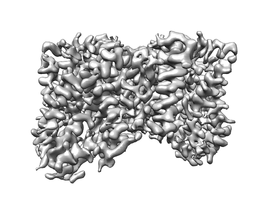 |
Particle stacks for VcINDY in Choline Chloride [multiple data sets in MRC format] | Sauer DB, Marden JJ, Song JM, Wang DN [Pubmed: 35551191] [DOI: 10.1038/s41467-022-30406-4] |
383.6 GB | 3.23 Å |
| 2023-03-15 | 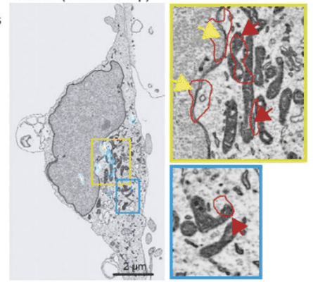 |
Performing Correlative Light and Electron Microscopy to reveal the structural organization and location of alpha-synuclein aggregation hotspots inside the neuron. [multiple data sets in DM4 and TIFF formats] | Choi ML, Chappard A, Singh BP, Maclachlan C, Rodrigues M, Fedotova E, Berezhnov AV, De S, Peddie C, Athauda D, Viridi GS, Zhang W, Evans JR, Wernick A, Zanjani ZS, Angelova PR, Esteras N, Vinikurov A, Morris K, Jeacock K, Tosatto L, Little D, Gissen P, Collinson L, Clarke DJ, Kunath T, Klenerman D, Abramov AY, Horrocks MH, Gandhi S [DOI: 10.1101/2022.06.07.494932] |
88.4 GB | — |
| 2017-12-18 |  |
Phase plate cryoET of T20S proteasome single particle [multiple data sets in MRC format] | Noble AJ, Dandey VP, Wei H, Brasch J, Chase J, Acharya P, Tan YZ, Zhang Z, Kim LY, Scapin G, Rapp M, Eng ET, Rice MJ, Cheng A, Negro CJ, Shapiro L, Kwong PD, Jeruzalmi D, des Georges A, Potter CS, Carragher B [Pubmed: 29809143] [DOI: 10.7554/eLife.34257] |
23.3 GB | — |
| 2020-07-06 |  |
Phase-plate cryo-EM of human TFIIH [multiple data sets in TIFF format] | Greber BJ, Toso DB, Fang J, Nogales E [Pubmed: 30860024] [DOI: 10.7554/eLife.44771] |
9.2 TB | 3.7 Å |
| 2022-12-12 |  |
Phencyclidine-bound GluN1a-GluN2B NMDA receptors [multiple data sets in TIFF format] | Chou THC, Furukawa HF [Pubmed: 35637422] [DOI: 10.1038/s41594-022-00772-0] |
1.6 TB | 4.3 Å |
| 2024-02-13 |  |
Plant SBF-SEM - Tobacco Leaf Chloroplast [130 micrographs in TIFF format] | Wickramanayake JS, Czymmek KJ [Pubmed: 37451777] [DOI: 10.1016/bs.mcb.2023.04.008] |
544.4 MB | — |
| 2021-11-16 |  |
Poly-alanine backbone model of E. coli BcsA bound to BcsB [multiple data sets in TIFF format] | Acheson JF, Ho R, Goularte NF, Cegelski L, Zimmer J [Pubmed: 33712813] [DOI: 10.1038/s41594-021-00569-7] |
3.2 TB | 3.4 - 4.2 Å |
| 2019-10-14 |  |
PolyA polymerase module of the cleavage and polyadenylation factor (CPF) from Saccharomyces cerevisiae [multiple data sets in MRCS format] | Casanal A, Kumar A, Hill CH, Emsley P, Passmore LA [Pubmed: 29074584] [DOI: 10.1126/science.aao6535] |
15.4 TB | 3.55 Å |
| 2023-09-14 |  |
Porcine uroplakin complex [multiple data sets in TIFF format] | Oda T, Yanagisawa H, Kikkawa M | 1.4 TB | 3.5 Å |
| 2013-10-30 |  |
Pre-fusion structure of trimeric HIV-1 envelope glycoprotein determined by cryo-electron microscopy [3755 micrographs in MRCS format] | Bartesaghi A, Merk A, Borgnia MJ, Milne JLS, Subramaniam S [Pubmed: 24154805] [DOI: 10.1038/nsmb.2711] |
117.8 GB | 6.0 Å |
| 2023-02-17 | 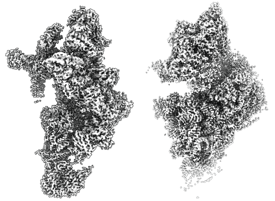 |
Principles of mitoribosomal small subunit assembly in eukaryotes [multiple data sets in TIFF format] | Harper NJ, Burnside C, Klinge S [Pubmed: 36482135] [DOI: 10.1038/s41586-022-05621-0] |
73.6 TB | 2.36 - 3.8 Å |
| 2019-10-07 | 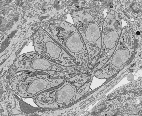 |
Processed FIB SEM images of a parasitophorous vacuole containing Toxoplasma gondii ∆CAP parasites, complemented with CAP. [1 multi-frame micrographs composed of 1 frames each in MRC format] | Hunt A, Russell MRG, Wagener J, Kent R, Carmeille R, Peddie CJ, Collinson L, Heaslip A, Ward GE, Treeck M [Pubmed: 31577230] [DOI: 10.7554/elife.50598] |
583.8 MB | — |
| 2019-10-07 | 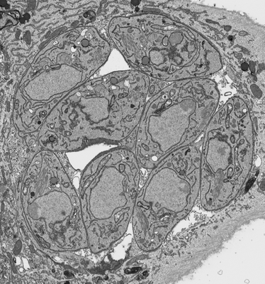 |
Processed FIB SEM images of a parasitophorous vacuole containing Toxoplasma gondii ∆CAP parasites. [1 multi-frame micrographs composed of 1 frames each in MRC format] | Hunt A, Russell MRG, Wagener J, Kent R, Carmeille R, Peddie CJ, Collinson L, Heaslip A, Ward GE, Treeck M [Pubmed: 31577230] [DOI: 10.7554/elife.50598] |
898.1 MB | — |
| 2022-06-22 | 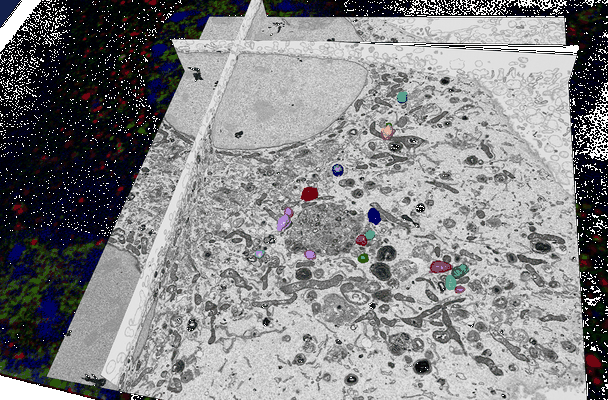 |
Processed FIB-SEM images of murine hypothalamus-derived GT1-7 neuronal cells [multiple data sets in TIFF format] | Peddie CJ, Collinson LM [DOI: 10.3389/fcell.2022.819571] |
140.5 GB | — |
