Electron Microscopy Public Image Archive
Electron Microscopy Public Image Archive
오사카 대학의 EMPIAR-PDBj 팀은, 아시아의 EM 연구자가 용량이 큰 EM 이미지를 EMPIAR 데이터베이스에 전송하는 것을 돕고 있습니다. 인터넷을 통하여 EBI (UK)에 직>접 데이터를 전송하는 대신, 이용자는 우편이나 택배를 통하여 하드 디스크를 오사카 대학으로 보내실 수 있습니다. 혹은 인터넷을 이용하여 오사카 대학의 서버로 전>송 하실 수 있습니다. 오사카 대학에 데이터 전송 서비스를 희망하시는 분은 데이터를 보내시기 전에 먼저 이메일 통하여 등록하시고 싶은 EM데이터에 관하여 상담하십시오.
| Release date | Imageset | Title | Authors and references | Size | Resolution |
|---|---|---|---|---|---|
| 2022-07-27 | 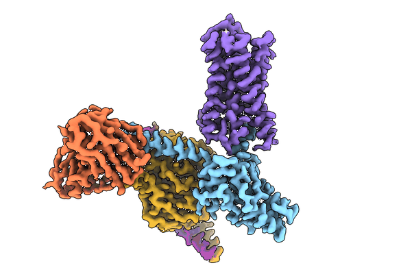 |
CryoEM structure of Go-coupled 5-HT5AR in complex with Lisuride [4633 micrographs in MRC format] | Fay JF, Roth BL, Zhang S [Pubmed: 35835867] [DOI: 10.1038/s41594-022-00796-6] |
406.8 GB | 2.79 Å |
| 2020-10-16 | 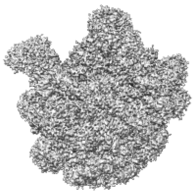 |
E. coli 50S ribosome bound to compounds 46 and VS1 [1095 multi-frame micrographs composed of 53 frames each in TIFF format] | Pellegrino J, Lee DJ, Fraser JS, Seiple IB [Pubmed: 32968273] [DOI: 10.1038/s41586-020-2761-3] |
403.8 GB | 2.7 Å |
| 2023-10-23 | 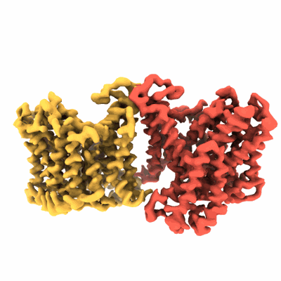 |
Cryo-EM structure of Dipyridamole-bound human Anion Exchanger 1 [4570 micrographs in MRC format] | Capper MJ, Yang S, Stone AC, Vatansever S, Zilberg G, Mathiharan YK, Habib R, Hutchinson K, Zhao Y, Schlessinger A, Mezei M, Osman R, Zhang B, Wacker D [Pubmed: 37679563] [DOI: 10.1038/s41594-023-01085-6] |
401.3 GB | 3.13 Å |
| 2022-11-16 | 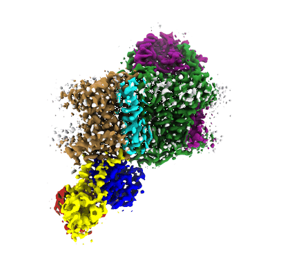 |
Single particle cryo-EM of KdpFABC WT (KdpB-Ser162-P) in presence of orthovanadate [multiple data sets in TIFF format] | Silberberg JM, Stock C, Hielkema L, Corey RA, Rheinberger J, Wunnicke D, Dubach VRA, Stansfeld PJ, Hänelt I, Paulino C [Pubmed: 36255052] [DOI: 10.7554/eLife.80988] |
399.3 GB | 3.3 - 7.4 Å |
| 2022-08-12 |  |
Staphylococcal self-loading helicases couple the staircase mechanism with inter domain high flexibility [3121 multi-frame micrographs composed of 40 frames each in TIFF format] | Qiao C, Debiasi-Anders G, Mir-Sanchis I [Pubmed: 35871290] [DOI: 10.1093/nar/gkac625] |
395.0 GB | 3.14 Å |
| 2022-07-27 |  |
PMCA-amplified α-synuclein fibrils, Multiple System Atrophy patient-derived seeds [4474 micrographs in MRC format] | Frieg B, Geraets JA, Schröder GF | 392.8 GB | 3.02 Å |
| 2020-07-27 |  |
Saccharomyces cerevisiae fatty acid synthase complex [multiple data sets in MRC and MRCS formats] | Singh K, Graf B, Stark H, Chari A [Pubmed: 32160528] [DOI: 10.1016/j.cell.2020.02.034] |
391.7 GB | 2.9 Å |
| 2020-11-27 |  |
CryoEM SPA of Holo-SrpI Encapsulin Complex (Raw Frames) [1023 multi-frame micrographs composed of 33 frames each in TIFF format] | Nichols RJ, LaFrance BJ, Phillips NR, Oltrogge LM, Valentin-Alvarado LE, Bischoff AJ, Nogales E, Savage DF [Pubmed: 33821786] [DOI: 10.7554/eLife.59288] |
390.3 GB | 2.2 Å |
| 2023-07-14 |  |
Mouse apoferritin, Dataset B taken with CRYO ARM 300 [1246 multi-frame micrographs composed of 50 frames each in TIFF format] | Maki-Yonekura S, Kawakami K, Takaba K, Hamaguchi T, Yonekura K [Pubmed: 37258702] [DOI: 10.1038/s42004-023-00900-x] |
384.7 GB | 1.49 Å |
| 2019-04-18 | 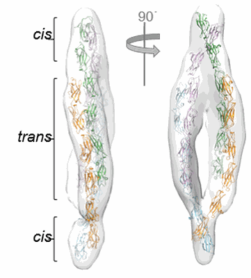 |
Single particle cryo electron tomography of mouse protocadherin gamma B6 [multiple data sets in MRC format] | Brasch J, Goodman KM, Noble AJ, Mannepalli S, Bahna F, Rapp M, Dandey VP, Bepler T, Berger B, Maniatis T, Potter CS, Carragher B, Honig B, Shapiro L [Pubmed: 30971825] [DOI: 10.1038/s41586-019-1089-3] |
384.1 GB | 35.0 Å |
| 2022-06-13 | 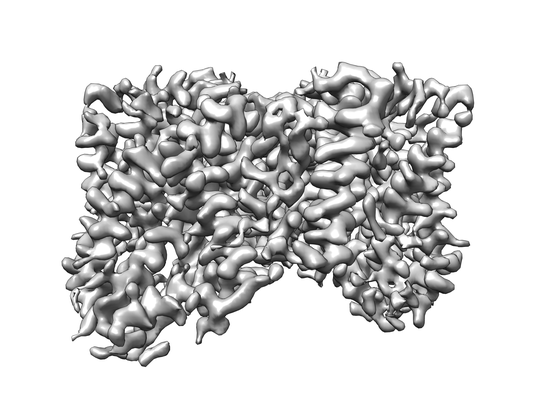 |
Particle stacks for VcINDY in Choline Chloride [multiple data sets in MRC format] | Sauer DB, Marden JJ, Song JM, Wang DN [Pubmed: 35551191] [DOI: 10.1038/s41467-022-30406-4] |
383.6 GB | 3.23 Å |
| 2024-04-21 |  |
Cryo-electron tomograms of Host - S. Typhi interaction [multiple data sets in MRC format] | Lian H, Park D, Chen M, Schueder F, Lara-Tejero M, Liu J, Galan J [Pubmed: 37640963] [DOI: 10.1038/s41564-023-01459-y] |
381.6 GB | 25.0 Å |
| 2023-05-22 | 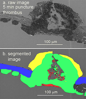 |
Venous puncture wound thrombi, 1, 5, 20 min post-puncture, 100 nm XY raw images, 20 nm XY pixels every 20 micons, 3 nm wide area TEM montages at selected depth [15 multi-frame micrographs composed of 1 frames each in DM4 format] | Storrie B, Leapman RD [Pubmed: 34531522] [DOI: 10.1038/s42003-021-02615-y] |
381.2 GB | — |
| 2019-04-18 |  |
CryoET of cis-mutated mouse protocadherin gamma B6 on membranes [multiple data sets in MRC format] | Brasch J, Bepler T, Berger B, Maniatis T, Potter CS, Carragher B, Honig B, Shapiro L [Pubmed: 30971825] [DOI: 10.1038/s41586-019-1089-3] |
379.7 GB | — |
| 2024-02-13 |  |
Cryo electron microscopy of Virus-like Particle based on PVY coat protein [502 multi-frame micrographs composed of 40 frames each in TIFF format] | Kavcic L, Kezar A [Pubmed: 38233506] [DOI: 10.1038/s42004-024-01100-x] |
379.4 GB | 2.99 - 3.34 Å |
| 2024-02-13 |  |
Cryo electron microscopy of Virus-like Particle based on PVY coat protein with dC79 deletion [491 multi-frame micrographs composed of 41 frames each in TIFF format] | Kavcic L, Kezar A [Pubmed: 38233506] [DOI: 10.1038/s42004-024-01100-x] |
377.4 GB | 3.2 Å |
| 2023-06-30 |  |
Cryo electron tomography of the Bacterial Voltage-Gated Sodium Channel NaChBac in Liposomes [123 tilt series in TIFF format] | Chang SYS, Kudryashev M | 377.3 GB | 16.3 Å |
| 2022-07-12 |  |
In situ cryo-electron tomography of T. kivui cells [multiple data sets in MRC and TIFF formats] | Dietrich HM, Righetto RD, Kumar A, Wietrzynski W, Trischler R, Schuller SK, Wagner J, Schwarz FM, Engel BD, Müller V, Schuller JM [Pubmed: 35859174] [DOI: 10.1038/s41586-022-04971-z] |
373.4 GB | 17.0 Å |
| 2021-03-19 |  |
1.93 A cryo-EM structure of streptavidin [2277 multi-frame micrographs composed of 70 frames each in TIFF format] | Hiraizumi M, Yamashita K, Nisihzawa T, Kikkawa M, Nureki O | 373.1 GB | 1.93 Å |
| 2023-07-10 |  |
Cryo electron tomography of Cytochalasin D-induced protrusions of Drosophila S2 cells treated with DMSO or thapsigargin - Datasets 5 - 7 [multiple data sets in TIFF and MRC formats] | Ventura Santos C, Carter AP, Rogers SL [Pubmed: 37034688] [DOI: 10.1101/2023.03.31.535077] |
373.1 GB | — |
| 2023-02-28 |  |
SARS-CoV-2 spike protein (1-up RBD) on EG-grid [1495 multi-frame micrographs composed of 60 frames each in TIFF format] | Fujita J, Makino F, Asahara H, Moriguchi M, Kumano S, Anzai I, Kishikawa J, Matsuura Y, Kato T, Namba K, Inoue T [Pubmed: 36755111] [DOI: 10.1038/s41598-023-29396-0] |
373.0 GB | 3.1 Å |
| 2020-08-19 | 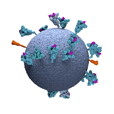 |
Subtomogram averaging and classification of SARS-CoV-2 Spike Proteins on intact virions [multiple data sets in TIFF and MRC formats] | Ke Z, Oton J, Cortese M, Zila V, Zivanov J, Lu JM, Peukes J, Scheres SHW, Briggs JAG [Pubmed: 32805734] [DOI: 10.1038/s41586-020-2665-2] |
372.4 GB | 7.7 - 9.9 Å |
| 2023-02-28 | 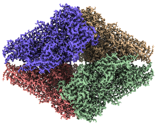 |
Beta-galactosidase on EG-grid [3500 multi-frame micrographs composed of 40 frames each in TIFF format] | Fujita J, Makino F, Asahara H, Moriguchi M, Kumano S, Anzai I, Kishikawa J, Matsuura Y, Kato T, Namba K, Inoue T [Pubmed: 36755111] [DOI: 10.1038/s41598-023-29396-0] |
369.6 GB | 1.81 Å |
| 2020-08-12 | 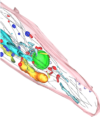 |
TEM tomograms of Drosophila tracheal terminal cells during subcellular tube formation [multiple data sets in TIFF and MRC formats] | Mathew R, Rios-Barrera LD, Machado P, Schwab Y, Leptin M [Pubmed: 32657472] [DOI: 10.15252/embj.2020105332] |
366.7 GB | — |
| 2021-11-02 | 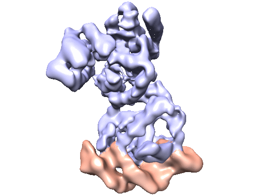 |
CryoEM single particle dataset for NanR dimer-DNA hetero-complex. [3465 multi-frame micrographs composed of 32 frames each in MRC format] | Venugopal H, Horne CR, Ramm G, Dobson RCJ [Pubmed: 33790291] [DOI: 10.1038/s41467-021-22253-6] |
364.8 GB | 3.9 Å |
