Electron Microscopy Public Image Archive
Electron Microscopy Public Image Archive
The EMPIAR-PDBj team at Osaka University assists Asian EM researchers with the transfer of big EM image data to EMPIAR. Instead of sending the data directly to the EBI (UK) via the internet, hard drives can also be sent to Osaka University by postal mail or via a courier service. As an alternative, internet transfer to our server in Osaka is also available. If you would like to take advantage of our submission services, please contact us first by e-mail before sending the data to us.
| Release date | Imageset | Title | Authors and references | Size | Resolution |
|---|---|---|---|---|---|
| 2022-01-12 |  |
CryoEM of Mycobacterium tuberculosis WT RNAP holoenzyme/RbpA [1381 multi-frame micrographs composed of 50 frames each in TIFF format] | Boyaci H, Chen J, Darst SA, Campbell EA [Pubmed: 29480804] [DOI: 10.7554/eLife.34823] |
838.9 GB | 3.38 Å |
| 2021-11-16 |  |
Cryo EM Structure of the E. coli BcsB Hexamer [multiple data sets in TIFF format] | Acheson JF, Ho R, Goularte NF, Cegelski L, Zimmer J [Pubmed: 33712813] [DOI: 10.1038/s41594-021-00569-7] |
837.2 GB | 3.4 Å |
| 2023-06-29 |  |
E. coli 50S ribosome bound to L-linker solithromycin conjugate [multiple data sets in MRC and TIFF formats] | Seiple IB, Pellegrino J, Fraser JS, Lee DJ | 835.4 GB | 2.25 Å |
| 2019-12-19 |  |
3.2 Å Single-Particle Cryo-EM Reconstruction of 49 kDa Membrane-Bound PfCRT Complexed with Fab [multiple data sets in MRCS and MRC formats] | Kim JK, Tan YZT, Wicht KJW, Erramilli SKE, Dhingra SKD, Okombo JO, Vendome JV, Hagenah LMH, Giacometti SIG, Warren ALW, Nosol KN, Roepe PDR, Potter CSP, Carragher BC, Kossiakoff AAK, Quick MQ, Fidock DAF, Mancia FM [Pubmed: 31776516] [DOI: 10.1038/s41586-019-1795-x] |
830.4 GB | 3.3 Å |
| 2022-09-07 |  |
Single-particle cryo-electron microscopy final particle stacks and .star files from TSHR complexes [multiple data sets in MRCS format] | Faust B, Manglik A [Pubmed: 35940205] [DOI: 10.1038/s41586-022-05159-1] |
828.7 GB | 2.9 Å |
| 2021-09-22 |  |
Di nucleosome fraction from metaphase chromosome in Xenopus egg extract lot1 [1386 multi-frame micrographs composed of 50 frames each in TIFF format] | Arimura YA, Funabiki HF [Pubmed: 34478647] [DOI: 10.1016/j.molcel.2021.08.010] |
825.4 GB | 8.1 Å |
| 2024-09-06 |  |
Human PRC2 - RvLEAM (short) (1:6 molar ratio), cross-linked 2 min [2981 multi-frame micrographs composed of 51 frames each in TIFF format] | Abe KM, Li G, He Q, Grant T, Lim CJ | 824.4 GB | 3.47 Å |
| 2023-01-16 |  |
Structure of RecT protein from Listeria innoccua phage A118 in complex with 83-mer single stranded DNA [1619 multi-frame micrographs composed of 45 frames each in TIFF format] | Bell CE [Pubmed: 36543802] [DOI: 10.1038/s41467-022-35572-z] |
822.3 GB | 4.5 Å |
| 2024-05-10 |  |
Structure of human calcium-sensing receptor in complex with chimeric Gq (miniGisq) protein in nanodiscs [9336 multi-frame micrographs composed of 60 frames each in MRC format] | Zuo H, Park J, Frangaj A, Ye J, Lu G, Manning JJ, Asher WB, Lu Z, Hu G, Wang L, Mendez J, Eng E, Zhang Z, Lin X, Grasucci R, Hendrickson WA, Clarke OB, Javitch JA, Conigrave AD, Fan QR [Pubmed: 38632411] [DOI: 10.1038/s41586-024-07331-1] |
820.1 GB | 3.4 Å |
| 2023-06-16 |  |
HIV-1 capsid-like particles assembled from purified capsid protein and assembly cofactor IP6 [1487 multi-frame micrographs composed of 50 frames each in TIFF format] | Highland CMH, Dick RAD [Pubmed: 37094124] [DOI: 10.1073/pnas.2220545120] |
818.8 GB | 3.6 Å |
| 2022-04-29 |  |
Cryo electron microscopy of wild-type hyaluronan synthase with UDP [3062 multi-frame micrographs composed of 40 frames each in TIFF format] | Maloney FP, Kuklewicz J, Zimmer J [Pubmed: 35355017] [DOI: 10.1038/s41586-022-04534-2] |
818.1 GB | 3.1 Å |
| 2024-09-06 |  |
Human PRC2 - cross-linked 10 min [3274 multi-frame micrographs composed of 40 frames each in TIFF format] | Abe KM, Li G, He Q, Grant T, Lim CJ | 817.0 GB | 4.29 Å |
| 2022-12-13 |  |
Single particle cryo-EM dataset of the Vairimorpha necatrix 20S proteasome from spores [7240 multi-frame micrographs composed of 30 frames each in TIFF format] | Jespersen N, Ehrenbolger K, Winiger RR, Svedberg D, Vossbrinck CR, Barandun J [Pubmed: 36379934] [DOI: 10.1038/s41467-022-34691-x] |
814.1 GB | 2.77 Å |
| 2024-06-07 | 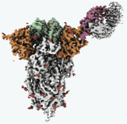 |
Single-particle cryo-EM unaligned micrographs of NTD-directed neutralizing antibody 1-87 in complex with prefusion SARS-CoV-2 spike glycoprotein [3135 multi-frame micrographs composed of 60 frames each in TIFF format] | Cerutti G, Guo Y, Zhou T, Gorman J, Lee M, Rapp M, Reddem ER, Yu J, Bahna F, Bimela J, Huang Y, Katsamba PS, Liu L, Nair MS, Rawi R, Olia AS, Wang P, Zhang B, Chuang GY, Ho DD, Sheng Z, Kwong PD, Shapiro L [Pubmed: 33789084] [DOI: 10.1016/j.chom.2021.03.005] |
812.5 GB | 3.55 Å |
| 2021-09-24 | 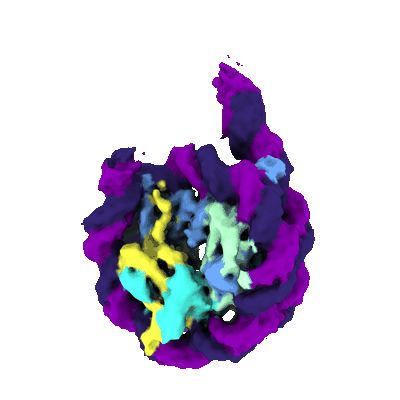 |
Di nucleosome fraction from interphase chromosome in Xenopus egg extract lot1 [1656 multi-frame micrographs composed of 50 frames each in TIFF format] | Arimura YA, Funabiki HF [Pubmed: 34478647] [DOI: 10.1016/j.molcel.2021.08.010] |
805.9 GB | 4.74 Å |
| 2021-11-16 |  |
Cryo-electron microscopy reconstruction of apo bovine MRP1 [2226 multi-frame micrographs composed of 50 frames each in TIFF format] | Johnson ZL, Chen J [Pubmed: 28238471] [DOI: 10.1016/j.cell.2017.01.041] |
804.5 GB | 3.49 Å |
| 2023-07-06 |  |
CryoEM micrographs of slipper limpet (Crepidula fornicata) hemocyanin [856 multi-frame micrographs composed of 30 frames each in MRC format] | Young MT, Pasqualetto G, Clare D [Pubmed: 37347755] [DOI: 10.1371/journal.pone.0287294] |
802.5 GB | 4.7 - 7.0 Å |
| 2022-07-15 | 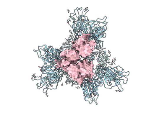 |
CryoEM single particle dataset for psNb 1-23 with spike protein [1872 multi-frame micrographs composed of 40 frames each in TIFF format] | Xiang Y, Huang W, Liu H, Sang Z, Nambulli S, Tubiana J, Williams Jr KL, Duprex WP, Schneidman-Duhovny D, Wilson IA, Taylor DJ, Shi Y [Pubmed: 35738279] [DOI: 10.1016/j.celrep.2022.111004] |
802.4 GB | 2.9 Å |
| 2024-06-06 |  |
Single-particle cryo-EM unaligned micrographs of antibody SKT05 in complex with Western Equine Encephalitis Virus-Like Particles [5133 multi-frame micrographs composed of 50 frames each in TIFF format] | Sutton MS, Pletnev S, Callahan V, Ko S, Tsybovsky Y, Bylund T, Casner RG, Cerutti G, Gardner CL, Guirguis V, Verardi R, Zhang B, Ambrozak D, Beddall M, Lei H, Yang ES, Liu T, Henry AR, Rawi R, Schon A, Schramm CA, Shen CH, Shi W, Stephens T, Yang Y, Florez MB, Ledgerwood JE, Burke CW, Shapiro L, Fox JM, Kwong PD, Roederer M [Pubmed: 37295404] [DOI: 10.1016/j.cell.2023.05.019] |
796.0 GB | 6.1 Å |
| 2023-08-18 |  |
KpFtsZ–Mb double helical tube [3096 multi-frame micrographs composed of 60 frames each in TIFF format] | Fujita J, Amesaka H, Yoshizawa T, Hibino K, Kamimura N, Kuroda N, Konishi T, Kato Y, Hara M, Inoue T, Namba K, Tanaka SI, Matsumura H [Pubmed: 37429870] [DOI: 10.1038/s41467-023-39807-5] |
794.4 GB | 2.67 Å |
| 2021-11-12 |  |
WT MDA5-dsRNA filaments in complex with ADP [4680 multi-frame micrographs composed of 40 frames each in TIFF format] | Yu Q, Modis Y [Pubmed: 34795277] [DOI: 10.1038/s41467-021-27062-5] |
791.8 GB | 3.4 - 3.9 Å |
| 2022-07-12 |  |
CryoEM single particle dataset for psNb 2-38 with spike protein [1901 multi-frame micrographs composed of 40 frames each in TIFF format] | Xiang Y, Huang W, Liu H, Sang Z, Nambulli S, Tubiana J, Williams Jr KL, Duprex WP, Schneidman-Duhovny D, Wilson IA, Taylor DJ, Shi Y [Pubmed: 35738279] [DOI: 10.1016/j.celrep.2022.111004] |
791.0 GB | 2.9 Å |
| 2024-10-09 |  |
Cryo-EM structure of in-vitro alpha-synuclein fibril [5193 multi-frame micrographs composed of 40 frames each in TIFF format] | Sanchez JC, Borcik CG, Tonelli M, Sibert B, Rienstra CM, Wright ER | 789.6 GB | 2.04 Å |
| 2024-03-18 |  |
Cryo electron tomography image- and volume-series subtomograms of chloramphenicol-treated mycoplasma pneumoniae [multiple data sets in MRCS and MRC formats] | Powell B.M., Davis J.H. [Pubmed: 37398315] [DOI: 10.1101/2023.05.31.542975] |
788.5 GB | 26.0 Å |
| 2020-08-18 |  |
Mechanism of Ribosome Rescue by Alternative Release Factor B [multiple data sets in MRC format] | Chan K-H, Petrychenko V, Mueller C, Maracci C, Holtkamp W, Wilson DN, Fischer N, Rodnina MV [Pubmed: 32796827] [DOI: 10.1038/s41467-020-17853-7] |
788.2 GB | 2.6 - 3.7 Å |
