Electron Microscopy Public Image Archive
Electron Microscopy Public Image Archive
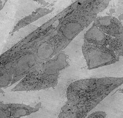
FAST-EM array tomography: a workflow for multibeam volume electron microscopy
Kievits AJ, Duinkerken BHP, Lane R, de Heus C, van Beijeren Bergen en Henegouwen D, Höppener T, Wolters AHG, Liv N, Giepmans BNG, Hoogenboom JP
Methods in Microscopy (---)