3D reconstructions of parasite development and the intracellular niche of the microsporidian pathogen E. intestinalis [multiple data sets in DM4 format]
Publication:
3D reconstructions of parasite development and the intracellular niche of the microsporidian pathogenE. intestinalis
Riggi MR, Antao NV, Lam C, Davydov A, Sall J, Petzold C, Liang F, Iwasa J, Ekiert DC, Bhabha G
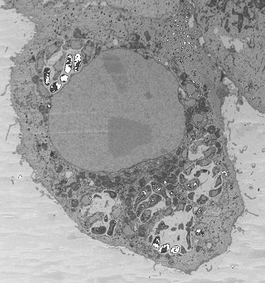
Unaligned and unprocessed micrograph stack of a Vero cell at 24 hours post infection with E. intestinalis. Slice thickness=50nm. Images were processed in Fiji using the Gaussian blur filter with a sigma (radius=2) prior to analysis in Dragonfly.
Unaligned and unprocessed micrograph stack of a Vero cell at 24 hours post infection with E. intestinalis. Slice thickness=50nm. Images were processed in Fiji using the Gaussian blur filter with a sigma (radius=2) prior to analysis in Dragonfly.
Unaligned and unprocessed micrograph stack of a Vero cell at 48 hours post infection with E. intestinalis. Slice thickness=30nm. Images were processed in Fiji using the Gaussian blur filter with a sigma (radius=2) prior to analysis in Dragonfly.
Unaligned and unprocessed micrograph stack of a Vero cell at 48 hours post infection with E. intestinalis. There are two ROIs associated with this infected cell: ROI00 (22000x23000) and ROI01 (22000x23000). ROI00 and ROI01 were stitched together for segmentation analysis. Slice thickness=50nm. Images were processed in Fiji using the Gaussian blur filter with a sigma (radius=2) prior to analysis in Dragonfly.
Unaligned and unprocessed micrograph stack of a parasitophorous vacuole in a Vero cell at 48 hours post infection with E. intestinalis. Slice thickness=50nm. Images were processed in Fiji using the Gaussian blur filter with a sigma (radius=2) prior to analysis in Dragonfly.
Unaligned and unprocessed micrograph stack of a parasitophorous vacuole in a Vero cell at 48 hours post infection with E. intestinalis. Slice thickness=50nm. Images were processed in Fiji using the Gaussian blur filter with a sigma (radius=2) prior to analysis in Dragonfly.
Unaligned and unprocessed micrograph stack of a parasitophorous vacuole in a Vero cell at 48 hours post infection with E. intestinalis. Slice thickness=50nm. Images were processed in Fiji using the Gaussian blur filter with a sigma (radius=2) prior to analysis in Dragonfly.
E. intestinalis spores purified from Vero cells. Slice thickness=50nm. Images were processed in Fiji using the Gaussian blur filter with a sigma (radius=2) prior to analysis in Dragonfly.
E. intestinalis spores purified from Vero cells. Slice thickness=50nm. Images were processed in Fiji using the Gaussian blur filter with a sigma (radius=2) prior to analysis in Dragonfly.
Partial volume of an uninfected Vero cell. Slice thickness=60nm. Images were processed in Fiji using the Gaussian blur filter with a sigma (radius=2) prior to analysis in Dragonfly.
Partial volume of an uninfected Vero cell. Slice thickness=60nm. Images were processed in Fiji using the Gaussian blur filter with a sigma (radius=2) prior to analysis in Dragonfly.