Electron Microscopy Public Image Archive
Electron Microscopy Public Image Archive
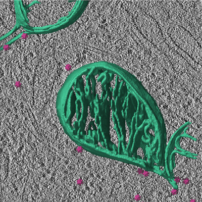
Genetically encoded multimeric tags for subcellular protein localisation in cryo-EM
Fung HKH, Hayashi Y, Salo VT, Babenko A, Zagoriy I, Brunner A, Ellenberg J, Müller CW, Cuylen-Haering S, Mahamid J
(---)