Unaligned multi-frame micrographs (frames/*.tif) corresponding to 2 tilt series with corresponding gain reference, defect map and metadata (mdoc/*.mdoc). Data acquired with defocus only, no Volta phase plate, on a K3 BioQuantum detector. See README.md under All Files for more information (also copied below).
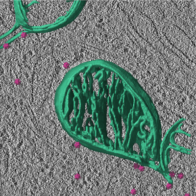
# Cryo-electron tomography of GEM2-labelled Mito-EGFP in HeLa cells
Five independent cryo-ET datasets were collected. Each dataset is organised under the following directory structure, with the directories "data/", "alignment/", and "tomograms/" each designated as an Image Set.
- data/ - CountRef.mrc gain reference - defects.tif defect map (K3 data only) - frames/ unaligned, unbinned multi-frame micrographs - mdoc/ MDOC files listing exposure dose, pixel size and tilt angle information for each tilt - alignment/ - TS_###.mrc/ - TS_###.mrc.st sorted, motion-corrected tilt series from WARP - TS_###.mrc.rawtlt tilt angle information corresponding to tilt series - TS_###.mrc.aln AreTomo tilt series alignment solution - TS_###.mrc.xf XF file from AreTomo alignment - mdoc_edited/ MDOC files with entries for discarded tilts during alignment removed - tomograms/ - *.mrc tomograms reconstructed by weighted back-projection in AreTomo, 4-times binned - ######.star combined STAR file listing the refined coordinates of GEM2 particles at 6.85 A/px in this dataset
Acquisition parameters differ for each dataset and are specified below. Tilt series were aligned in AreTomo 1.3.1 with the following parameters: 8 by 6 patches (K3 data) or 6 by 6 patches (K2 data), VolZ 2000, AlignZ 1000, TiltAxis ## -1 (as calibrated for the magnification and microsco pe used), TiltCor 0.
## Dataset 1 (211206) - Image Sets 1 to 3
Data acquired with defocus only, no Volta phase plate, on a K3 BioQuantum detector. 2 tilt series. Pixel size, 3.425 A/px.
## Dataset 2 (220330) - Image Sets 4 to 6
Data acquired with defocus only, no Volta phase plate, on a K3 BioQuantum detector. 3 tilt series. Pixel size, 3.425 A/px.
## Dataset 3 (220504) - Image Sets 7 to 9
Data acquired with Volta phase plate on a K2 Summit detector. 1 tilt series. Pixel size, 3.3701 A/px.
## Dataset 4 (220506) - Image Sets 10 to 12
Data acquired with Volta phase plate on a K3 BioQuantum detector. 5 tilt series. Pixel size, 3.425 A/px.
## Dataset 5 (220720) - Image Sets 13 to 15
Data acquired with Volta phase plate on a K3 BioQuantum detector. 6 tilt series. Pixel size, 3.425 A/px.
Files:
Available download options:
Archives and downloads the selected files into an uncompressed zip file.
Depending on the number and size of files to be downloaded, this can take an enormous amount of time.
Also, this feature is unstable and may not work properly, and checksums of downloaded files are not verified.
We recommend using rsync, aspera, globus, etc.
Download a list of selected files.
It is possible to download files by specifying the file list with rsync command, etc.
Tilt series following motion correction in WARP (*.st), with tilt angle information (*.rawtlt) for tilt series alignment in AreTomo and alignment solutions outputted by AreTomo (*.aln and *.xf). Data acquired with defocus only, no Volta phase plate, on a K3 BioQuantum detector. See README.md under All Files for more information (also copied below).
# Cryo-electron tomography of GEM2-labelled Mito-EGFP in HeLa cells
Five independent cryo-ET datasets were collected. Each dataset is organised under the following directory structure, with the directories "data/", "alignment/", and "tomograms/" each designated as an Image Set.
- data/ - CountRef.mrc gain reference - defects.tif defect map (K3 data only) - frames/ unaligned, unbinned multi-frame micrographs - mdoc/ MDOC files listing exposure dose, pixel size and tilt angle information for each tilt - alignment/ - TS_###.mrc/ - TS_###.mrc.st sorted, motion-corrected tilt series from WARP - TS_###.mrc.rawtlt tilt angle information corresponding to tilt series - TS_###.mrc.aln AreTomo tilt series alignment solution - TS_###.mrc.xf XF file from AreTomo alignment - mdoc_edited/ MDOC files with entries for discarded tilts during alignment removed - tomograms/ - *.mrc tomograms reconstructed by weighted back-projection in AreTomo, 4-times binned - ######.star combined STAR file listing the refined coordinates of GEM2 particles at 6.85 A/px in this dataset
Acquisition parameters differ for each dataset and are specified below. Tilt series were aligned in AreTomo 1.3.1 with the following parameters: 8 by 6 patches (K3 data) or 6 by 6 patches (K2 data), VolZ 2000, AlignZ 1000, TiltAxis ## -1 (as calibrated for the magnification and microsco pe used), TiltCor 0.
## Dataset 1 (211206) - Image Sets 1 to 3
Data acquired with defocus only, no Volta phase plate, on a K3 BioQuantum detector. 2 tilt series. Pixel size, 3.425 A/px.
## Dataset 2 (220330) - Image Sets 4 to 6
Data acquired with defocus only, no Volta phase plate, on a K3 BioQuantum detector. 3 tilt series. Pixel size, 3.425 A/px.
## Dataset 3 (220504) - Image Sets 7 to 9
Data acquired with Volta phase plate on a K2 Summit detector. 1 tilt series. Pixel size, 3.3701 A/px.
## Dataset 4 (220506) - Image Sets 10 to 12
Data acquired with Volta phase plate on a K3 BioQuantum detector. 5 tilt series. Pixel size, 3.425 A/px.
## Dataset 5 (220720) - Image Sets 13 to 15
Data acquired with Volta phase plate on a K3 BioQuantum detector. 6 tilt series. Pixel size, 3.425 A/px.
Files:
Available download options:
Archives and downloads the selected files into an uncompressed zip file.
Depending on the number and size of files to be downloaded, this can take an enormous amount of time.
Also, this feature is unstable and may not work properly, and checksums of downloaded files are not verified.
We recommend using rsync, aspera, globus, etc.
Download a list of selected files.
It is possible to download files by specifying the file list with rsync command, etc.
Tomograms (*.mrc) reconstructed by weighted back-projection in AreTomo and 4-times binned, and coordinates from subtomogram averaging of GEM2 particles (*.star). Data acquired with defocus only, no Volta phase plate, on a K3 BioQuantum detector. See README.md under All Files for more information (also copied below).
# Cryo-electron tomography of GEM2-labelled Mito-EGFP in HeLa cells
Five independent cryo-ET datasets were collected. Each dataset is organised under the following directory structure, with the directories "data/", "alignment/", and "tomograms/" each designated as an Image Set.
- data/ - CountRef.mrc gain reference - defects.tif defect map (K3 data only) - frames/ unaligned, unbinned multi-frame micrographs - mdoc/ MDOC files listing exposure dose, pixel size and tilt angle information for each tilt - alignment/ - TS_###.mrc/ - TS_###.mrc.st sorted, motion-corrected tilt series from WARP - TS_###.mrc.rawtlt tilt angle information corresponding to tilt series - TS_###.mrc.aln AreTomo tilt series alignment solution - TS_###.mrc.xf XF file from AreTomo alignment - mdoc_edited/ MDOC files with entries for discarded tilts during alignment removed - tomograms/ - *.mrc tomograms reconstructed by weighted back-projection in AreTomo, 4-times binned - ######.star combined STAR file listing the refined coordinates of GEM2 particles at 6.85 A/px in this dataset
Acquisition parameters differ for each dataset and are specified below. Tilt series were aligned in AreTomo 1.3.1 with the following parameters: 8 by 6 patches (K3 data) or 6 by 6 patches (K2 data), VolZ 2000, AlignZ 1000, TiltAxis ## -1 (as calibrated for the magnification and microsco pe used), TiltCor 0.
## Dataset 1 (211206) - Image Sets 1 to 3
Data acquired with defocus only, no Volta phase plate, on a K3 BioQuantum detector. 2 tilt series. Pixel size, 3.425 A/px.
## Dataset 2 (220330) - Image Sets 4 to 6
Data acquired with defocus only, no Volta phase plate, on a K3 BioQuantum detector. 3 tilt series. Pixel size, 3.425 A/px.
## Dataset 3 (220504) - Image Sets 7 to 9
Data acquired with Volta phase plate on a K2 Summit detector. 1 tilt series. Pixel size, 3.3701 A/px.
## Dataset 4 (220506) - Image Sets 10 to 12
Data acquired with Volta phase plate on a K3 BioQuantum detector. 5 tilt series. Pixel size, 3.425 A/px.
## Dataset 5 (220720) - Image Sets 13 to 15
Data acquired with Volta phase plate on a K3 BioQuantum detector. 6 tilt series. Pixel size, 3.425 A/px.
Files:
Available download options:
Archives and downloads the selected files into an uncompressed zip file.
Depending on the number and size of files to be downloaded, this can take an enormous amount of time.
Also, this feature is unstable and may not work properly, and checksums of downloaded files are not verified.
We recommend using rsync, aspera, globus, etc.
Download a list of selected files.
It is possible to download files by specifying the file list with rsync command, etc.
Unaligned multi-frame micrographs (frames/*.tif) corresponding to 3 tilt series with corresponding gain reference, defect map and metadata (mdoc/*.mdoc). Data acquired with defocus only, no Volta phase plate, on a K3 BioQuantum detector. See README.md under 'All Files' for more information (also copied below).
# Cryo-electron tomography of GEM2-labelled Mito-EGFP in HeLa cells
Five independent cryo-ET datasets were collected. Each dataset is organised under the following directory structure, with the directories "data/", "alignment/", and "tomograms/" each designated as an Image Set.
- data/ - CountRef.mrc gain reference - defects.tif defect map (K3 data only) - frames/ unaligned, unbinned multi-frame micrographs - mdoc/ MDOC files listing exposure dose, pixel size and tilt angle information for each tilt - alignment/ - TS_###.mrc/ - TS_###.mrc.st sorted, motion-corrected tilt series from WARP - TS_###.mrc.rawtlt tilt angle information corresponding to tilt series - TS_###.mrc.aln AreTomo tilt series alignment solution - TS_###.mrc.xf XF file from AreTomo alignment - mdoc_edited/ MDOC files with entries for discarded tilts during alignment removed - tomograms/ - *.mrc tomograms reconstructed by weighted back-projection in AreTomo, 4-times binned - ######.star combined STAR file listing the refined coordinates of GEM2 particles at 6.85 A/px in this dataset
Acquisition parameters differ for each dataset and are specified below. Tilt series were aligned in AreTomo 1.3.1 with the following parameters: 8 by 6 patches (K3 data) or 6 by 6 patches (K2 data), VolZ 2000, AlignZ 1000, TiltAxis ## -1 (as calibrated for the magnification and microsco pe used), TiltCor 0.
## Dataset 1 (211206) - Image Sets 1 to 3
Data acquired with defocus only, no Volta phase plate, on a K3 BioQuantum detector. 2 tilt series. Pixel size, 3.425 A/px.
## Dataset 2 (220330) - Image Sets 4 to 6
Data acquired with defocus only, no Volta phase plate, on a K3 BioQuantum detector. 3 tilt series. Pixel size, 3.425 A/px.
## Dataset 3 (220504) - Image Sets 7 to 9
Data acquired with Volta phase plate on a K2 Summit detector. 1 tilt series. Pixel size, 3.3701 A/px.
## Dataset 4 (220506) - Image Sets 10 to 12
Data acquired with Volta phase plate on a K3 BioQuantum detector. 5 tilt series. Pixel size, 3.425 A/px.
## Dataset 5 (220720) - Image Sets 13 to 15
Data acquired with Volta phase plate on a K3 BioQuantum detector. 6 tilt series. Pixel size, 3.425 A/px.
Files:
Available download options:
Archives and downloads the selected files into an uncompressed zip file.
Depending on the number and size of files to be downloaded, this can take an enormous amount of time.
Also, this feature is unstable and may not work properly, and checksums of downloaded files are not verified.
We recommend using rsync, aspera, globus, etc.
Download a list of selected files.
It is possible to download files by specifying the file list with rsync command, etc.
Tilt series following motion correction in WARP (*.st), with tilt angle information (*.rawtlt) for tilt series alignment in AreTomo and alignment solutions outputted by AreTomo (*.aln and *.xf). Data acquired with defocus only, no Volta phase plate, on a K3 BioQuantum detector. See README.md under All Files for more information (also copied below).
# Cryo-electron tomography of GEM2-labelled Mito-EGFP in HeLa cells
Five independent cryo-ET datasets were collected. Each dataset is organised under the following directory structure, with the directories "data/", "alignment/", and "tomograms/" each designated as an Image Set.
- data/ - CountRef.mrc gain reference - defects.tif defect map (K3 data only) - frames/ unaligned, unbinned multi-frame micrographs - mdoc/ MDOC files listing exposure dose, pixel size and tilt angle information for each tilt - alignment/ - TS_###.mrc/ - TS_###.mrc.st sorted, motion-corrected tilt series from WARP - TS_###.mrc.rawtlt tilt angle information corresponding to tilt series - TS_###.mrc.aln AreTomo tilt series alignment solution - TS_###.mrc.xf XF file from AreTomo alignment - mdoc_edited/ MDOC files with entries for discarded tilts during alignment removed - tomograms/ - *.mrc tomograms reconstructed by weighted back-projection in AreTomo, 4-times binned - ######.star combined STAR file listing the refined coordinates of GEM2 particles at 6.85 A/px in this dataset
Acquisition parameters differ for each dataset and are specified below. Tilt series were aligned in AreTomo 1.3.1 with the following parameters: 8 by 6 patches (K3 data) or 6 by 6 patches (K2 data), VolZ 2000, AlignZ 1000, TiltAxis ## -1 (as calibrated for the magnification and microsco pe used), TiltCor 0.
## Dataset 1 (211206) - Image Sets 1 to 3
Data acquired with defocus only, no Volta phase plate, on a K3 BioQuantum detector. 2 tilt series. Pixel size, 3.425 A/px.
## Dataset 2 (220330) - Image Sets 4 to 6
Data acquired with defocus only, no Volta phase plate, on a K3 BioQuantum detector. 3 tilt series. Pixel size, 3.425 A/px.
## Dataset 3 (220504) - Image Sets 7 to 9
Data acquired with Volta phase plate on a K2 Summit detector. 1 tilt series. Pixel size, 3.3701 A/px.
## Dataset 4 (220506) - Image Sets 10 to 12
Data acquired with Volta phase plate on a K3 BioQuantum detector. 5 tilt series. Pixel size, 3.425 A/px.
## Dataset 5 (220720) - Image Sets 13 to 15
Data acquired with Volta phase plate on a K3 BioQuantum detector. 6 tilt series. Pixel size, 3.425 A/px.
Files:
Available download options:
Archives and downloads the selected files into an uncompressed zip file.
Depending on the number and size of files to be downloaded, this can take an enormous amount of time.
Also, this feature is unstable and may not work properly, and checksums of downloaded files are not verified.
We recommend using rsync, aspera, globus, etc.
Download a list of selected files.
It is possible to download files by specifying the file list with rsync command, etc.
Tomograms (*.mrc) reconstructed by weighted back-projection in AreTomo and 4-times binned, and coordinates from subtomogram averaging of GEM2 particles (*.star). Data acquired with defocus only, no Volta phase plate, on a K3 BioQuantum detector. See README.md under All Files for more information (also copied below).
# Cryo-electron tomography of GEM2-labelled Mito-EGFP in HeLa cells
Five independent cryo-ET datasets were collected. Each dataset is organised under the following directory structure, with the directories "data/", "alignment/", and "tomograms/" each designated as an Image Set.
- data/ - CountRef.mrc gain reference - defects.tif defect map (K3 data only) - frames/ unaligned, unbinned multi-frame micrographs - mdoc/ MDOC files listing exposure dose, pixel size and tilt angle information for each tilt - alignment/ - TS_###.mrc/ - TS_###.mrc.st sorted, motion-corrected tilt series from WARP - TS_###.mrc.rawtlt tilt angle information corresponding to tilt series - TS_###.mrc.aln AreTomo tilt series alignment solution - TS_###.mrc.xf XF file from AreTomo alignment - mdoc_edited/ MDOC files with entries for discarded tilts during alignment removed - tomograms/ - *.mrc tomograms reconstructed by weighted back-projection in AreTomo, 4-times binned - ######.star combined STAR file listing the refined coordinates of GEM2 particles at 6.85 A/px in this dataset
Acquisition parameters differ for each dataset and are specified below. Tilt series were aligned in AreTomo 1.3.1 with the following parameters: 8 by 6 patches (K3 data) or 6 by 6 patches (K2 data), VolZ 2000, AlignZ 1000, TiltAxis ## -1 (as calibrated for the magnification and microsco pe used), TiltCor 0.
## Dataset 1 (211206) - Image Sets 1 to 3
Data acquired with defocus only, no Volta phase plate, on a K3 BioQuantum detector. 2 tilt series. Pixel size, 3.425 A/px.
## Dataset 2 (220330) - Image Sets 4 to 6
Data acquired with defocus only, no Volta phase plate, on a K3 BioQuantum detector. 3 tilt series. Pixel size, 3.425 A/px.
## Dataset 3 (220504) - Image Sets 7 to 9
Data acquired with Volta phase plate on a K2 Summit detector. 1 tilt series. Pixel size, 3.3701 A/px.
## Dataset 4 (220506) - Image Sets 10 to 12
Data acquired with Volta phase plate on a K3 BioQuantum detector. 5 tilt series. Pixel size, 3.425 A/px.
## Dataset 5 (220720) - Image Sets 13 to 15
Data acquired with Volta phase plate on a K3 BioQuantum detector. 6 tilt series. Pixel size, 3.425 A/px.
Files:
Available download options:
Archives and downloads the selected files into an uncompressed zip file.
Depending on the number and size of files to be downloaded, this can take an enormous amount of time.
Also, this feature is unstable and may not work properly, and checksums of downloaded files are not verified.
We recommend using rsync, aspera, globus, etc.
Download a list of selected files.
It is possible to download files by specifying the file list with rsync command, etc.
Unaligned multi-frame micrographs (frames/*.tif) corresponding to 1 tilt series with corresponding gain reference, defect map and metadata (mdoc/*.mdoc). Data acquired with Volta phase plate on a K2 Summit detector. See README.md under All Files for more information (also copied below).
# Cryo-electron tomography of GEM2-labelled Mito-EGFP in HeLa cells
Five independent cryo-ET datasets were collected. Each dataset is organised under the following directory structure, with the directories "data/", "alignment/", and "tomograms/" each designated as an Image Set.
- data/ - CountRef.mrc gain reference - defects.tif defect map (K3 data only) - frames/ unaligned, unbinned multi-frame micrographs - mdoc/ MDOC files listing exposure dose, pixel size and tilt angle information for each tilt - alignment/ - TS_###.mrc/ - TS_###.mrc.st sorted, motion-corrected tilt series from WARP - TS_###.mrc.rawtlt tilt angle information corresponding to tilt series - TS_###.mrc.aln AreTomo tilt series alignment solution - TS_###.mrc.xf XF file from AreTomo alignment - mdoc_edited/ MDOC files with entries for discarded tilts during alignment removed - tomograms/ - *.mrc tomograms reconstructed by weighted back-projection in AreTomo, 4-times binned - ######.star combined STAR file listing the refined coordinates of GEM2 particles at 6.85 A/px in this dataset
Acquisition parameters differ for each dataset and are specified below. Tilt series were aligned in AreTomo 1.3.1 with the following parameters: 8 by 6 patches (K3 data) or 6 by 6 patches (K2 data), VolZ 2000, AlignZ 1000, TiltAxis ## -1 (as calibrated for the magnification and microsco pe used), TiltCor 0.
## Dataset 1 (211206) - Image Sets 1 to 3
Data acquired with defocus only, no Volta phase plate, on a K3 BioQuantum detector. 2 tilt series. Pixel size, 3.425 A/px.
## Dataset 2 (220330) - Image Sets 4 to 6
Data acquired with defocus only, no Volta phase plate, on a K3 BioQuantum detector. 3 tilt series. Pixel size, 3.425 A/px.
## Dataset 3 (220504) - Image Sets 7 to 9
Data acquired with Volta phase plate on a K2 Summit detector. 1 tilt series. Pixel size, 3.3701 A/px.
## Dataset 4 (220506) - Image Sets 10 to 12
Data acquired with Volta phase plate on a K3 BioQuantum detector. 5 tilt series. Pixel size, 3.425 A/px.
## Dataset 5 (220720) - Image Sets 13 to 15
Data acquired with Volta phase plate on a K3 BioQuantum detector. 6 tilt series. Pixel size, 3.425 A/px.
Files:
Available download options:
Archives and downloads the selected files into an uncompressed zip file.
Depending on the number and size of files to be downloaded, this can take an enormous amount of time.
Also, this feature is unstable and may not work properly, and checksums of downloaded files are not verified.
We recommend using rsync, aspera, globus, etc.
Download a list of selected files.
It is possible to download files by specifying the file list with rsync command, etc.
Tilt series following motion correction in WARP (*.st), with tilt angle information (*.rawtlt) for tilt series alignment in AreTomo and alignment solutions outputted by AreTomo (*.aln and *.xf). Data acquired with Volta phase plate on a K2 Summit detector. See README.md under All Files for more information (also copied below).
# Cryo-electron tomography of GEM2-labelled Mito-EGFP in HeLa cells
Five independent cryo-ET datasets were collected. Each dataset is organised under the following directory structure, with the directories "data/", "alignment/", and "tomograms/" each designated as an Image Set.
- data/ - CountRef.mrc gain reference - defects.tif defect map (K3 data only) - frames/ unaligned, unbinned multi-frame micrographs - mdoc/ MDOC files listing exposure dose, pixel size and tilt angle information for each tilt - alignment/ - TS_###.mrc/ - TS_###.mrc.st sorted, motion-corrected tilt series from WARP - TS_###.mrc.rawtlt tilt angle information corresponding to tilt series - TS_###.mrc.aln AreTomo tilt series alignment solution - TS_###.mrc.xf XF file from AreTomo alignment - mdoc_edited/ MDOC files with entries for discarded tilts during alignment removed - tomograms/ - *.mrc tomograms reconstructed by weighted back-projection in AreTomo, 4-times binned - ######.star combined STAR file listing the refined coordinates of GEM2 particles at 6.85 A/px in this dataset
Acquisition parameters differ for each dataset and are specified below. Tilt series were aligned in AreTomo 1.3.1 with the following parameters: 8 by 6 patches (K3 data) or 6 by 6 patches (K2 data), VolZ 2000, AlignZ 1000, TiltAxis ## -1 (as calibrated for the magnification and microsco pe used), TiltCor 0.
## Dataset 1 (211206) - Image Sets 1 to 3
Data acquired with defocus only, no Volta phase plate, on a K3 BioQuantum detector. 2 tilt series. Pixel size, 3.425 A/px.
## Dataset 2 (220330) - Image Sets 4 to 6
Data acquired with defocus only, no Volta phase plate, on a K3 BioQuantum detector. 3 tilt series. Pixel size, 3.425 A/px.
## Dataset 3 (220504) - Image Sets 7 to 9
Data acquired with Volta phase plate on a K2 Summit detector. 1 tilt series. Pixel size, 3.3701 A/px.
## Dataset 4 (220506) - Image Sets 10 to 12
Data acquired with Volta phase plate on a K3 BioQuantum detector. 5 tilt series. Pixel size, 3.425 A/px.
## Dataset 5 (220720) - Image Sets 13 to 15
Data acquired with Volta phase plate on a K3 BioQuantum detector. 6 tilt series. Pixel size, 3.425 A/px.
Files:
Available download options:
Archives and downloads the selected files into an uncompressed zip file.
Depending on the number and size of files to be downloaded, this can take an enormous amount of time.
Also, this feature is unstable and may not work properly, and checksums of downloaded files are not verified.
We recommend using rsync, aspera, globus, etc.
Download a list of selected files.
It is possible to download files by specifying the file list with rsync command, etc.
Tomograms (*.mrc) reconstructed by weighted back-projection in AreTomo and 4-times binned, and coordinates from subtomogram averaging of GEM2 particles (*.star). Data acquired with Volta phase plate on a K2 Summit detector. See README.md under All Files for more information (also copied below).
# Cryo-electron tomography of GEM2-labelled Mito-EGFP in HeLa cells
Five independent cryo-ET datasets were collected. Each dataset is organised under the following directory structure, with the directories "data/", "alignment/", and "tomograms/" each designated as an Image Set.
- data/ - CountRef.mrc gain reference - defects.tif defect map (K3 data only) - frames/ unaligned, unbinned multi-frame micrographs - mdoc/ MDOC files listing exposure dose, pixel size and tilt angle information for each tilt - alignment/ - TS_###.mrc/ - TS_###.mrc.st sorted, motion-corrected tilt series from WARP - TS_###.mrc.rawtlt tilt angle information corresponding to tilt series - TS_###.mrc.aln AreTomo tilt series alignment solution - TS_###.mrc.xf XF file from AreTomo alignment - mdoc_edited/ MDOC files with entries for discarded tilts during alignment removed - tomograms/ - *.mrc tomograms reconstructed by weighted back-projection in AreTomo, 4-times binned - ######.star combined STAR file listing the refined coordinates of GEM2 particles at 6.85 A/px in this dataset
Acquisition parameters differ for each dataset and are specified below. Tilt series were aligned in AreTomo 1.3.1 with the following parameters: 8 by 6 patches (K3 data) or 6 by 6 patches (K2 data), VolZ 2000, AlignZ 1000, TiltAxis ## -1 (as calibrated for the magnification and microsco pe used), TiltCor 0.
## Dataset 1 (211206) - Image Sets 1 to 3
Data acquired with defocus only, no Volta phase plate, on a K3 BioQuantum detector. 2 tilt series. Pixel size, 3.425 A/px.
## Dataset 2 (220330) - Image Sets 4 to 6
Data acquired with defocus only, no Volta phase plate, on a K3 BioQuantum detector. 3 tilt series. Pixel size, 3.425 A/px.
## Dataset 3 (220504) - Image Sets 7 to 9
Data acquired with Volta phase plate on a K2 Summit detector. 1 tilt series. Pixel size, 3.3701 A/px.
## Dataset 4 (220506) - Image Sets 10 to 12
Data acquired with Volta phase plate on a K3 BioQuantum detector. 5 tilt series. Pixel size, 3.425 A/px.
## Dataset 5 (220720) - Image Sets 13 to 15
Data acquired with Volta phase plate on a K3 BioQuantum detector. 6 tilt series. Pixel size, 3.425 A/px.
Files:
Available download options:
Archives and downloads the selected files into an uncompressed zip file.
Depending on the number and size of files to be downloaded, this can take an enormous amount of time.
Also, this feature is unstable and may not work properly, and checksums of downloaded files are not verified.
We recommend using rsync, aspera, globus, etc.
Download a list of selected files.
It is possible to download files by specifying the file list with rsync command, etc.
Unaligned multi-frame micrographs (frames/*.tif) corresponding to 5 tilt series with corresponding gain reference, defect map and metadata (mdoc/*.mdoc). Data acquired with Volta phase plate on a K3 BioQuantum detector. See README.md under All Files for more information (also copied below).
# Cryo-electron tomography of GEM2-labelled Mito-EGFP in HeLa cells
Five independent cryo-ET datasets were collected. Each dataset is organised under the following directory structure, with the directories "data/", "alignment/", and "tomograms/" each designated as an Image Set.
- data/ - CountRef.mrc gain reference - defects.tif defect map (K3 data only) - frames/ unaligned, unbinned multi-frame micrographs - mdoc/ MDOC files listing exposure dose, pixel size and tilt angle information for each tilt - alignment/ - TS_###.mrc/ - TS_###.mrc.st sorted, motion-corrected tilt series from WARP - TS_###.mrc.rawtlt tilt angle information corresponding to tilt series - TS_###.mrc.aln AreTomo tilt series alignment solution - TS_###.mrc.xf XF file from AreTomo alignment - mdoc_edited/ MDOC files with entries for discarded tilts during alignment removed - tomograms/ - *.mrc tomograms reconstructed by weighted back-projection in AreTomo, 4-times binned - ######.star combined STAR file listing the refined coordinates of GEM2 particles at 6.85 A/px in this dataset
Acquisition parameters differ for each dataset and are specified below. Tilt series were aligned in AreTomo 1.3.1 with the following parameters: 8 by 6 patches (K3 data) or 6 by 6 patches (K2 data), VolZ 2000, AlignZ 1000, TiltAxis ## -1 (as calibrated for the magnification and microsco pe used), TiltCor 0.
## Dataset 1 (211206) - Image Sets 1 to 3
Data acquired with defocus only, no Volta phase plate, on a K3 BioQuantum detector. 2 tilt series. Pixel size, 3.425 A/px.
## Dataset 2 (220330) - Image Sets 4 to 6
Data acquired with defocus only, no Volta phase plate, on a K3 BioQuantum detector. 3 tilt series. Pixel size, 3.425 A/px.
## Dataset 3 (220504) - Image Sets 7 to 9
Data acquired with Volta phase plate on a K2 Summit detector. 1 tilt series. Pixel size, 3.3701 A/px.
## Dataset 4 (220506) - Image Sets 10 to 12
Data acquired with Volta phase plate on a K3 BioQuantum detector. 5 tilt series. Pixel size, 3.425 A/px.
## Dataset 5 (220720) - Image Sets 13 to 15
Data acquired with Volta phase plate on a K3 BioQuantum detector. 6 tilt series. Pixel size, 3.425 A/px.
Files:
Available download options:
Archives and downloads the selected files into an uncompressed zip file.
Depending on the number and size of files to be downloaded, this can take an enormous amount of time.
Also, this feature is unstable and may not work properly, and checksums of downloaded files are not verified.
We recommend using rsync, aspera, globus, etc.
Download a list of selected files.
It is possible to download files by specifying the file list with rsync command, etc.
Tilt series following motion correction in WARP (*.st), with tilt angle information (*.rawtlt) for tilt series alignment in AreTomo and alignment solutions outputted by AreTomo (*.aln and *.xf). Data acquired with Volta phase plate on a K3 BioQuantum detector. See README.md under All Files for more information (also copied below).
# Cryo-electron tomography of GEM2-labelled Mito-EGFP in HeLa cells
Five independent cryo-ET datasets were collected. Each dataset is organised under the following directory structure, with the directories "data/", "alignment/", and "tomograms/" each designated as an Image Set.
- data/ - CountRef.mrc gain reference - defects.tif defect map (K3 data only) - frames/ unaligned, unbinned multi-frame micrographs - mdoc/ MDOC files listing exposure dose, pixel size and tilt angle information for each tilt - alignment/ - TS_###.mrc/ - TS_###.mrc.st sorted, motion-corrected tilt series from WARP - TS_###.mrc.rawtlt tilt angle information corresponding to tilt series - TS_###.mrc.aln AreTomo tilt series alignment solution - TS_###.mrc.xf XF file from AreTomo alignment - mdoc_edited/ MDOC files with entries for discarded tilts during alignment removed - tomograms/ - *.mrc tomograms reconstructed by weighted back-projection in AreTomo, 4-times binned - ######.star combined STAR file listing the refined coordinates of GEM2 particles at 6.85 A/px in this dataset
Acquisition parameters differ for each dataset and are specified below. Tilt series were aligned in AreTomo 1.3.1 with the following parameters: 8 by 6 patches (K3 data) or 6 by 6 patches (K2 data), VolZ 2000, AlignZ 1000, TiltAxis ## -1 (as calibrated for the magnification and microsco pe used), TiltCor 0.
## Dataset 1 (211206) - Image Sets 1 to 3
Data acquired with defocus only, no Volta phase plate, on a K3 BioQuantum detector. 2 tilt series. Pixel size, 3.425 A/px.
## Dataset 2 (220330) - Image Sets 4 to 6
Data acquired with defocus only, no Volta phase plate, on a K3 BioQuantum detector. 3 tilt series. Pixel size, 3.425 A/px.
## Dataset 3 (220504) - Image Sets 7 to 9
Data acquired with Volta phase plate on a K2 Summit detector. 1 tilt series. Pixel size, 3.3701 A/px.
## Dataset 4 (220506) - Image Sets 10 to 12
Data acquired with Volta phase plate on a K3 BioQuantum detector. 5 tilt series. Pixel size, 3.425 A/px.
## Dataset 5 (220720) - Image Sets 13 to 15
Data acquired with Volta phase plate on a K3 BioQuantum detector. 6 tilt series. Pixel size, 3.425 A/px.
Files:
Available download options:
Archives and downloads the selected files into an uncompressed zip file.
Depending on the number and size of files to be downloaded, this can take an enormous amount of time.
Also, this feature is unstable and may not work properly, and checksums of downloaded files are not verified.
We recommend using rsync, aspera, globus, etc.
Download a list of selected files.
It is possible to download files by specifying the file list with rsync command, etc.
Tomograms (*.mrc) reconstructed by weighted back-projection in AreTomo and 4-times binned, and coordinates from subtomogram averaging of GEM2 particles (*.star). Data acquired with Volta phase plate on a K3 BioQuantum detector. See README.md under All Files for more information (also copied below).
# Cryo-electron tomography of GEM2-labelled Mito-EGFP in HeLa cells
Five independent cryo-ET datasets were collected. Each dataset is organised under the following directory structure, with the directories "data/", "alignment/", and "tomograms/" each designated as an Image Set.
- data/ - CountRef.mrc gain reference - defects.tif defect map (K3 data only) - frames/ unaligned, unbinned multi-frame micrographs - mdoc/ MDOC files listing exposure dose, pixel size and tilt angle information for each tilt - alignment/ - TS_###.mrc/ - TS_###.mrc.st sorted, motion-corrected tilt series from WARP - TS_###.mrc.rawtlt tilt angle information corresponding to tilt series - TS_###.mrc.aln AreTomo tilt series alignment solution - TS_###.mrc.xf XF file from AreTomo alignment - mdoc_edited/ MDOC files with entries for discarded tilts during alignment removed - tomograms/ - *.mrc tomograms reconstructed by weighted back-projection in AreTomo, 4-times binned - ######.star combined STAR file listing the refined coordinates of GEM2 particles at 6.85 A/px in this dataset
Acquisition parameters differ for each dataset and are specified below. Tilt series were aligned in AreTomo 1.3.1 with the following parameters: 8 by 6 patches (K3 data) or 6 by 6 patches (K2 data), VolZ 2000, AlignZ 1000, TiltAxis ## -1 (as calibrated for the magnification and microsco pe used), TiltCor 0.
## Dataset 1 (211206) - Image Sets 1 to 3
Data acquired with defocus only, no Volta phase plate, on a K3 BioQuantum detector. 2 tilt series. Pixel size, 3.425 A/px.
## Dataset 2 (220330) - Image Sets 4 to 6
Data acquired with defocus only, no Volta phase plate, on a K3 BioQuantum detector. 3 tilt series. Pixel size, 3.425 A/px.
## Dataset 3 (220504) - Image Sets 7 to 9
Data acquired with Volta phase plate on a K2 Summit detector. 1 tilt series. Pixel size, 3.3701 A/px.
## Dataset 4 (220506) - Image Sets 10 to 12
Data acquired with Volta phase plate on a K3 BioQuantum detector. 5 tilt series. Pixel size, 3.425 A/px.
## Dataset 5 (220720) - Image Sets 13 to 15
Data acquired with Volta phase plate on a K3 BioQuantum detector. 6 tilt series. Pixel size, 3.425 A/px.
Files:
Available download options:
Archives and downloads the selected files into an uncompressed zip file.
Depending on the number and size of files to be downloaded, this can take an enormous amount of time.
Also, this feature is unstable and may not work properly, and checksums of downloaded files are not verified.
We recommend using rsync, aspera, globus, etc.
Download a list of selected files.
It is possible to download files by specifying the file list with rsync command, etc.
Unaligned multi-frame micrographs (frames/*.tif) corresponding to 6 tilt series with corresponding gain reference, defect map and metadata (mdoc/*.mdoc). Data acquired with Volta phase plate on a K3 BioQuantum detector. See README.md under All Files for more information (also copied below).
# Cryo-electron tomography of GEM2-labelled Mito-EGFP in HeLa cells
Five independent cryo-ET datasets were collected. Each dataset is organised under the following directory structure, with the directories "data/", "alignment/", and "tomograms/" each designated as an Image Set.
- data/ - CountRef.mrc gain reference - defects.tif defect map (K3 data only) - frames/ unaligned, unbinned multi-frame micrographs - mdoc/ MDOC files listing exposure dose, pixel size and tilt angle information for each tilt - alignment/ - TS_###.mrc/ - TS_###.mrc.st sorted, motion-corrected tilt series from WARP - TS_###.mrc.rawtlt tilt angle information corresponding to tilt series - TS_###.mrc.aln AreTomo tilt series alignment solution - TS_###.mrc.xf XF file from AreTomo alignment - mdoc_edited/ MDOC files with entries for discarded tilts during alignment removed - tomograms/ - *.mrc tomograms reconstructed by weighted back-projection in AreTomo, 4-times binned - ######.star combined STAR file listing the refined coordinates of GEM2 particles at 6.85 A/px in this dataset
Acquisition parameters differ for each dataset and are specified below. Tilt series were aligned in AreTomo 1.3.1 with the following parameters: 8 by 6 patches (K3 data) or 6 by 6 patches (K2 data), VolZ 2000, AlignZ 1000, TiltAxis ## -1 (as calibrated for the magnification and microsco pe used), TiltCor 0.
## Dataset 1 (211206) - Image Sets 1 to 3
Data acquired with defocus only, no Volta phase plate, on a K3 BioQuantum detector. 2 tilt series. Pixel size, 3.425 A/px.
## Dataset 2 (220330) - Image Sets 4 to 6
Data acquired with defocus only, no Volta phase plate, on a K3 BioQuantum detector. 3 tilt series. Pixel size, 3.425 A/px.
## Dataset 3 (220504) - Image Sets 7 to 9
Data acquired with Volta phase plate on a K2 Summit detector. 1 tilt series. Pixel size, 3.3701 A/px.
## Dataset 4 (220506) - Image Sets 10 to 12
Data acquired with Volta phase plate on a K3 BioQuantum detector. 5 tilt series. Pixel size, 3.425 A/px.
## Dataset 5 (220720) - Image Sets 13 to 15
Data acquired with Volta phase plate on a K3 BioQuantum detector. 6 tilt series. Pixel size, 3.425 A/px.
Files:
Available download options:
Archives and downloads the selected files into an uncompressed zip file.
Depending on the number and size of files to be downloaded, this can take an enormous amount of time.
Also, this feature is unstable and may not work properly, and checksums of downloaded files are not verified.
We recommend using rsync, aspera, globus, etc.
Download a list of selected files.
It is possible to download files by specifying the file list with rsync command, etc.
Tilt series following motion correction in WARP (*.st), with tilt angle information (*.rawtlt) for tilt series alignment in AreTomo and alignment solutions outputted by AreTomo (*.aln and *.xf). Data acquired with Volta phase plate on a K3 BioQuantum detector. See README.md under All Files for more information (also copied below).
# Cryo-electron tomography of GEM2-labelled Mito-EGFP in HeLa cells
Five independent cryo-ET datasets were collected. Each dataset is organised under the following directory structure, with the directories "data/", "alignment/", and "tomograms/" each designated as an Image Set.
- data/ - CountRef.mrc gain reference - defects.tif defect map (K3 data only) - frames/ unaligned, unbinned multi-frame micrographs - mdoc/ MDOC files listing exposure dose, pixel size and tilt angle information for each tilt - alignment/ - TS_###.mrc/ - TS_###.mrc.st sorted, motion-corrected tilt series from WARP - TS_###.mrc.rawtlt tilt angle information corresponding to tilt series - TS_###.mrc.aln AreTomo tilt series alignment solution - TS_###.mrc.xf XF file from AreTomo alignment - mdoc_edited/ MDOC files with entries for discarded tilts during alignment removed - tomograms/ - *.mrc tomograms reconstructed by weighted back-projection in AreTomo, 4-times binned - ######.star combined STAR file listing the refined coordinates of GEM2 particles at 6.85 A/px in this dataset
Acquisition parameters differ for each dataset and are specified below. Tilt series were aligned in AreTomo 1.3.1 with the following parameters: 8 by 6 patches (K3 data) or 6 by 6 patches (K2 data), VolZ 2000, AlignZ 1000, TiltAxis ## -1 (as calibrated for the magnification and microsco pe used), TiltCor 0.
## Dataset 1 (211206) - Image Sets 1 to 3
Data acquired with defocus only, no Volta phase plate, on a K3 BioQuantum detector. 2 tilt series. Pixel size, 3.425 A/px.
## Dataset 2 (220330) - Image Sets 4 to 6
Data acquired with defocus only, no Volta phase plate, on a K3 BioQuantum detector. 3 tilt series. Pixel size, 3.425 A/px.
## Dataset 3 (220504) - Image Sets 7 to 9
Data acquired with Volta phase plate on a K2 Summit detector. 1 tilt series. Pixel size, 3.3701 A/px.
## Dataset 4 (220506) - Image Sets 10 to 12
Data acquired with Volta phase plate on a K3 BioQuantum detector. 5 tilt series. Pixel size, 3.425 A/px.
## Dataset 5 (220720) - Image Sets 13 to 15
Data acquired with Volta phase plate on a K3 BioQuantum detector. 6 tilt series. Pixel size, 3.425 A/px.
Files:
Available download options:
Archives and downloads the selected files into an uncompressed zip file.
Depending on the number and size of files to be downloaded, this can take an enormous amount of time.
Also, this feature is unstable and may not work properly, and checksums of downloaded files are not verified.
We recommend using rsync, aspera, globus, etc.
Download a list of selected files.
It is possible to download files by specifying the file list with rsync command, etc.
Tomograms (*.mrc) reconstructed by weighted back-projection in AreTomo and 4-times binned, and coordinates from subtomogram averaging of GEM2 particles (*.star). Data acquired with Volta phase plate on a K3 BioQuantum detector. See README.md under All Files for more information (also copied below).
# Cryo-electron tomography of GEM2-labelled Mito-EGFP in HeLa cells
Five independent cryo-ET datasets were collected. Each dataset is organised under the following directory structure, with the directories "data/", "alignment/", and "tomograms/" each designated as an Image Set.
- data/ - CountRef.mrc gain reference - defects.tif defect map (K3 data only) - frames/ unaligned, unbinned multi-frame micrographs - mdoc/ MDOC files listing exposure dose, pixel size and tilt angle information for each tilt - alignment/ - TS_###.mrc/ - TS_###.mrc.st sorted, motion-corrected tilt series from WARP - TS_###.mrc.rawtlt tilt angle information corresponding to tilt series - TS_###.mrc.aln AreTomo tilt series alignment solution - TS_###.mrc.xf XF file from AreTomo alignment - mdoc_edited/ MDOC files with entries for discarded tilts during alignment removed - tomograms/ - *.mrc tomograms reconstructed by weighted back-projection in AreTomo, 4-times binned - ######.star combined STAR file listing the refined coordinates of GEM2 particles at 6.85 A/px in this dataset
Acquisition parameters differ for each dataset and are specified below. Tilt series were aligned in AreTomo 1.3.1 with the following parameters: 8 by 6 patches (K3 data) or 6 by 6 patches (K2 data), VolZ 2000, AlignZ 1000, TiltAxis ## -1 (as calibrated for the magnification and microsco pe used), TiltCor 0.
## Dataset 1 (211206) - Image Sets 1 to 3
Data acquired with defocus only, no Volta phase plate, on a K3 BioQuantum detector. 2 tilt series. Pixel size, 3.425 A/px.
## Dataset 2 (220330) - Image Sets 4 to 6
Data acquired with defocus only, no Volta phase plate, on a K3 BioQuantum detector. 3 tilt series. Pixel size, 3.425 A/px.
## Dataset 3 (220504) - Image Sets 7 to 9
Data acquired with Volta phase plate on a K2 Summit detector. 1 tilt series. Pixel size, 3.3701 A/px.
## Dataset 4 (220506) - Image Sets 10 to 12
Data acquired with Volta phase plate on a K3 BioQuantum detector. 5 tilt series. Pixel size, 3.425 A/px.
## Dataset 5 (220720) - Image Sets 13 to 15
Data acquired with Volta phase plate on a K3 BioQuantum detector. 6 tilt series. Pixel size, 3.425 A/px.
Files:
Available download options:
Archives and downloads the selected files into an uncompressed zip file.
Depending on the number and size of files to be downloaded, this can take an enormous amount of time.
Also, this feature is unstable and may not work properly, and checksums of downloaded files are not verified.
We recommend using rsync, aspera, globus, etc.
Download a list of selected files.
It is possible to download files by specifying the file list with rsync command, etc.