File S1: Volume EM of Plasmodium falciparum 3D7 early schizont 1. Aligned, denoised, binned, and cropped serial slices of an early schizont from the [+]E64-treated 48-50hpi block. Voxels are 10nm x 10nm x 20nm. Additional ultrastructural features are present in the raw data that were not rendered (e.g. PVM).
File S2: Volume EM of Plasmodium falciparum 3D7 early schizont 2. Aligned, denoised, binned, and cropped serial slices of an early schizont from the [+]E64-treated 48-50hpi block. Voxels are 10nm x 10nm x 20nm. Additional ultrastructural features are present in the raw data that were not rendered (e.g. PVM).
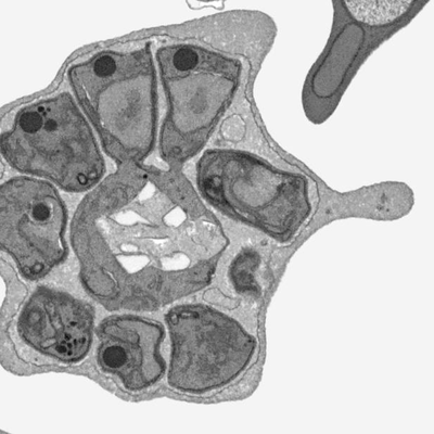
File S3: Volume EM of Plasmodium falciparum 3D7 early schizont A. Aligned, denoised, binned, and cropped serial slices of an early schizont from the [-]E64 46-48hpi block. Voxels are 10nm x 10nm x 20nm. Additional ultrastructural features are present in the raw data that were not rendered (e.g. PVM).
File S4: Volume EM of Plasmodium falciparum 3D7 early schizont B. Aligned, denoised, binned, and cropped serial slices of an early schizont from the [-]E64 46-48hpi block. Voxels are 10nm x 10nm x 20nm. Additional ultrastructural features are present in the raw data that were not rendered (e.g. PVM).
File S5: Volume EM of Plasmodium falciparum 3D7 mid-segmentation schizont 1. Aligned, denoised, binned, and cropped serial slices of a mid-segmentation schizont from the [+]E64-treated 48-50hpi block. Voxels are 10nm x 10nm x 20nm. Additional ultrastructural features are present in the raw data that were not rendered (e.g. PVM).
File S6: Volume EM of Plasmodium falciparum 3D7 mid-segmentation schizont 2. Aligned, denoised, binned, and cropped serial slices of a mid-segmentation schizont from the [+]E64-treated 48-50hpi block. Voxels are 10nm x 10nm x 20nm. Additional ultrastructural features are present in the raw data that were not rendered (e.g. PVM).
File S7: Volume EM of Plasmodium falciparum 3D7 mid-segmentation schizont A. Aligned, denoised, binned, and cropped serial slices of a mid-segmentation schizont from the [-]E64 46-48hpi block. Voxels are 10nm x 10nm x 20nm. Additional ultrastructural features are present in the raw data that were not rendered (e.g. PVM).
File S8: Volume EM of Plasmodium falciparum 3D7 mid-segmentation schizont B. Aligned, denoised, binned, and cropped serial slices of a mid-segmentation schizont from the [-]E64 46-48hpi block. Voxels 10nm x 10nm x 20nm. Additional ultrastructural features are present in the raw data that were not rendered (e.g. PVM).
File S9: Volume EM of Plasmodium falciparum 3D7 schizont at PVM rupture. Aligned, denoised, binned, and cropped serial slices of a schizont at the PVM rupture stage from the [+]E64 48-50hpi block. Voxels are 10nm x 10nm x 20nm. Additional ultrastructural features are present in the raw data that were not rendered (e.g. PVM).
File S10: Volume EM of Plasmodium falciparum 3D7 post-PVM rupture schizont. Aligned, denoised, binned, and cropped serial slices of a post-PVM rupture schizont from the [+]E64-treated block. Voxels are 10nm x 10nm x 20nm. Additional ultrastructural features are present in the raw data that were not rendered (e.g. cellular debris).
File S11: Volume EM of Plasmodium falciparum 3D7 post-PVM rupture schizont. Aligned, denoised, binned, and cropped serial slices of a post-PVM rupture schizont from the [-]E64-treated block. Voxels are 10nm x 10nm x 20nm. The image quality of this cell is relatively lower; thus, it was not rendered.