Electron Microscopy Public Image Archive
Electron Microscopy Public Image Archive
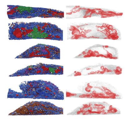
Quantitative subcellular reconstruction reveals a lipid mediated inter-organelle biogenesis network
Lee RG, Rudler DL, Raven SA, Peng L, Chopin A, Moh ESX, McCubbin T, Siira SJ, Fagan SV, DeBono NJ, Stentenbach M, Browne J, Rackham FF, Li J, Simpson KJ, Marcellin E, Packer NH, Reid GE, Padman BS, Rackham O, Filipovska A
Nature cell Biology (---)