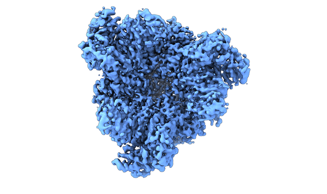
EMPIAR-11671
Structure of the peroxisomal Pex1/Pex6 ATPase complex bound to a substrate [multiple data sets in TIFF format]
Publication:

Structure of the peroxisomal Pex1/Pex6 ATPase complex bound to a substrate

Rüttermann M, Koci M, Lill P, Geladas ED, Kaschani F, Klink BU, Erdmann R, Gatsogiannis C

Nature communications 14 (2023)

PMID: 37741838

Related EMDB entry:
Deposited:
2023-07-13
Released:
2023-10-13
Last modified:
2023-10-13
Imageset size:
4.90 TB
Imageset DOI:
Experimental metadata:
Download xml json
Contains:
  • micrographs - multiframe
1. Unaligned multi-frame micrographs in super resolution of the peroxisomal AAA ATPase complex Pex1/Pex6 from Saccharomyces cerevisiae in 'single-seam' state (class3)
Category:
micrographs - multiframe
Image format:
TIFF
No. of images or tilt series:
13949
Image size:
(11520, 8184)
Pixel type:
UNSIGNED BYTE
Pixel spacing:
(0.34, 0.34)
Details:
Each raw movie is composed of 60 raw frames (prior alignment, accumulating 60 electron per squared Angstrom in a 1.0 second exposure). Final particle positions are in the respective .box files. Final 3D refinement star file is deposited in the particles_class3 directory.
Files:
Loading...
2. Unaligned multi-frame micrographs in super resolution of the peroxisomal AAA ATPase complex Pex1/Pex6 from Saccharomyces cerevisiae in 'twin-seam' state (class4)
Category:
micrographs - multiframe
Image format:
TIFF
No. of images or tilt series:
13949
Image size:
(11520, 8184)
Pixel type:
UNSIGNED BYTE
Pixel spacing:
(0.34, 0.34)
Details:
Each raw movie is composed of 60 raw frames (prior alignment, accumulating 60 electron per squared Angstrom in a 1.0 second exposure). Final particle positions are in the respective .box files. Final 3D refinement star file is deposited in the particles_class4 directory.
Files:
Loading...