EMPIAR-10420
Single-Particle Cryo-EM of Arabinosyltransferase EmbB from Mycobacterium smegmatis, Collected using both Falcon III and K2 Detectors [multiple data sets in MRCS, TIFF and MRC formats]
Publication:

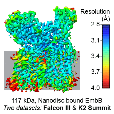
Cryo-EM structure of arabinosyltransferase EmbB from Mycobacterium smegmatis

Tan YZ, Rodrigues J, Keener JE, Zheng RB, Brunton R, Kloss B, Giacometti SI, Rosário AL, Zhang L, Niederweis M, Clarke OB, Lowary TL, Marty MT, Archer M, Potter CS, Carragher B, Mancia F, Tan YZ, Rodrigues J, Keener JE, Zheng RB, Brunton R, Kloss B, Giacometti SI, Rosário AL, Zhang L, Niederweis M, Clarke OB, Lowary TL, Marty MT, Archer M, Potter CS, Carragher B, Mancia F

Nature communications 11 (2020) 3396-3396

PMID: 32636380

Related EMDB entry:
Deposited:
2020-05-14
Released:
2020-07-30
Last modified:
2020-07-30
Imageset size:
4.51 TB
Imageset DOI:
Experimental metadata:
Download xml json
Contains:
  • micrographs - multiframe
  • micrographs - single frame
  • picked particles - single frame - processed
1. Unaligned and compressed multiframe movies from Falcon III Detector
Category:
micrographs - multiframe
Image format:
MRCS
No. of images or tilt series:
2159
Image size:
(4096, 4096)
Pixel type:
SIGNED 16 BIT INTEGER
Pixel spacing:
(0.665, 0.655)
Details:
Images were recorded in two sessions. This first session was on a Titan Krios electron microscope (FEI) equipped with a Falcon III direct detector operating at 0.665 Å per pixel in electron counting mode using the Leginon software package. Pixel size was calibrated after obtaining a preliminary map by docking with a crystal structure of a homolog of the soluble part of EmbB (PDB ID: 3PTY). Data collection was performed using a dose of ~78.02 e-/Å2 across 80 frames (1080 msec per frame) at a dose rate of ~0.40 e–/pix/sec, using a set defocus range of -0.5 μm to -2.5 μm. 100 micron objective aperture was used. The movies were recorded over three days using an image beam shift data collection strategy.

These files have been CTF corrected using the parameters in: data/02_Aligned_Micrographs/micrographs_ctf_18nov21d_FalconIII.star
Files:
Loading...
2. Unaligned and compressed multiframe movies from K2 Summit Detector
Category:
micrographs - multiframe
Image format:
TIFF
No. of images or tilt series:
7943
Image size:
(3710, 3838)
Pixel type:
UNSIGNED 16 BIT INTEGER
Pixel spacing:
(0.667089, 0.667089)
Details:
Images were recorded in two sessions. This second session was on a Titan Krios electron microscope (FEI) equipped with a K2 summit direct detector operating at 0.667 Å per pixel in counting mode using the Leginon software package. Pixel size was calibrated in-house using a proteasome test sample. Data collection was performed using a dose of ~77.53 e-/Å2 across 80 frames (100 msec per frame) at a dose rate of ~4.3 e–/pix/sec, using a set defocus range of -0.3 μm to -2.9 μm. 100 micron objective aperture was used. The movies were recorded over three days using an image beam shift data collection strategy.

These files have been CTF corrected using the parameters in: data/02_Aligned_Micrographs/micrographs_ctf_19may24c_K2.star
Files:
Loading...
3. Aligned and dose-weighted micrographs from Falcon III Detector
Category:
micrographs - single frame
Image format:
MRC
No. of images or tilt series:
1955
Image size:
(4096, 4096)
Pixel type:
32 BIT FLOAT
Pixel spacing:
(0.665, 0.665)
Details:
For this first Falcon III dataset, movie frames were aligned using MotionCor2 with 5 by 5 patches and B-factor of 500 for global alignment and 100 for local alignment through the Relion package.
Files:
Loading...
4. Aligned and dose-weighted micrographs from K2 Summit Detector
Category:
micrographs - single frame
Image format:
MRC
No. of images or tilt series:
6025
Image size:
(3710, 3838)
Pixel type:
32 BIT FLOAT
Pixel spacing:
(0.667089, 0.667089)
Details:
For the second K2 summit dataset, movie frames were aligned using MotionCor2 with 5 by 5 patches and B-factor of 500 for global alignment and 100 for local alignment through the Relion package.
Files:
Loading...
5. Final Particle Stack with Refined Euler Angles and Shifts
Category:
picked particles - single frame - processed
Image format:
MRCS
No. of images or tilt series:
1
Image size:
(256, 256)
Pixel type:
32 BIT FLOAT
Pixel spacing:
(1.0, 1.0)
Details:
Combined particle stack from both datasets.
Files:
Loading...
Files:
Loading...

Ito F, Alvarez-Cabrera AL, Liu S, Yang H, Shiriaeva A, Zhou ZH, Chen XS. (2023)
Rigden DJ, Fernández XM. (2023)
Iudin A, Korir PK, Somasundharam S, Weyand S, Cattavitello C, Fonseca N, Salih O, Kleywegt GJ, Patwardhan A. (2023)
Serra Lleti JM, Steyer AM, Schieber NL, Neumann B, Tischer C, Hilsenstein V, Holtstrom M, Unrau D, Kirmse R, Lucocq JM, Pepperkok R, Schwab Y. (2023)
Caldwell BJ, Norris AS, Karbowski CF, Wiegand AM, Wysocki VH, Bell CE. (2022)