Micrographs were recorded as movies on a Titan Krios at 300 keV equipped with a K3 direct detector and an energy filter, with an exposure rate of 1 e-/Å2 and a total exposure of 50 e-/Å2.
Files:
Available download options:
Archives and downloads the selected files into an uncompressed zip file.
Depending on the number and size of files to be downloaded, this can take an enormous amount of time.
Also, this feature is unstable and may not work properly, and checksums of downloaded files are not verified.
We recommend using rsync, aspera, globus, etc.
Download a list of selected files.
It is possible to download files by specifying the file list with rsync command, etc.
Maximum intensity projections (MIPs) of the observed SNR values, scaled by subtracting the average SNR, and dividing by the standard deviation of the SNR per pixel. The MIPs display peaks in places where large ribosomal subunits were detected by 2D template matching. Peaks are associated with specific x,y locations, defocus and orientations of the targets that correspond with the matching template.
Files:
Available download options:
Archives and downloads the selected files into an uncompressed zip file.
Depending on the number and size of files to be downloaded, this can take an enormous amount of time.
Also, this feature is unstable and may not work properly, and checksums of downloaded files are not verified.
We recommend using rsync, aspera, globus, etc.
Download a list of selected files.
It is possible to download files by specifying the file list with rsync command, etc.
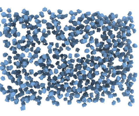
3D density maps used as templates in 2D template matching to detect large ribosomal subunits in the 2D images of lamellae of S. cerevisiae cells. The density maps were generated with the cryo-EM simulator implemented in cisTEM.
Files:
Available download options:
Archives and downloads the selected files into an uncompressed zip file.
Depending on the number and size of files to be downloaded, this can take an enormous amount of time.
Also, this feature is unstable and may not work properly, and checksums of downloaded files are not verified.
We recommend using rsync, aspera, globus, etc.
Download a list of selected files.
It is possible to download files by specifying the file list with rsync command, etc.