Archives and downloads the selected files into an uncompressed zip file.
Depending on the number and size of files to be downloaded, this can take an enormous amount of time.
Also, this feature is unstable and may not work properly, and checksums of downloaded files are not verified.
We recommend using rsync, aspera, globus, etc.
Download a list of selected files.
It is possible to download files by specifying the file list with rsync command, etc.
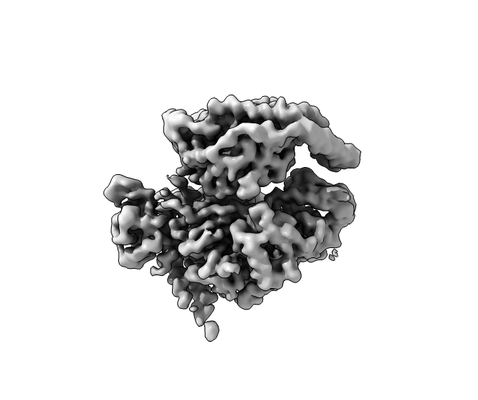
Particle coordinate files (*.star) for P-Rex1-Gbg can be found in the directory data/matching
Files:
Available download options:
Archives and downloads the selected files into an uncompressed zip file.
Depending on the number and size of files to be downloaded, this can take an enormous amount of time.
Also, this feature is unstable and may not work properly, and checksums of downloaded files are not verified.
We recommend using rsync, aspera, globus, etc.
Download a list of selected files.
It is possible to download files by specifying the file list with rsync command, etc.