
EMPIAR-11216
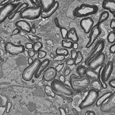
Focused ion beam-scanning electron microscopy links pathological myelin outfoldings to axonal changes in mice lacking Plp1 or Mag [1447 micrographs in TIFF format]
Publication:

Focused ion beam-scanning electron microscopy links pathological myelin outfoldings to axonal changes in mice lacking Plp1 or Mag

Steyer AM, Buscham TJ, Lorenz C, Hümmert S, Eichel-Vogel MA, Schadt LC, Edgar JM, Köster S, Möbius W, Nave KA, Werner HB

Glia (2022)
PMID: 36354016

Related EMDB entry:
Deposited:
2022-09-15
Released:
2022-12-13
Last modified:
2022-12-13
Imageset size:
34.56 GB
Imageset DOI:
Experimental metadata:
Download xml json
Contains:
  • micrographs - single frame
1. FIB-SEM of mouse optic nerve of Plp knock-out (Plp-/Y-4007)
Category:
micrographs - single frame
Image format:
TIFF
No. of images or tilt series:
1447
Image size:
(6624, 3872)
Pixel type:
UNSIGNED BYTE
Pixel spacing:
(50.0, 50.0)
Details:
Optic nerve of mice harboring null-alleles of the Plp/Plp1 (Plp-/Y) sarificed by cervical dislocation at postnatal day 75 (P75). The optic nerves were dissected and immersion fixed and prepared using the reduced osmium tetroxide – thiocarbohydrazide - osmium (OTO) method as previously described in detail (Weil et al. 2018; Steyer et al. 2020) upon implementing modifications to the original protocol first introduced by (Deerinck et al. 2010). The micrographs are obtained by FIB-SEM (Zeiss Crossbeam 540): SEM: 1.5 kV (700 pA), ESB grid 450 V, pixel size x/y 5 nm, continuous mill-and-acquire mode; FIB: 3 nA , z-step 25 nm. Postprocessing: alignment with Fiji/ImageJ SIFT, cropped, inverted and blurred (Gaussian blur, sigma 1) and local contrast enhancement (127, 127, 1.5) applied.
Files:
Loading...