Raw image frames were not gain or defect corrected. 10 frames were acquired per tilt. The gain reference is provided as gainref.dm4. The defects file is provided as defects.txt. The dose per tilt was 1.85 e/Å^2. SerialEM Mdoc files are provided in a separate folder (Mdoc_files) and can be used to generate stacks from the raw image frames. The pixel spacing of the raw image frames is 2.75 Å/pixel. The pixel spacing in the header of the raw image frames and in the Mdoc files (for stacks binned by 4) is inaccurate.
Files:
Available download options:
Archives and downloads the selected files into an uncompressed zip file.
Depending on the number and size of files to be downloaded, this can take an enormous amount of time.
Also, this feature is unstable and may not work properly, and checksums of downloaded files are not verified.
We recommend using rsync, aspera, globus, etc.
Download a list of selected files.
It is possible to download files by specifying the file list with rsync command, etc.
Corrected, aligned, dose-filtered and order-sorted tilt series. The tilt series were generated from the raw frame images by correction for gain, defects and motion. In addition, tilt images were dose-filtered. Gain-correction and defects files are provided in the Raw_frames folder. Motion-correction was performed with alignframes (IMOD) operated in subTOM.
Files:
Available download options:
Archives and downloads the selected files into an uncompressed zip file.
Depending on the number and size of files to be downloaded, this can take an enormous amount of time.
Also, this feature is unstable and may not work properly, and checksums of downloaded files are not verified.
We recommend using rsync, aspera, globus, etc.
Download a list of selected files.
It is possible to download files by specifying the file list with rsync command, etc.
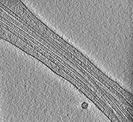
Reconstructed bin4 tomograms. Volumes were filtered using the Wiener-like deconvolution filter implemented in WARP (Tegunov 2018, https://github.com/dtegunov/tom_deconv). Tomograms were reconstructed in IMOD by weighted back projection.
Files:
Available download options:
Archives and downloads the selected files into an uncompressed zip file.
Depending on the number and size of files to be downloaded, this can take an enormous amount of time.
Also, this feature is unstable and may not work properly, and checksums of downloaded files are not verified.
We recommend using rsync, aspera, globus, etc.
Download a list of selected files.
It is possible to download files by specifying the file list with rsync command, etc.