EMPIAR-10006
2D crystal images of the potassium channel MloK1 with and without cAMP ligand [multiple data sets in TIFF format]
Publication:

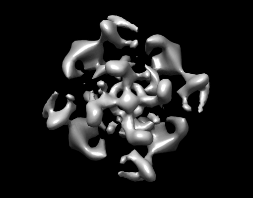
Ligand-induced structural changes in the cyclic nucleotide-modulated potassium channel MloK1

Kowal J, Chami M, Baumgartner P, Arheit M, Chiu P-L, Rangl M, Scheuring S, Schroeder GF, Nimigean CM, Stahlberg H

Nat Commun 5 (2014) 3106

PMID: 24469021

Related EMDB entry:
Deposited:
2013-12-05
Released:
2014-01-03
Last modified:
2020-05-21
Imageset size:
11.64 GB
Imageset DOI:
Experimental metadata:
Download xml json
Version history:
1
2015-10-27
REL
Restructured the entry directory to have "data" and ".private".
Contains:
  • micrographs - single frame
1. Potassium channel MloK1 with cAMP ligand
Category:
micrographs - single frame
Image format:
TIFF
No. of images or tilt series:
78
Image size:
(5600, 5600)
Pixel type:
UNSIGNED 16 BIT INTEGER
Pixel spacing:
(1.0, 1.0)
Details:
Some micrographs have pixel width: 0.83A. In a recent work, Julia Kowal and others from our lab studied the MloK1 bacterial potassium channel, which has putative voltage sensors and cAMP ligand binding sites, in the presence and absence of the ligand, by electron crystallography.

For the two configurations, we recorded a total of 78 respectively 67 images of 2D crystals tilted up to 45 degrees on photographic film and digitized them with a film scanner. Image processing in 2dx of the real-space images led to 3D maps of the protein in the two configurations at 7A resolution in plane, and 12A vertical resolution. We have deposited the used 145 raw micrographs (TIFF format) with the corresponding 2dx processing file structure, including 2dx_image.cfg parameter files, merged APH files, phase origins, tilt angles, lattice line data, and final volumes, in short the entire 2dx image processing project, in one 12 GB ZIP file. This file contains an entire 2dx processing file structure.

To open, extract the file structure onto your harddrive.

Then launch 2dx_merge, direct it to the base of either the cAMP-free or cAMP-bound dataset.

In 2dx_merge, run the script "repair project links" to re-create the symbolic links for your local directory structure.

Now you should be able to directly "merge" the included APH (Amplitude and Phase) files into a 2D or 3D structure, or you can open individual entries by double-clicking on the corresponding image line in 2dx_merge, so that you can re-process or insepct them with 2dx_image. If questions, please contact Henning.Stahlberg@unibas.ch. 2dx is available under the GPL at http://2dx.org
Files:
Loading...
2. Potassium channel MloK1 without cAMP ligand
Category:
micrographs - single frame
Image format:
TIFF
No. of images or tilt series:
67
Image size:
(5600, 5600)
Pixel type:
UNSIGNED 16 BIT INTEGER
Pixel spacing:
(1.0, 1.0)
Details:
Some micrographs have pixel width: 0.83A. In a recent work, Julia Kowal and others from our lab studied the MloK1 bacterial potassium channel, which has putative voltage sensors and cAMP ligand binding sites, in the presence and absence of the ligand, by electron crystallography.

For the two configurations, we recorded a total of 78 respectively 67 images of 2D crystals tilted up to 45 degrees on photographic film and digitized them with a film scanner. Image processing in 2dx of the real-space images led to 3D maps of the protein in the two configurations at 7A resolution in plane, and 12A vertical resolution. We have deposited the used 145 raw micrographs (TIFF format) with the corresponding 2dx processing file structure, including 2dx_image.cfg parameter files, merged APH files, phase origins, tilt angles, lattice line data, and final volumes, in short the entire 2dx image processing project, in one 12 GB ZIP file. This file contains an entire 2dx processing file structure.

To open, extract the file structure onto your harddrive.

Then launch 2dx_merge, direct it to the base of either the cAMP-free or cAMP-bound dataset.

In 2dx_merge, run the script "repair project links" to re-create the symbolic links for your local directory structure.

Now you should be able to directly "merge" the included APH (Amplitude and Phase) files into a 2D or 3D structure, or you can open individual entries by double-clicking on the corresponding image line in 2dx_merge, so that you can re-process or insepct them with 2dx_image. If questions, please contact Henning.Stahlberg@unibas.ch. 2dx is available under the GPL at http://2dx.org
Files:
Loading...
Files:
Loading...

Ito F, Alvarez-Cabrera AL, Liu S, Yang H, Shiriaeva A, Zhou ZH, Chen XS. (2023)
Rigden DJ, Fernández XM. (2023)
Iudin A, Korir PK, Somasundharam S, Weyand S, Cattavitello C, Fonseca N, Salih O, Kleywegt GJ, Patwardhan A. (2023)
Serra Lleti JM, Steyer AM, Schieber NL, Neumann B, Tischer C, Hilsenstein V, Holtstrom M, Unrau D, Kirmse R, Lucocq JM, Pepperkok R, Schwab Y. (2023)
Caldwell BJ, Norris AS, Karbowski CF, Wiegand AM, Wysocki VH, Bell CE. (2022)