EMPIAR-10459
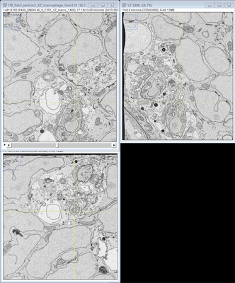
FIB SEM images of a Zebrafish hindbrain macrophage containing 2 Toxoplasma gondii tachizoites [multiple data sets in TIFF format]
Publication:

The zebrafish as a novel model for the in vivo study of Toxoplasma gondii replication and interaction with macrophages

Yoshida N, Domart MC, Peddie CJ, Yakimovich A, Mazon-Moya MJ, Hawkins TA, Collinson L, Mercer J, Frickel EM, Mostowy S

Disease models & mechanisms 13 (2020)

PMID: 32461265

Related EMDB entry:
Deposited:
2020-07-01
Released:
2020-10-14
Last modified:
2020-10-14
Imageset size:
1.11 TB
Imageset DOI:
Experimental metadata:
Download xml json
Contains:
  • micrographs - multiframe
  • micrographs - single frame
1. Aligned FIB image stack of a macrophage containing 2 Toxoplasma Gondii tachizoites
Category:
micrographs - multiframe
Image format:
TIFF
No. of images or tilt series:
3200
Image size:
(3437, 3800)
Pixel type:
UNSIGNED BYTE
Pixel spacing:
(50.0, 50.0)
Details:
Images were acquired at 5 nm isotropic resolution throughout the region of interest, using a 4.2 us dwell time. During acquisition the SEM was operated at an accelerating voltage of 1.5 kV with 1.5 nA current.
Files:
Loading...
2. Raw FIB image stack of a macrophage containing 2 Toxoplasma Gondii tachizoites - part 1
Category:
micrographs - single frame
Image format:
TIFF
No. of images or tilt series:
2062
Image size:
(12032, 12032)
Pixel type:
UNSIGNED 16 BIT INTEGER
Pixel spacing:
(50.0, 50.0)
Details:
First half of the raw data. See part 2 for second half. Images were acquired at 5 nm isotropic resolution throughout the region of interest, using a 4.2 us dwell time. During acquisition the SEM was operated at an accelerating voltage of 1.5 kV with 1.5 nA current.
Files:
Loading...
3. Raw FIB image stack of a macrophage containing 2 Toxoplasma Gondii tachizoites - part 2
Category:
micrographs - single frame
Image format:
TIFF
No. of images or tilt series:
1963
Image size:
(12032, 12032)
Pixel type:
UNSIGNED 16 BIT INTEGER
Pixel spacing:
(50.0, 50.0)
Details:
Second half of the raw data. See part 1 for first half. Images were acquired at 5 nm isotropic resolution throughout the region of interest, using a 4.2 us dwell time. During acquisition the SEM was operated at an accelerating voltage of 1.5 kV with 1.5 nA current.
Files:
Loading...