EMPIAR-10413
Soft X-ray Tomography of mock-infected U2OS cells [1 tilt series in MRC format]
Publication:

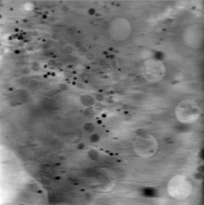
3D Correlative Cryo-Structured Illumination Fluorescence and Soft X-ray Microscopy Elucidates Reovirus Intracellular Release Pathway

Kounatidis I, Stanifer ML, Phillips MA, Paul-Gilloteaux P, Heiligenstein X, Wang H, Okolo CA, Fish TM, Spink MC, Stuart DI, Davis I, Boulant S, Grimes JM, Dobbie IM, Harkiolaki M

Cell 182 (2020) 515-530.e17

PMID: 32610083

Related EMDB entry:
Deposited:
2020-05-11
Released:
2020-06-30
Last modified:
2022-02-08
Imageset size:
2.51 GB
Imageset DOI:
Experimental metadata:
Download xml json
Contains:
  • tilt series
1. Tomogram of reovirus mock-infected U2OS cells (Area 2)
Category:
tilt series
Image format:
MRC
No. of images or tilt series:
1
Image size:
(946, 946)
Pixel type:
32 BIT FLOAT
Pixel spacing:
(160.0, 160.0)
Details:
Tomogram of reovirus mock-infected U2OS cells (Area2)
EMPIAR entry 10413: U2OS_Reo_MockInfected_Area2_Reconstruction (S-BIAD17)


Note:
Associated with EMPIAR entry 10412 (adjacent/overlapping FOVs)

Sample characteristics:
Substrate = Quantifoil F1 2/2
Fiducials = Au 250nm nanospheres

Data collection:
Data collected at beamline B24 at Diamond Light source
Data collected 18th Feb 2019
Allocation award ID MX21046

Tilt series:
-65deg to 65deg
0.5deg step
Total frames 261
0.5sec exposure

Microscope make:
Zeiss UltraXRM-S220C

Data collection software:
XRM Controller

Microscope setup:
Energy: 500eV
Magnification = x812.5
FOV = 16microns
pixel size = 16x16nm
Zoneplate = 40nm

Data processing:
Type = Automated
Software = Batchruntomo
Alignment protocol = SIRT
Files:
Loading...