EMPIAR-10406
Motion-corrected micrographs and extracted particle images of the 70S ribosome from the human pathogen Acinetobacter baumannii in complex with amikacin [multiple data sets in MRC and MRCS formats]
Publication:

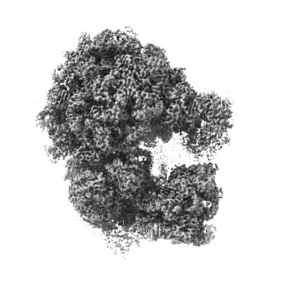
Structure of the 70S Ribosome from the Human Pathogen Acinetobacter baumannii in Complex with Clinically Relevant Antibiotics

Nicholson D, Edwards TA, O'Neill AJ, Ranson NA

Structure (London, England : 1993) 28 (2020) 1087-1100.e3

PMID: 32857965

Related EMDB entry:
Deposited:
2020-05-05
Released:
2020-11-24
Last modified:
2022-02-08
Imageset size:
177.68 GB
Imageset DOI:
Experimental metadata:
Download xml json
Contains:
  • micrographs - single frame
  • picked particles - multiframe - processed
1. Motion-corrected micrographs of the 70S ribosome from the human pathogen Acinetobacter baumannii in complex with amikacin
Category:
micrographs - single frame
Image format:
MRC
No. of images or tilt series:
2717
Image size:
(3838, 3710)
Pixel type:
32 BIT FLOAT
Pixel spacing:
(1.07, 1.07)
Details:
Drift-corrected and dose-corrected averages of movies collected at an accumulated dose of 58 e-/A^2 across 50 frames over a 10 s exposure, recorded by a Gatan K2 Summit detector. Motion correction was performed using MotionCor2 within RELION 3.0, by using all frames, applying a B-factor of 150, and using 5 patches in both the X and Y directions. Coordinates corresponding to extracted particle images are in the particles.star file.
Files:
Loading...
2. Extracted particle images of the 70S ribosome from the human pathogen Acinetobacter baumannii in complex with amikacin
Category:
picked particles - multiframe - processed
Image format:
MRCS
No. of images or tilt series:
554
Image size:
(416, 416)
Pixel type:
32 BIT FLOAT
Pixel spacing:
(1.07, 1.07)
Details:
Particles were extracted from motion-corrected, CTF-estimated micrographs into 416 square pixel boxes with re-centring of refined coordinates. Images of particles extracted from each micrograph are stacked into separate mrcs files. Coordinates and further metadata corresponding to extracted particle images are in the particle.star file.
Files:
Loading...
Files:
Loading...

Ito F, Alvarez-Cabrera AL, Liu S, Yang H, Shiriaeva A, Zhou ZH, Chen XS. (2023)
Rigden DJ, Fernández XM. (2023)
Iudin A, Korir PK, Somasundharam S, Weyand S, Cattavitello C, Fonseca N, Salih O, Kleywegt GJ, Patwardhan A. (2023)
Serra Lleti JM, Steyer AM, Schieber NL, Neumann B, Tischer C, Hilsenstein V, Holtstrom M, Unrau D, Kirmse R, Lucocq JM, Pepperkok R, Schwab Y. (2023)
Caldwell BJ, Norris AS, Karbowski CF, Wiegand AM, Wysocki VH, Bell CE. (2022)