Electron Microscopy Public Image Archive
Electron Microscopy Public Image Archive
The EMPIAR-PDBj team at Osaka University assists Asian EM researchers with the transfer of big EM image data to EMPIAR. Instead of sending the data directly to the EBI (UK) via the internet, hard drives can also be sent to Osaka University by postal mail or via a courier service. As an alternative, internet transfer to our server in Osaka is also available. If you would like to take advantage of our submission services, please contact us first by e-mail before sending the data to us.
| Release date | Imageset | Title | Authors and references | Size | Resolution |
|---|---|---|---|---|---|
| 2014-01-03 |  |
2D crystal images of the potassium channel MloK1 with and without cAMP ligand [multiple data sets in TIFF format] | Kowal J, Chami M, Baumgartner P, Arheit M, Chiu P-L, Rangl M, Scheuring S, Schroeder GF, Nimigean CM, Stahlberg H [Pubmed: 24469021] [DOI: 10.1038/ncomms4106] |
11.6 GB | 7.0 Å |
| 2019-05-08 |  |
Integrative structure and functional anatomy of a nuclear pore complex [multiple data sets in TIFF format] | Kim SJ, Fernandez-Martinez J, Nudelman I, Shi Y, Zhang W, Raveh B, Herricks T, Slaughter BD, Hogan JA, Upla P, Chemmama IE, Pellarin R, Echeverria I, Shivaraju M, Chaudhury AS, Wang J, Williams R, Unruh JR, Greenberg CH, Jacobs EY, Yu Z, de la Cruz MJ, Mironska R, Stokes DL, Aitchison JD, Jarrold MF, Gerton JL, Ludtke SJ, Akey CW, Chait BT, Sali A, Rout MP [Pubmed: 29539637] [DOI: 10.1038/nature26003] |
11.8 GB | 28.0 Å |
| 2019-10-04 |  |
Single particle cryo-EM dataset of clathrin cages with phase flipping suitable for refinement [stack of 12785 particles in MRCS format] | Morris KL, Jones JR, Halebian M, Wu S, Baker M, Armache JP, Avila Ibarra A, Sessions RB, Cameron AD, Cheng Y, Smith CJ [Pubmed: 31582853] [DOI: 10.1038/s41594-019-0292-0] |
11.9 GB | 9.07 - 23.68 Å |
| 2022-11-11 |  |
Cryo-electron tomography of microtubules assembled in Xenopus egg cytoplasmic extracts [6 tilt series in MRC format] | Guyomar C, Bousquet C, Ku S, Heumann J, Guilloux G, Gaillard N, Heichette C, Duchesne L, Steinmetz MO, Gibeaux R, Chrétien D [Pubmed: 36503602] [DOI: 10.7554/eLife.83021] |
12.1 GB | 43.3 Å |
| 2017-12-18 | 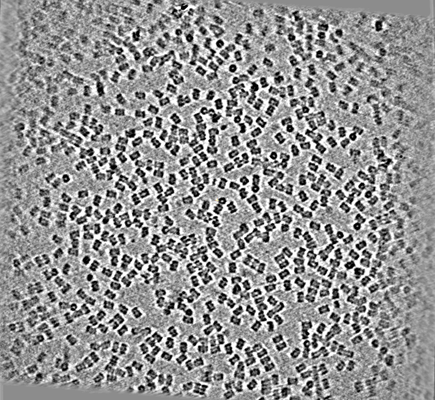 |
CryoET of T20S proteasome single particle [multiple data sets in MRC format] | Dandey VP, Wei H, Brasch J, Chase J, Acharya P, Tan YZ, Zhang Z, Kim LY, Scapin G, Rapp M, Eng ET, Rice MJ, Cheng A, Negro CJ, Shapiro L, Kwong PD, Jeruzalmi D, des Georges A, Potter CS, Carragher B [Pubmed: 29809143] [DOI: 10.7554/eLife.34257] |
12.4 GB | — |
| 2022-12-13 | 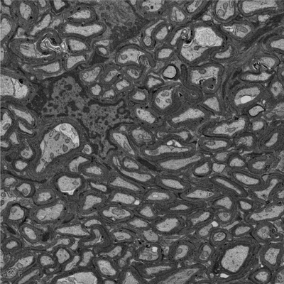 |
Focused ion beam-scanning electron microscopy links pathological myelin outfoldings to axonal changes in mice lacking Plp1 or Mag [940 micrographs in TIFF format] | Steyer AM, Möbius W [Pubmed: 36354016] [DOI: 10.1002/glia.24290] |
12.5 GB | — |
| 2019-11-18 |  |
cryo-ET of cryo-FIB milled yeast cell in which scs2/22 ist2 are deleted [multiple data sets in MRC format] | Hoffmann PC, Bharat TAM, Wozny MR, Boulanger J, Miller EA, Kukulski W [Pubmed: 31743663] [DOI: 10.1016/j.devcel.2019.09.019] |
12.8 GB | — |
| 2016-06-28 |  |
Designer nanoscale DNA assemblies programmed from the top down [177 micrographs in MRC format] | Veneziano R, Ratanalert S, Zhang K, Zhang F, Yan H, Chiu W, Bathe M [Pubmed: 27229143] [DOI: 10.1126/science.aaf4388] |
13.0 GB | 20.0 Å |
| 2021-09-01 | 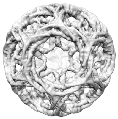 |
Multi-modal adaptor-clathrin contacts drive coated vesicle assembly [stack of 4782 particles in MRCS format] | Smith SM, Larocque G, Wood KM, Morris KL, Roseman AM, Sessions RB, Royle SJ, Smith CJ [Pubmed: 34487371] [DOI: 10.15252/embj.2021108795] |
13.0 GB | 9.1 - 15.0 Å |
| 2016-01-27 | 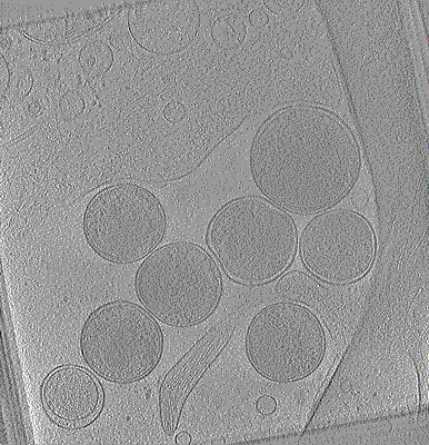 |
Cryo-electron tomogram of Chlamydia trachomatis with type III secretion system in contact with HeLa cell [1 class averages in MRC format] | Nans A, Kudryashev M, Saibil HR, Hayward RD [Pubmed: 26656452] [DOI: 10.1038/ncomms10114] |
13.6 GB | 38.0 Å |
| 2022-12-13 | 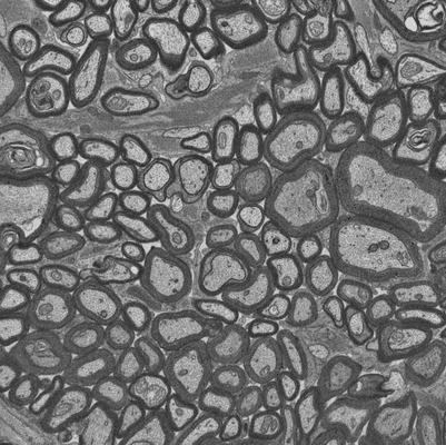 |
Focused ion beam-scanning electron microscopy links pathological myelin outfoldings to axonal changes in mice lacking Plp1 or Mag [910 micrographs in TIFF format] | Steyer AM, Möbius W [Pubmed: 36354016] [DOI: 10.1002/glia.24290] |
13.7 GB | — |
| 2018-04-27 | 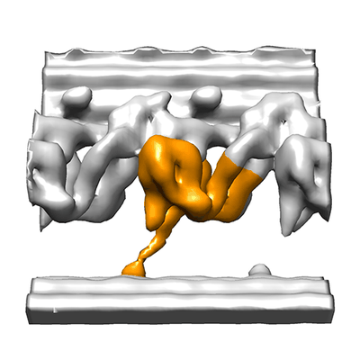 |
Cryo electron tomography of immotile sea urchin sperm flagella [27 tilt series in MRC format] | Lin J, Nicastro D [Pubmed: 29700238] [DOI: 10.1126/science.aar1968] |
13.8 GB | 33.0 - 41.0 Å |
| 2017-11-30 |  |
FIB-SEM of a dividing cell at 4.3 min after anaphase onset [1358 multi-frame micrographs composed of 1 frames each in TIFF format] | Otsuka S, Steyer AM, Schorb M, Hériché JK, Hossain MJ, Sethi S, Kueblbeck M, Schwab Y, Beck M, Ellenberg J [Pubmed: 29323269] [DOI: 10.1038/s41594-017-0001-9] |
14.0 GB | — |
| 2019-11-18 |  |
cryo-ET of cryo-FIB milled yeast cell in which scs2/22 ist2 are deleted with high intracellular calcium [121 tilt series in MRC format] | Hoffmann PC, Bharat TAM, Wozny MR, Boulanger J, Miller EA, Kukulski W [Pubmed: 31743663] [DOI: 10.1016/j.devcel.2019.09.019] |
14.3 GB | — |
| 2022-07-26 | 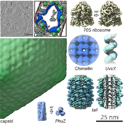 |
In situ cryo-electron tomography of P. chlororaphis infected by 201phi2-1 [269 multi-frame micrographs composed of 12 frames each in TIFF format] | Laughlin TG, Deep A, Prichard AM, Seitz C, Gu Y, Enustun E, Suslov S, Khanna K, Birkholz EA, Armbruster E, McCammon JA, Amaro RE, Pogliano J, Corbett KD, Villa E [Pubmed: 35922510] [DOI: 10.1038/s41586-022-05013-4] |
14.5 GB | 10.2 - 24.0 Å |
| 2023-07-18 |  |
Targeted volume Correlative Light and Electron Microscopy of environmental marine microorganisms [1937 micrographs in TIFF format] | Mocaer K, Mizzon G, Gunkel M, Halavatyi A, Steyer AM, Oorschot V, Schorb M, Le Kieffre C, Yee DP, Chevalier F, Gallet B, Decelle J, Schwab Y, Ronchi P [DOI: 10.1101/2023.01.27.525698] |
14.5 GB | — |
| 2021-11-01 |  |
Cryo Soft X-ray data for tetraspeck correlation [multiple data sets in MRC format] | Groen J, Pereiro E [Pubmed: 33990802] [DOI: 10.1038/s41596-021-00522-4] |
15.1 GB | — |
| 2020-12-11 |  |
Cryo electron tomography after FIB-milling of Planctomycetes species Tuwongella immobilis (33k magnification) [multiple data sets in MRC format] | Seeger C, Andersson SG [Pubmed: 32761170] [DOI: 10.1093/gbe/evaa159] |
15.5 GB | — |
| 2020-08-06 |  |
Cropped regions from Serial Block Face SEM of HeLa cell pellet with 10 nm pixels and 50 nm slices (benchmark dataset) [18 multi-frame micrographs composed of 300 frames each in TIFF format] | Peddie CP, Jones ML, Collinson LM | 15.6 GB | — |
| 2015-01-16 |  |
Tobacco Mosaic Virus K2 Summit dataset including manually boxed helix coordinates [14 multi-frame micrographs composed of 22 frames each in MRC format] | Fromm SA, Bharat TAM, Jakobi AJ, Hagen WJH, Sachse C [Pubmed: 25528571] [DOI: 10.1016/j.jsb.2014.12.002] |
15.7 GB | 4.0 Å |
| 2018-04-27 | 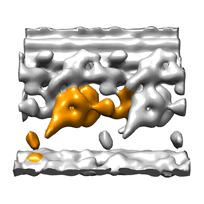 |
Cryo electron tomography of sea urchin sperm flagella [31 tilt series in MRC format] | Lin J, Nicastro D [Pubmed: 29700238] [DOI: 10.1126/science.aar1968] |
15.8 GB | 30.0 - 31.0 Å |
| 2022-01-12 | 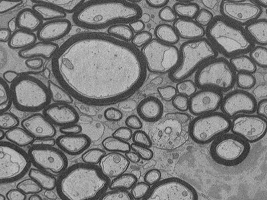 |
FIB-SEM of mouse optic nerve of an inducible conditional Mbp knock-out 16 weeks after induction [696 micrographs in TIFF format] | Meschkat M, Steyer AM, Ruhwedel T, Möbius W [Pubmed: 35246535] [DOI: 10.1038/s41467-022-28720-y] |
15.9 GB | — |
| 2020-02-18 | 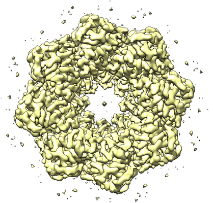 |
Structure of an undocked hemichannel of the N-terminal-deleted INX-6 in a nanodisc [300 micrographs in MRC format] | Burendei B, Shinozaki R, Watanabe M, Terada T, Tani K, Fujiyoshi Y, Oshima A [Pubmed: 32095518] [DOI: 10.1126/sciadv.aax3157] |
15.9 GB | 3.6 Å |
| 2023-06-02 | 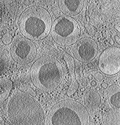 |
Cryo-ET tilt series from mouse islets lift-out sample [multiple data sets in TIFF format] | Wu Y, Qin C, Du W, Guo Z, Chen L, Guo Q [Pubmed: 37201639] [DOI: 10.1016/j.jsb.2023.107971] |
16.0 GB | — |
| 2021-01-22 | 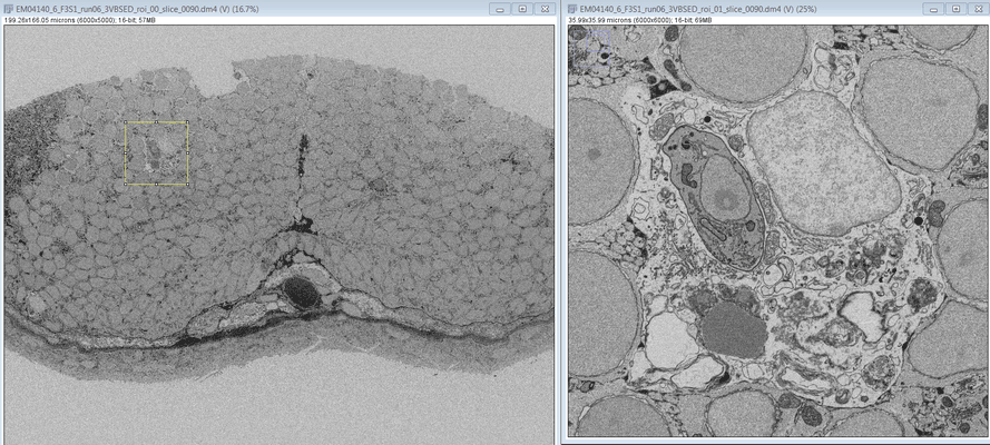 |
SBF SEM images of a Zebrafish hindbrain macrophage containing 2 Toxoplasma gondii tachizoites [multiple data sets in DM4 format] | Peddie CJ, Domart MC, Collinson L [Pubmed: 32461265] [DOI: 10.1242/dmm.043091] |
16.5 GB | — |
