Electron Microscopy Public Image Archive
Electron Microscopy Public Image Archive
The EMPIAR-PDBj team at Osaka University assists Asian EM researchers with the transfer of big EM image data to EMPIAR. Instead of sending the data directly to the EBI (UK) via the internet, hard drives can also be sent to Osaka University by postal mail or via a courier service. As an alternative, internet transfer to our server in Osaka is also available. If you would like to take advantage of our submission services, please contact us first by e-mail before sending the data to us.
| Release date | Imageset | Title | Authors and references | Size | Resolution |
|---|---|---|---|---|---|
| 2023-02-28 | 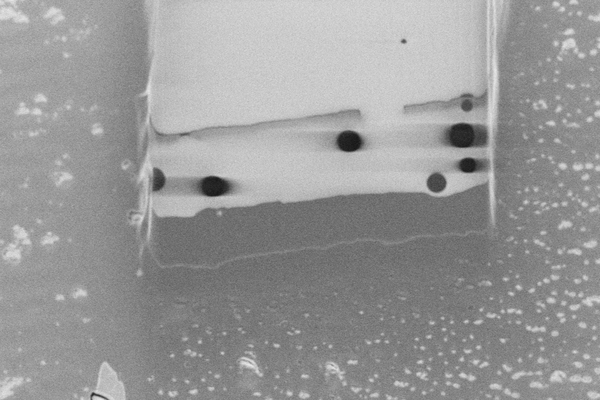 |
Cryo pFIB/SEM of PEG beads (test sample) [193 micrographs in TIFF format] | Dumoux M, Glen T, Smith JLR, Ho EML, Perdigão LMA, Pennington A, Klumpe S, Yee NBY, Farmer DA, Lai PYA, Bowles W, Kelley R, Plitzko JM, Wu L, Basham M, Clare DK, Siebert CA, Darrow MC, Naismith JH, Grange M [Pubmed: 36805107] [DOI: 10.7554/elife.83623] |
5.3 GB | — |
| 2023-01-16 | 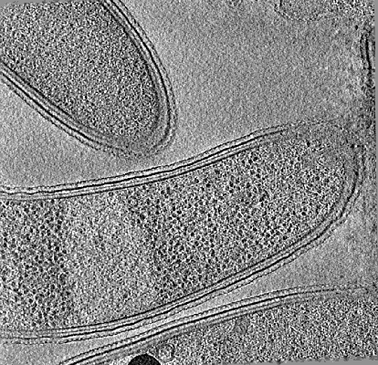 |
Cryo-electron tomography of FIB-milled Caulobacter crescentus expressing PopZ with IDR-156 [multiple data sets in MRC and TIFF formats] | Lasker K, Lam V, Villa E [Pubmed: 36163138] [DOI: 10.1038/s41467-022-33221-z] |
5.5 GB | — |
| 2018-03-09 | 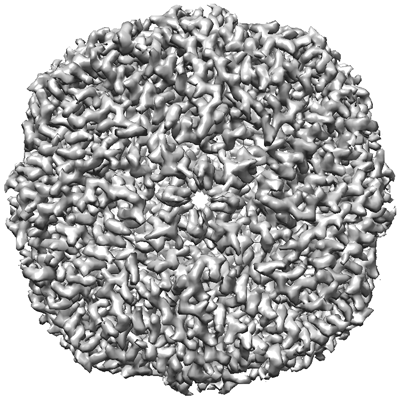 |
Apoferritin tutorial dataset for cisTEM [20 multi-frame micrographs composed of 50 frames each in MRC format] | Grant T, Rohou A, Grigorieff N [Pubmed: 29513216] [DOI: 10.7554/elife.35383] |
5.5 GB | — |
| 2022-05-31 | 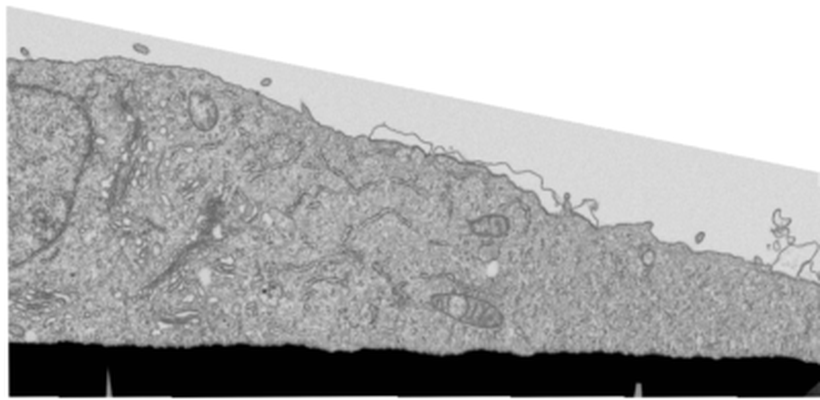 |
FIB-SEM of U2OS cell at leading edge of a wound assay, non-LPA stimulated, non-polarized [998 multi-frame micrographs composed of 1 frames each in TIFF format] | Costa J, Pinto A, Machado P | 5.7 GB | — |
| 2023-02-17 | 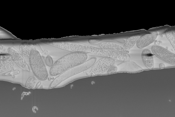 |
Cryo serial FIB/SEM of Rhodospirillum rubrum [43 micrographs in TIFF format] | Dumoux M, Glen T, Smith JLR, Ho EML, Perdigão LMA, Pennington A, Klumpe S, Yee NBY, Farmer DA, Lai PYA, Bowles W, Kelley R, Plitzko JM, Wu L, Basham M, Clare DK, Siebert CA, Darrow MC, Naismith JH, Grange M [Pubmed: 36805107] [DOI: 10.7554/elife.83623] |
5.7 GB | — |
| 2021-04-14 |  |
In situ cryo-electron tomogram of a pyrenoid inside a Chlamydomonas reinhardtii cell (tilt series) [1 tilt series in MRC format] | Cuellar LK, Schaffer M, Martinez-Sanchez A, Plitzko JM, Foerster F, Engel BD [Pubmed: 28938114] [DOI: 10.1016/j.cell.2017.08.008] |
5.8 GB | — |
| 2023-01-16 |  |
Cryo-electron tomography of FIB-milled Caulobacter crescentus expressing PopZ with IDR-156 and pentavalent OD [multiple data sets in MRC and TIFF formats] | Lasker K, Lam V, Villa E [Pubmed: 36163138] [DOI: 10.1038/s41467-022-33221-z] |
5.9 GB | — |
| 2021-09-10 |  |
Structural analysis of receptors and actin polarity in platelet protrusions [3 reconstructed volumes in EM format] | Sorrentino S, Conesa JJ, Cuervo A, Melero R, Martins B, Fernandez-Gimenez E, de Isidro-Gomez FP, de la Morena J, Studt JD, Sorzano COS, Eibauer M, Carazo JM, Medalia O [Pubmed: 34504018] [DOI: 10.1073/pnas.2105004118] |
6.0 GB | 26.6 Å |
| 2021-04-14 | 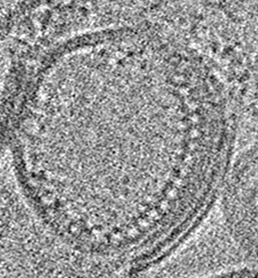 |
Cryo electron tomography of the reconstituted TRIM72-proteoliposomes on both positive and negative curvatures. [multiple data sets in MRC format] | Park SH, Song HK | 6.2 GB | — |
| 2018-07-05 |  |
Cryo-EM structure of alpha-synuclein fibrils [118 multi-frame micrographs composed of 50 frames each in MRC format] | Guerrero-Ferreira R, Taylor NM, Mona D, Ringler P, Lauer ME, Riek R, Britschgi M, Stahlberg H [Pubmed: 29969391] [DOI: 10.7554/elife.36402] |
6.3 GB | — |
| 2021-08-20 | 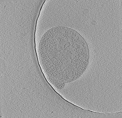 |
Cryo-electron tomography of JCVI-Syn3A (small cell) [multiple data sets in MRC and TIFF formats] | Lam V, Villa E, Thornburg ZR [Pubmed: 34368224] [DOI: 10.3389/fmolb.2021.644133] |
6.4 GB | — |
| 2021-11-30 |  |
Entropy Regularized Deconvolution of Cellular Cryo-Transmission Electron Tomograms [3 tilt series in MRC format] | Croxford MW, Elbaum ME, Arigovindan M, Kam Z, Agard DA, Villa E, Sedat J [Pubmed: 34876518] [DOI: 10.1073/pnas.2108738118] |
6.4 GB | — |
| 2022-01-12 | 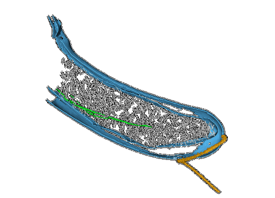 |
In vivo architecture of the polar organizing protein Z (PopZ) meshwork in the Alphaproteobacteria Magnetospirillum gryphiswaldense and Caulobacter crescentus [multiple data sets in MRC format] | Toro-Nahuelpan M, Plitzko JM, Schüler D, Pfeiffer D [Pubmed: 34971672] [DOI: 10.1016/j.jmb.2021.167423] |
6.4 GB | — |
| 2021-08-27 |  |
Cryo-electron tomography of JCVI-Syn3A (large cell) [multiple data sets in MRC and TIFF formats] | Lam V, Villa E, Thornburg ZR [Pubmed: 34368224] [DOI: 10.3389/fmolb.2021.644133] |
6.5 GB | — |
| 2018-01-24 |  |
Tilt-series of e. coli carrying the ple7 plasmid carrying YFP-MreB induced with 20 uM IPTG [7 tilt series in MRC format] | Swulius MT, Jensen GJ [Pubmed: 22904287] [DOI: 10.1128/JB.00505-12] |
6.6 GB | — |
| 2023-02-28 |  |
Cryo serial FIB/SEM of HeLa cells [46 micrographs in TIFF format] | Dumoux M, Glen T, Smith JLR, Ho EML, Perdigão LMA, Pennington A, Klumpe S, Yee NBY, Farmer DA, Lai PYA, Bowles W, Kelley R, Plitzko JM, Wu L, Basham M, Clare DK, Siebert CA, Darrow MC, Naismith JH, Grange M [Pubmed: 36805107] [DOI: 10.7554/elife.83623] |
6.7 GB | — |
| 2023-03-17 |  |
Serial section electron tomography of a Leishmania haptomonad on the stomodeal valve in the sand fly [670 multi-frame micrographs composed of 1 frames each in MRC format] | Yanase R, Sunter JD [DOI: 10.1101/2022.10.28.514187] |
6.7 GB | — |
| 2021-04-30 |  |
FIB-SEM of Tuwongella immobilis - a species of the Planctomycetes phylum [1 multi-frame micrographs composed of 464 frames each in TIFF format] | Andersson SGE, Odelgard A, Dyrhage K, Mahajan M, Seeger C [DOI: 10.3389/fmicb.2021.643045] |
6.7 GB | — |
| 2019-09-27 |  |
Cryo-EM structure of TMV in water [62 multi-frame micrographs composed of 20 frames each in TIFF format] | Weis F, Beckers M, von der Hocht I, Sachse C [Pubmed: 31535454] [DOI: 10.15252/embr.201948451] |
6.8 GB | 1.9 Å |
| 2021-02-12 |  |
Cryo Soft X-ray data for tetraspeck correlation [multiple data sets in MRC format] | Groen J, Pereiro E [Pubmed: 33990802] [DOI: 10.1038/s41596-021-00522-4] |
6.8 GB | — |
| 2015-10-09 |  |
GroEL dataset - NRAMM (06jul12a) [186 micrographs in MRC format] | Stagg SM, Lander GC, Pulokas J, Fellmann D, Cheng A, Quispe JD, Mallick SP, Avila RM, Carragher B, Potter CS [Pubmed: 16762565] [DOI: 10.1016/j.jsb.2006.04.005] |
7.0 GB | 7.8 Å |
| 2022-11-28 |  |
Cryo-electron tomography of FIB-milled Caulobacter crescentus expressing WT PopZ [multiple data sets in MRC and TIFF formats] | Lasker K, Lam V, Villa E [Pubmed: 36163138] [DOI: 10.1038/s41467-022-33221-z] |
7.1 GB | — |
| 2023-02-17 |  |
Cryo serial FIB/SEM of Vero cells [46 micrographs in TIFF format] | Dumoux M, Glen T, Smith JLR, Ho EML, Perdigão LMA, Pennington A, Klumpe S, Yee NBY, Farmer DA, Lai PYA, Bowles W, Kelley R, Plitzko JM, Wu L, Basham M, Clare DK, Siebert CA, Darrow MC, Naismith JH, Grange M [Pubmed: 36805107] [DOI: 10.7554/elife.83623] |
7.1 GB | — |
| 2016-06-28 | 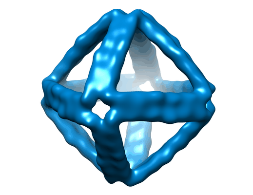 |
Designer nanoscale DNA assemblies programmed from the top down [100 micrographs in MRC format] | Veneziano R, Ratanalert S, Zhang K, Zhang F, Yan H, Chiu W, Bathe M [Pubmed: 27229143] [DOI: 10.1126/science.aaf4388] |
7.3 GB | 25.0 Å |
| 2018-08-09 |  |
Three-dimensional nanostructure of an intact microglia cell [multiple data sets in TIFF and IMOD formats] | Bolasco G, Weinhard L, Boissonnet T, Neujahr R, Gross CT [DOI: 10.3389/fnana.2018.00105] |
7.4 GB | — |
