Electron Microscopy Public Image Archive
Electron Microscopy Public Image Archive
The EMPIAR-PDBj team at Osaka University assists Asian EM researchers with the transfer of big EM image data to EMPIAR. Instead of sending the data directly to the EBI (UK) via the internet, hard drives can also be sent to Osaka University by postal mail or via a courier service. As an alternative, internet transfer to our server in Osaka is also available. If you would like to take advantage of our submission services, please contact us first by e-mail before sending the data to us.
| Release date | Imageset | Title | Authors and references | Size | Resolution |
|---|---|---|---|---|---|
| 2023-08-18 |  |
Unaligned cryo-EM micrographs of AL55 amyloid fibrils extracted from the kidney of an AL amyloidosis patient [1819 multi-frame micrographs composed of 40 frames each in MRC format] | Chaves-Sanjuan A, Puri S, Schulte T, Ricagno S [Pubmed: 37516426] [DOI: 10.1016/j.jmb.2023.168215] |
1.0 TB | 4.0 Å |
| 2016-11-11 |  |
Electron cryotomography of cryosectioned budding yeast cells [29 class averages in MRC format] | Chen C, Lim HH, Shi J, Tamura S, Maeshima M, Surana U, Gan L [Pubmed: 27605704] [DOI: 10.1091/mbc.E16-07-0506] |
125.3 GB | — |
| 2023-06-05 |  |
Cryo electron microscopy of Mce1 transporter from Mycobacterium smegmatis [multiple data sets in TIFF format] | Chen J, Bhabha G, Ekiert D [Pubmed: 37495693] [DOI: 10.1038/s41586-023-06366-0] |
20.9 TB | 2.71 - 3.19 Å |
| 2018-10-25 |  |
CryoET of bacterial RNA polymerase with several detergents [multiple data sets in MRC and RAW TEXT formats] | Chen J, Noble AJ, Kang JY, Darst SA [DOI: 10.1016/j.yjsbx.2019.100005] |
207.5 GB | — |
| 2022-11-11 | 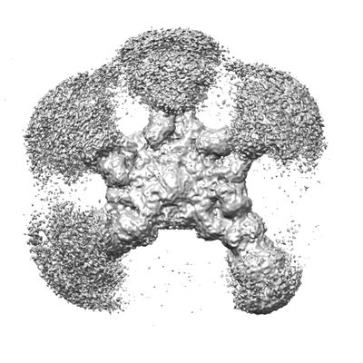 |
Cryo-EM structure of full-length human immunoglobulin M [43331 micrographs in MRC format] | Chen Q, Rosenthal PB, Tolar P [Pubmed: 36274064] [DOI: 10.1038/s41467-022-34090-2] |
2.6 TB | 4.4 Å |
| 2016-11-10 | 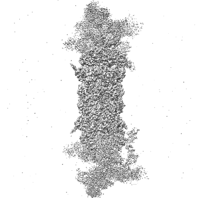 |
Structural basis for dynamic regulation of the human 26S proteasome [multiple data sets in SPIDER and MRC formats] | Chen S, Wu J, Lu Y, Ma YB, Lee BH, Yu Z, Ouyang Q, Finley D, Kirschner MW, Mao Y [Pubmed: 27791164] [DOI: 10.1073/pnas.1614614113] |
2.0 TB | 3.8 - 8.0 Å |
| 2020-03-20 | 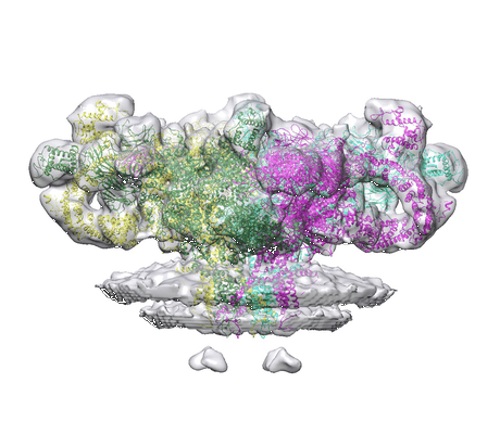 |
Cryo Electron Tomograms of Membrane Fractions of Rabbit Skeletal Muscle for Structural Determination of RyR1 in SR Vesicles [82 tilt series in MRC format] | Chen W, Kudryashev M [Pubmed: 32147968] [DOI: 10.15252/embr.201949891] |
254.2 GB | 12.6 - 38.0 Å |
| 2020-12-09 | 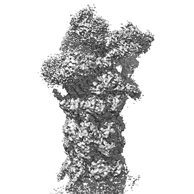 |
SA-like and SD-like states of human 26S Proteasome with non-cleavable M1-linked hexaubiquitin and E3 ubiquitin ligase E6AP/UBE3A [6216 micrographs in MRC format] | Chen X, Walters KJ [Pubmed: 32783951] [DOI: 10.1016/j.str.2020.07.011] |
330.3 GB | 4.1 - 5.7 Å |
| 2020-12-09 | 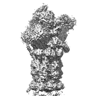 |
SA-like and SD-like states of human 26S Proteasome with non-cleavable M1-linked hexaubiquitin (dataset 2) [6220 micrographs in MRC format] | Chen X, Walters KJ [Pubmed: 32783951] [DOI: 10.1016/j.str.2020.07.011] |
330.2 GB | 5.8 - 5.96 Å |
| 2020-12-09 | 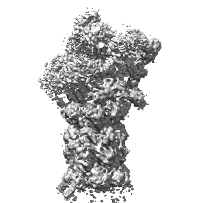 |
SA-like and SD-like states of human 26S Proteasome with non-cleavable M1-linked hexaubiquitin (dataset 1) [1645 micrographs in MRC format] | Chen X, Walters KJ [Pubmed: 32783951] [DOI: 10.1016/j.str.2020.07.011] |
87.3 GB | 6.47 - 6.75 Å |
| 2023-05-23 | 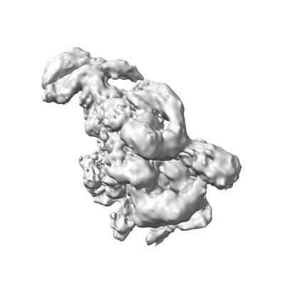 |
PIC-Mediator in complex with +1 nucleosome (T40N) [20082 micrographs in MRC format] | Chen X, Wang X, Liu W, Ren Y, Qu X, Li J, Yin X, Xu Y [Pubmed: 36201575] [DOI: 10.1126/science.abn8131] |
1.7 TB | 5.04 Å |
| 2023-02-01 | 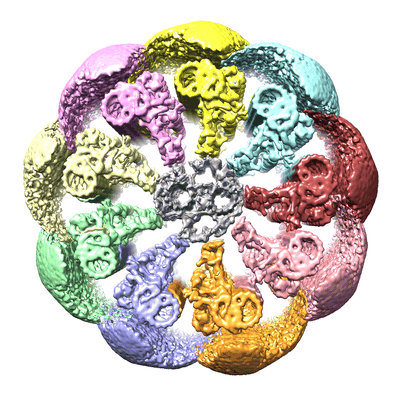 |
CryoET tilt series of mouse sperm flagella after FIB-SEM milling [69 tilt series in MRC format] | Chen Z, Greenan GA, Shiozaki M, Liu Y, Skinner WM, Zhao X, Zhao S, Yan R, Guo C, Yu Z, Lishko PV, Agard DA, Vale RD [Pubmed: 36593309] [DOI: 10.1038/s41594-022-00861-0] |
194.0 GB | 25.0 Å |
| 2024-02-16 | 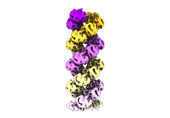 |
Cryo-EM reconstruction of the influenza A virus helical ribonucleoprotein-like [26515 multi-frame micrographs composed of 40 frames each in TIFF format] | Chenavier F, Ruigrok RWH, Schoehn G, Ballandras-Colas A, Crépin T [Pubmed: 38100595] [DOI: 10.1126/sciadv.adj9974] |
9.5 TB | 5.3 - 8.7 Å |
| 2022-11-23 | 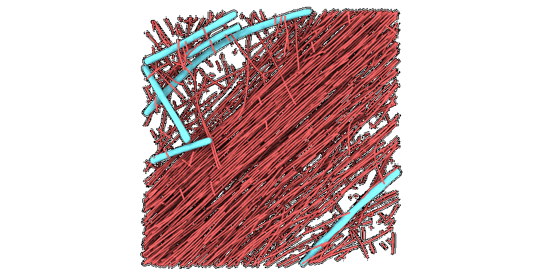 |
Cryo-electron tomograms of RPE1 cells with comprehensive annotation of actin filaments and microtubules [multiple data sets in TIFF and MRC formats] | Cheng DWC, Goetz SK, Mahamid J [Pubmed: 36690741] [DOI: 10.1038/s41592-022-01746-2] |
32.9 GB | — |
| 2015-09-01 | 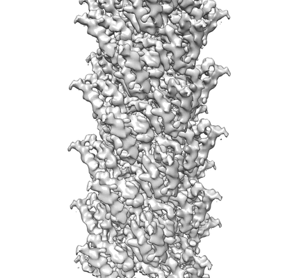 |
New movie data for MAVS CARD C1 filaments [512 multi-frame micrographs composed of 16 frames each in MRC format] | Chew PL, Ng TS, Lok SM, Xu H, He X, Zheng H, Huang LJ, Hou F, Yu Z, de la Cruz MJ, Borkowski B, Zhang X, Chen ZJ, Jiang QX [Pubmed: 26314863] [DOI: 10.7554/eLife.07546] |
512.1 GB | 4.2 Å |
| 2023-07-25 | 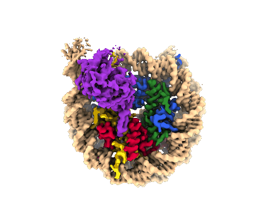 |
Cryo-EM structure of the human Sirtuin 6-nucleosome complex [11872 multi-frame micrographs composed of 40 frames each in TIFF format] | Chio US, Rechiche O, Bryll AR, Zhu J, Leith EM, Feldman JL, Peterson CL, Tan S, Armache JP [Pubmed: 37058572] [DOI: 10.1126/sciadv.adf7586] |
6.0 TB | 3.07 Å |
| 2023-03-15 |  |
Performing Correlative Light and Electron Microscopy to reveal the structural organization and location of alpha-synuclein aggregation hotspots inside the neuron. [multiple data sets in DM4 and TIFF formats] | Choi ML, Chappard A, Singh BP, Maclachlan C, Rodrigues M, Fedotova E, Berezhnov AV, De S, Peddie C, Athauda D, Viridi GS, Zhang W, Evans JR, Wernick A, Zanjani ZS, Angelova PR, Esteras N, Vinikurov A, Morris K, Jeacock K, Tosatto L, Little D, Gissen P, Collinson L, Clarke DJ, Kunath T, Klenerman D, Abramov AY, Horrocks MH, Gandhi S [DOI: 10.1101/2022.06.07.494932] |
88.4 GB | — |
| 2023-02-06 |  |
D-cycloserine and glutamate bound human GluN1a-GluN2C NMDA receptor [15350 multi-frame micrographs composed of 30 frames each in TIFF format] | Chou TH [Pubmed: 36309015] [DOI: 10.1016/j.molcel.2022.10.008] |
2.9 TB | 3.71 - 3.96 Å |
| 2023-01-31 |  |
PYD-106 bound human GluN1a-GluN2C NMDA receptor in the presence of D-cycloserine and glutamate [11714 multi-frame micrographs composed of 30 frames each in TIFF format] | Chou TH [Pubmed: 36309015] [DOI: 10.1016/j.molcel.2022.10.008] |
2.1 TB | 3.72 - 4.19 Å |
| 2023-02-06 |  |
D-cycloserine and glutamate bound human GluN1a-GluN2C NMDA receptor in nanodisc [4714 multi-frame micrographs composed of 30 frames each in TIFF format] | Chou TH [Pubmed: 36309015] [DOI: 10.1016/j.molcel.2022.10.008] |
1007.2 GB | 3.28 - 3.75 Å |
| 2023-02-06 | 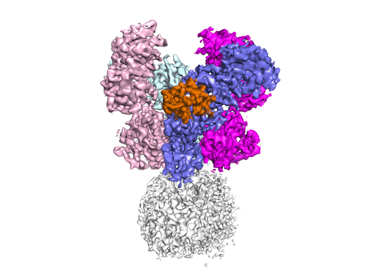 |
Human GluN1a-GluN2A-GluN2C triheteromeric NMDA receptor in complex with a nanobody [16468 multi-frame micrographs composed of 30 frames each in TIFF format] | Chou TH [Pubmed: 36309015] [DOI: 10.1016/j.molcel.2022.10.008] |
2.8 TB | 4.24 Å |
| 2023-07-11 | 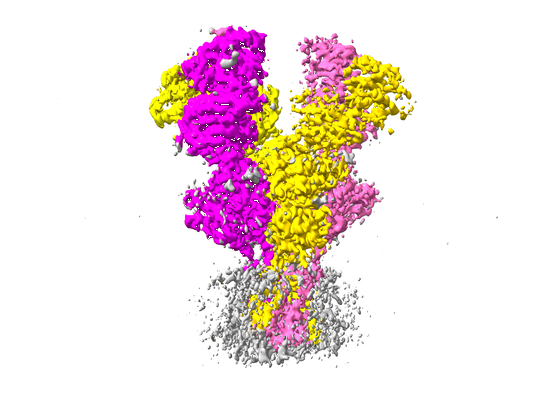 |
Movies of Glycine/Glutamate bound GluN1a-GluN2D NMDAR [17718 multi-frame micrographs composed of 30 frames each in TIFF format] | Chou TH, Kang H, Simorowski N, Traynelis SF, Furukawa H [Pubmed: 36309015] [DOI: 10.1016/j.molcel.2022.10.008] |
3.5 TB | 3.38 Å |
| 2022-11-14 | 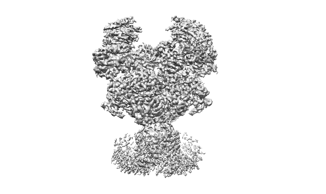 |
Glycine and glutamate bound GluN1a-GluN2B NMDA receptors in non-active 1 conformation at 2.97 Angstrom resolution [7942 multi-frame micrographs composed of 30 frames each in TIFF format] | Chou THC, Furukawa FH [Pubmed: 35637422] [DOI: 10.1038/s41594-022-00772-0] |
1.4 TB | 2.97 Å |
| 2022-12-09 | 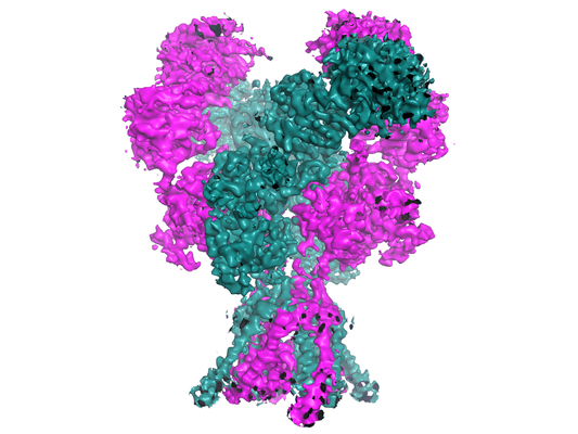 |
S-(+)-ketamine bound GluN1a-GluN2B NMDA receptors at 3.69 Angstrom resolution [multiple data sets in TIFF format] | Chou THC, Furukawa FH [Pubmed: 35637422] [DOI: 10.1038/s41594-022-00772-0] |
2.3 TB | 3.69 Å |
| 2022-11-14 | 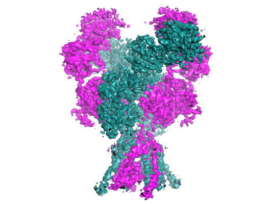 |
Memantine-bound GluN1a-GluN2B NMDA receptors [3657 multi-frame micrographs composed of 30 frames each in TIFF format] | Chou THC, Furukawa FH [Pubmed: 35637422] [DOI: 10.1038/s41594-022-00772-0] |
721.7 GB | 3.96 Å |
