Electron Microscopy Public Image Archive
Electron Microscopy Public Image Archive
The EMPIAR-PDBj team at Osaka University assists Asian EM researchers with the transfer of big EM image data to EMPIAR. Instead of sending the data directly to the EBI (UK) via the internet, hard drives can also be sent to Osaka University by postal mail or via a courier service. As an alternative, internet transfer to our server in Osaka is also available. If you would like to take advantage of our submission services, please contact us first by e-mail before sending the data to us.
| Release date | Imageset | Title | Authors and references | Size | Resolution |
|---|---|---|---|---|---|
| 2020-09-28 |  |
Single-particle cryo-EM of the human CDK-activating kinase in complex with THZ1 [4453 multi-frame micrographs composed of 69 frames each in TIFF format] | Greber BJ, Perez-Bertoldi JM, Lim K, Iavarone AT, Toso DB, Nogales E [Pubmed: 32855301] [DOI: 10.1073/pnas.2009627117] |
2.4 TB | 3.3 Å |
| 2023-08-18 |  |
Single particle cryo-EM dataset of Mus musculus mitochondrial complex I [2674 multi-frame micrographs composed of 40 frames each in TIFF format] | Grba DN, Chung I, Bridges HR, Agip AA, Hirst J [Pubmed: 37531432] [DOI: 10.1126/sciadv.adi1359] |
1.1 TB | 2.39 Å |
| 2023-08-18 |  |
Single particle cryo-EM dataset of Mus musculus mitochondrial complex I bound with inhibitor Piericidin A [1200 multi-frame micrographs composed of 25 frames each in MRC format] | Grba DN, Chung I, Bridges HR, Agip AA, Hirst J [Pubmed: 37531432] [DOI: 10.1126/sciadv.adi1359] |
1.6 TB | 2.84 Å |
| 2023-08-11 |  |
Single particle cryo-EM dataset of Mus musculus mitochondrial complex I bound with inhibitor Piericidin A [1565 multi-frame micrographs composed of 40 frames each in MRC format] | Grba DN, Chung I, Bridges HR, Agip AA, Hirst J [Pubmed: 37531432] [DOI: 10.1126/sciadv.adi1359] |
960.0 GB | 2.84 Å |
| 2022-04-25 |  |
Single particle cryo-EM dataset of Mus musculus mitochondrial complex I bound with an acetogenin inhibitor [1283 multi-frame micrographs composed of 50 frames each in MRC format] | Grba DN, Blaza JN, Bridges HR, Agip AA, Yin Z, Murai M, Miyoshi H, Hirst J [Pubmed: 35063503] [DOI: 10.1016/j.jbc.2022.101602] |
3.3 TB | 3.4 Å |
| 2018-03-09 | 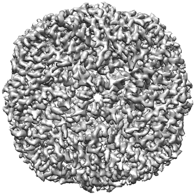 |
Apoferritin tutorial dataset for cisTEM [20 multi-frame micrographs composed of 50 frames each in MRC format] | Grant T, Rohou A, Grigorieff N [Pubmed: 29513216] [DOI: 10.7554/elife.35383] |
5.5 GB | — |
| 2023-02-15 | 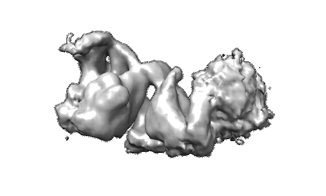 |
Structural basis of a transcription pre-initiation complex on a divergent promoter [51 tilt series in TIFF format] | Gorbea Colón JJ, Palao L, Chen SF, Kim HJ, Snyder L, Chang YW, Tsai KL, Murakami K [Pubmed: 36731470] [DOI: 10.1016/j.molcel.2023.01.011] |
151.3 GB | 26.0 Å |
| 2023-02-15 | 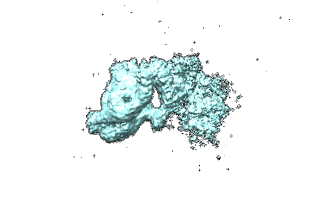 |
Structural basis of a transcription pre-initiation complex on a divergent promoter [55 tilt series in TIFF format] | Gorbea Colón JJ, Palao L, Chen SF, Kim HJ, Snyder L, Chang YW, Tsai KL, Murakami K [Pubmed: 36731470] [DOI: 10.1016/j.molcel.2023.01.011] |
105.8 GB | 36.0 Å |
| 2022-09-09 | 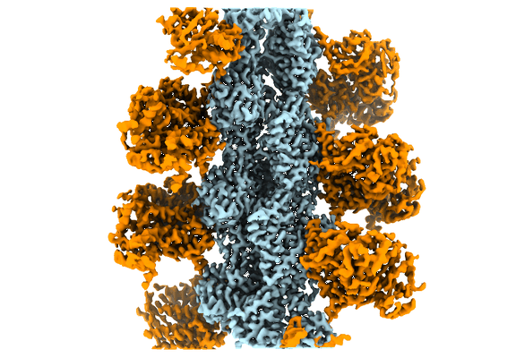 |
cryo-EM structure of the rigor state wild type myosin-15-F-actin complex [1485 multi-frame micrographs composed of 40 frames each in TIFF format] | Gong R, Bird JE, Alushin GM [Pubmed: 35857845] [DOI: 10.1126/sciadv.abl4733] |
506.7 GB | 2.83 - 3.17 Å |
| 2022-09-09 |  |
cryo-EM structure of the ADP state wild type myosin-15-F-actin complex [1624 multi-frame micrographs composed of 24 frames each in TIFF format] | Gong R, Bird JE, Alushin GM [Pubmed: 35857845] [DOI: 10.1126/sciadv.abl4733] |
313.4 GB | 3.63 - 4.15 Å |
| 2022-09-09 |  |
Cryo-EM structure of the rigor state Jordan myosin-15-F-actin complex [2641 multi-frame micrographs composed of 24 frames each in TIFF format] | Gong R, Bird JE, Alushin GM [Pubmed: 35857845] [DOI: 10.1126/sciadv.abl4733] |
519.5 GB | 3.76 - 4.18 Å |
| 2022-11-17 |  |
Defocus and Volta potential phase plate cryo-electron tomography of S. pombe cryo-FIB lamellae with comprehensive annotations of structures and macromolecules [multiple data sets in TIFF and MRC formats] | Goetz SK, Mahamid J [Pubmed: 36690741] [DOI: 10.1038/s41592-022-01746-2] |
305.5 GB | 9.3 - 34.0 Å |
| 2022-03-28 |  |
Cryo-EM structure of SARS-CoV-2 Main protease C145S in complex with N-terminal peptide [multiple data sets in MRCS and TIFF formats] | Godoy AS, Song Y, Noske GD, Oliva G | 3.3 TB | 3.5 Å |
| 2021-09-17 | 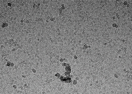 |
Cryo-EM structure of SARS-CoV-2 NSP15 NendoU at pH 6.0 [4080 micrographs in MRC format] | Godoy AS | 359.3 GB | 2.48 Å |
| 2016-11-18 | 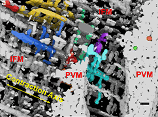 |
Focused Ion Beam-Scanning Electron Microscopy of mitochondrial reticulum in murine skeletal muscle [291 reconstructed volumes in MRC format] | Glancy B, Hartnell LM, Malide D, Yu ZX, Combs CA, Connelly PS, Subramaniam S, Balaban RS [Pubmed: 26223627] [DOI: 10.1038/nature14614] |
1.5 GB | — |
| 2021-05-07 | 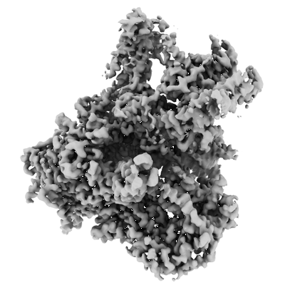 |
Cryo-EM structures of human RNA Polymerase III [multiple data sets in TIFF and MRCS formats] | Girbig M, Misiaszek AD, Vorlaender MK, Mueller CW [Pubmed: 33558764] [DOI: 10.1038/s41594-020-00555-5] |
3.3 TB | 2.8 - 3.4 Å |
| 2021-11-15 | 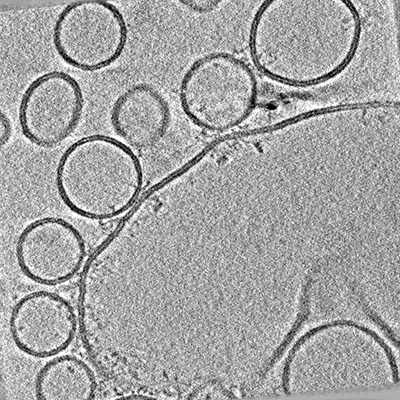 |
Arrangements of proteins at reconstituted synaptic vesicle fusion sites depend on membrane separation. [multiple data sets in MRC format] | Ginger L, Malsam J, Sonnen A.F.P., Morado D, Scheutzow A, Söllner T.H., Briggs J.A.G. [Pubmed: 32860428] [DOI: 10.1002/1873-3468.13916] |
70.0 GB | — |
| 2022-05-25 |  |
SARS-CoV-2 spike protein S:D614G + S:A222V variant [4841 micrographs in MRC format] | Ginex T, Marco-Marín C, Wieczór M, Mata CP, Krieger J, Ruiz-Rodriguez P, López-Redondo ML, Francés-Gómez C, Melero R, Sánchez-Sorzano CÓ, Martínez M, Gougeard N, Forcada-Nadal A, Zamora-Caballero S, Gozalbo-Rovira R, Sanz-Frasquet C, Arranz R, Bravo J, Rubio V, Marina A, Geller R, Comas I, Gil C, Coscolla M, Orozco M, Llácer JL, Carazo JM [Pubmed: 35816514] [DOI: 10.1371/journal.ppat.1010631] |
303.8 GB | 3.4 Å |
| 2022-01-10 |  |
Proton-powered peptide transporter SbmA in lipid nanodisc [7306 multi-frame micrographs composed of 40 frames each in TIFF format] | Ghilarov D, Inaba-Inoue S, Stepien P, Qu F, Michalczyk E, Pakosz Z, Nomura N, Ogasawara S, Walker GC, Rebuffat S, Iwata S, Heddle JG, Beis K [Pubmed: 34516884] [DOI: 10.1126/sciadv.abj5363] |
2.7 TB | 3.9 Å |
| 2021-11-02 |  |
Proton-powered peptide transporter SbmA in lipid nanodisc complexed with Fab S11-1 (SbmA-FabS11-1-MccB17) [7286 multi-frame micrographs composed of 40 frames each in TIFF format] | Ghilarov D, Inaba-Inoue S, Stepien P, Qu F, Michalczyk E, Pakosz Z, Nomura N, Ogasawara S, Walker GC, Rebuffat S, Iwata S, Heddle JG, Beis K [Pubmed: 34516884] [DOI: 10.1126/sciadv.abj5363] |
2.5 TB | 3.59 Å |
| 2021-07-30 |  |
Structure of human telomerase holoenzyme with bound telomeric DNA [43639 multi-frame micrographs composed of 48 frames each in TIFF format] | Ghanim GE, Fountain AJ, van Roon AM, Rangan R, Das R, Collins K, Nguyen THD [Pubmed: 33883742] [DOI: 10.1038/s41586-021-03415-4] |
11.2 TB | 3.4 - 6.6 Å |
| 2023-01-31 |  |
Cryo-EM structure of a delivery complex containing the SspB adaptor, an ssrA-tagged substrate, and the AAA+ ClpXP protease [9524 multi-frame micrographs composed of 40 frames each in TIFF format] | Ghanbarpour A, Fei X, Baker TA, Davis JH, Sauer RT [DOI: 10.1101/2022.11.06.515074] |
4.0 TB | 3.7 Å |
| 2024-01-15 |  |
Full-length ClpX AAA protein in a complex with ClpP peptidase [multiple data sets in TIFF and MRC formats] | Ghanbarpour A, Cohen SE, Fei X, Davis JH, Sauer RT [Pubmed: 37949857] [DOI: 10.1038/s41467-023-43145-x] |
1.9 TB | 3.12 Å |
| 2019-08-07 |  |
Identification of a druggable VP1-VP3 interprotomer pocket in the capsid of enteroviruses [2120 micrographs in MRC format] | Geraets JA, Flatt JW, Domanska A, Butcher SJ [Pubmed: 31185007] [DOI: 10.1371/journal.pbio.3000281] |
2.2 TB | 4.0 Å |
| 2023-11-06 |  |
Micrographs of ER-derived vesicles from HEK293F cells [893 multi-frame micrographs composed of 8 frames each in TIFF format] | Gemmer M, Chaillet ML, van Loenhout J, Cuevas Arenas R, Vismpas D, Gröllers-Mulderij M, Koh FA, Albanese P, Scheltema RA, Howes SC, Kotecha A, Fedry J, Förster F [Pubmed: 36697828] [DOI: 10.1038/s41586-022-05638-5] |
1.1 TB | 4.5 - 9.3 Å |
